Intraoperative video consultation expedites and improves surgical care. Here, we present a case where intraoperative video consultation between a rural hospital and an academic hepatobiliary surgeon facilitated immediate diagnosis of bile duct transection during laparoscopic cholecystectomy, direct to operating room transfer between centers, and injury repair with robotic-assisted Roux-en-Y hepaticojejunostomy.
Case Report
Intraoperative Video Consultation Following Bile Duct Transection Facilitates Direct OR Transfer for Robotic Hepaticojejunostomy at Tertiary Center
In This Article
Summary
Abstract
Bile duct injuries during cholecystectomy are most frequently attributed to misidentification of surgical anatomy. Initial management following a major bile duct injury is time-sensitive and critical to the patient's clinical course and overall outcome. We present a 63-year-old male patient who sustained a common bile duct transection during laparoscopic cholecystectomy at a resource-limited rural surgery center. Upon recognition of the injury, an intraoperative video consultation was made to hepatobiliary surgery at our academic referral institution, providing a real-time explanation of the dissection with direct visualization of surgical anatomy. This expedited sequence facilitated acquisition of diagnostic studies, including an intraoperative cholangiogram, confirming common bile duct transection, and a CT angiogram, demonstrating intact arterial anatomy. Direct to OR transfer to the tertiary facility was therefore prioritized, circumventing a prolonged bed wait the patient would have otherwise required while awaiting injury diagnosis and characterization. He underwent definitive repair with robotic-assisted Roux-en-Y hepaticojejunostomy on the same day that the injury occurred. This paper provides an operative video with a concise and adoptable method for performing a definitive hepaticojejunostomy following bile duct transection intended for fellowship-trained hepatobiliary surgeons.
Introduction
Cholecystectomy is a standard surgical procedure used to treat cholecystitis and cholelithiasis. Complications from cholecystectomy are usually rare but significant, including iatrogenic bile duct injury (BDI). BDI has historically been more likely to occur when the procedure is done laparoscopically1,2,3, though with increased experience in laparoscopic surgery over the past several decades, the incidence has gradually decreased and approached that of open cholecystectomy. In fact, recent reports have shown that the incidence of BDI is only 0.08% of laparoscopic cases4. These injuries typically occur when surgeons misidentify anatomical structures. Most commonly, the bile duct is mistakenly identified as the cystic duct5,6. Multiple classification systems are used to describe BDIs, including Strasberg-Bismuth7, McMahon8, and Stewart-Way9.
Surgical repair is critical for all major BDIs and frequently necessary for minor BDIs. BDIs requiring surgical repair are associated with 20.8% all-cause mortality, an 8.8% increase compared to the expected death rate10. They also affect quality of life beyond physical health, with noted negative psychological and emotional impact on patients11. Additionally, they can result in costly legal cases that are detrimental to both the patient and the surgeon12. It is crucial to do everything possible to avoid iatrogenic BDI during cholecystectomy. However, when it does occur, the World Society of Emergency Surgery (WSES) has published guidelines for management based on the timing of injury recognition -- intraoperatively versus postoperatively13. Regardless of the timing of BDI identification, it is recommended that the patient be managed by a center with hepatobiliary expertise, even if this requires the patient to be transferred to another facility. This clearly improves patient outcomes, as the success rate for BDI repairs is only 21% when performed by the primary surgeon, compared to 95% effectiveness when performed by a hepatobiliary surgeon14.
Not all geographical areas have access to a specialized hepatobiliary surgeon. Rural areas are especially limited in their availability of specialist care15. Telemedicine became ubiquitous in the wake of the COVID-19 pandemic and continues to improve connectivity in healthcare. Such approaches provide convenient, efficient care that reduces the burden of travel on patients and allows them access to a broader range of providers16. The use of telemedicine has also expanded to include consultation between clinicians in surgery. Remote intraoperative consultation, albeit a rare phenomenon, provides surgeons with expert opinion in real time. This has improved surgical care in a range of procedures, including laparoscopic cholecystectomy, during which surgeons are able to receive assistance in identifying critical biliary structures, thus reducing the chance of BDI17. Resource-limited rural surgeons especially stand to benefit most from this strategy, with 78% agreeing that telemedicine would be beneficial to their practice, through both acquisition of new skills and assistance when surgical complications arise18. Distinctly lacking from published literature are reports in which an iatrogenic injury occurred, and expert consultation was sought remotely for real-time video assessment and intraoperative management decisions. Relevant to this case, it has been shown that patients have improved quality of life after BDI if the injury is identified intraoperatively, repaired by a hepatobiliary specialist, and repaired quickly19,20. This is not always possible in rural centers. Thus, identification of strategies that support early detection, the presence of hepatobiliary expertise, and rapid repair in rural settings remains a crucial knowledge gap. The presented case supports intraoperative video consultation as an unrealized method for addressing this gap.
CASE PRESENTATION:
A 63-year-old male presented with a 3 month history of episodic right upper quadrant abdominal pain radiating to his right side. The pain typically occurred in the hours following meals, especially those with greasy and fatty foods. It was accompanied by nausea with occasional emesis. His past medical history included prior smoking (35 pack years), GERD, type 2 diabetes mellitus, chronic obstructive pulmonary disease, hyperlipidemia, hypertension, hypercholesterolemia, coronary artery disease, and non-ST elevation myocardial infarction with angioplasty and stent placement four years prior. Upon workup, his white blood cell count, hepatic function, and electrolytes were found to be within normal range. Imaging by CT and a prior ultrasound revealed evidence of cholelithiasis without cholecystitis and a common bile duct measurement of 5.5 mm. Given this, he elected to undergo a laparoscopic cholecystectomy at a rural hospital. Intraoperatively, the gallbladder was noted to be inflamed and contracted, with omental adhesions to the anterior surface. The triangle of Calot was exposed, and a tubular structure thought to be the cystic duct was circumferentially dissected and clipped. As dissection progressed proximally toward the fundus of the gallbladder, a very short structure suspected to be the patient's true cystic duct was then identified. The surgeon became concerned with iatrogenic transection of the common bile duct.
Diagnosis, Assessment, and Plan:
Upon recognition of a transected biliary structure, the operating surgeon sought an intraoperative consult with a hepatobiliary surgeon at our academic hospital. Via a video call, the consultant was shown the bile duct anatomy and discussed the case with the operating surgeon in real time. To confirm the suspected bile duct transection, an intraoperative cholangiogram was recommended. A Reddick catheter was used to cannulize the transected bile duct, and radio-opaque dye was injected. Cholangiogram confirmed the presence of a common bile duct transection 3 cm from the bifurcation (Figure 1). At this point, the operating surgeon completed the cholecystectomy but was advised not to perform a laparotomy in doing this. Clips were placed on the distal portion of the bile duct, and the proximal portion was left open. Placement of an abdominal drain and transfer of the patient to our academic center were recommended. However, this case occurred during the COVID-19 pandemic, and our academic hospital was at maximum capacity, with a 3-day bed wait for direct admissions. In addition, the rural setting of the operating hospital did not allow for immediate availability of ambulance transport. Given this, an abdominal CT angiogram was obtained, confirming no aberrant arterial anatomy or injury. Because a complete workup was completed during the intraoperative consult, the patient was able to be admitted as a direct transfer to the operating room at our academic hospital for robotic Roux-en-Y-hepaticojejunostomy.
Protocol
Per West Virginia University Office of Human Research Protections protocols and consistent with federal policy, this protocol is classified as non-human subjects research and does not require Institutional Review Board approval. Patients provided informed consent for intra-operative video recording as part of our standard surgical consent process.
1. Patient positioning and port placement
- The patient was positioned supine on a split leg table with his arms out. Sterile preparation and surgical drapes were applied in standard fashion. An incision in the left upper quadrant was made to allow for abdominal entry with an optical separator device. CO2 was insufflated and the peritoneal cavity explored. The previously placed abdominal drain was removed at this time.
- Four robotic ports were positioned in the upper abdomen, and two assistant ports were placed in the lower abdomen (Figure 2). The 12-mm port in the left lower quadrant was used for inserting the stapler.
- Once the ports were in place, the patient was positioned in reverse Trendelenburg with the right side elevated.
2. Identifying the transected biliary structure and docking the robot
- The liver was retracted cranially using a flexible right flank liver retractor and the robot was docked. The surgeon sat at the da Vinci Xi console, and a bedside laparoscopic assistant stood between the patient's legs.
- The recent cholecystectomy site was evaluated, and the transected biliary structure was identified (Figure 3, left). The distal bile duct stump had two laparoscopic clips placed at the time of prior cholecystectomy and was oversewn using 2-0 silk figure-of-eight sutures. The transected proximal duct was then identified and trimmed with cold scissors with active bleeding at the cut edge (signifying appropriate perfusion for anastomotic healing).
3. Division of the jejunal loop and its passage through the retrocolic tunnel
- The ligament of Treitz was identified, and the proximal jejunum was followed 40 cm distally, where a loop of jejunum was identified as the segment designated for the future anastomosis with the bile duct.
- A mesenteric window was created using a vessel-sealing device, and the bowel was divided with a 60-mm purple load Endo GIA stapler. The vessel-sealing device was then used to divide the mesentery, ensuring avoidance of mesenteric vessels.
- A retrocolic tunnel was fashioned within the transverse colon mesentery using blunt dissection. The Roux limb was then guided through this tunnel into the lesser sac, and the jejunal segment reached the anastomotic site without tension.
4. Creation of running end-to-side, duct-to-mucosa hepaticojejunostomy
- Using cauterized scissors, an enterotomy was created in the Roux limb at the site of the anastomosis. Two 4-0 V-Loc sutures were then anchored at the corners of the enterotomy on the anti-mesenteric aspect of the jejunum.
- The hepaticojejunostomy anastomosis was then performed in an end-to-side, duct-to-mucosa fashion as each V-Loc suture was run along either side of the enterotomy. A 4-French Hobbs stent was placed into the duct to facilitate reconstruction and ensure patency during creation of the anastomosis. The sutures were tied together at completion of the anastomosis (Figure 3, right).
5. Re-establishing GI continuity with jejunojejunostomy
- The reconstructed gastrointestinal anatomy was verified by tracing the Roux and biliopancreatic limbs. Small enterotomies were created near the previously stapled end of the jejunum and at a point roughly 50 cm distal along the Roux limb. A 60-mm purple load Endo GIA stapler was then used to form the side-to-side jejunojejunostomy.
- The common enterotomy was closed with 3-0 V-Loc sutures, followed by interrupted 3-0 silk Lembert sutures. The 3-0 silk sutures were also used to close the mesenteric defect in the colon created around the Roux limb.
6. Placement of drains and abdominal closure
- A 19-French round channel drain was placed using a previous robotic port-site. The drain was passed into right upper quadrant coursing anterior to the hepaticojejunostomy. The falciform was then placed around the anastomosis to act as a vascularized flap. The liver retractor was then removed.
- The abdomen was extensively irrigated with saline. The 12-mm assistant port site was closed with #1 Vicryl using a laparoscopic port-site closure device. The skin and subcutaneous tissues were subsequently irrigated and closed with 4-0 Monocryl.
Results
The patient underwent a successful Roux-en-Y-hepaticojejunostomy for BDI repair. The completed end-to-side, duct-to-mucosa running hepaticojejunostomy anastomosis is highlighted in the right panel of Figure 3. Due to intraoperative video consultation allowing for direct-to-OR transfer, this patient avoided a long wait time for admission to our tertiary center. BDI repair at our center was completed just 10 h after closure of the patient's initial cholecystectomy at the rural hospital (Figure 4). His post-operative course is summarized in Table 1. He tolerated the Roux-en-Y-hepaticojejunostomy well with no immediate complications. On postoperative day 3, he became febrile and was treated with intravenous antibiotics. He improved and was switched to oral antibiotics on the day of discharge, post-operative day five. He was followed up 2 weeks later with no apparent surgical complications, and his postprandial pain episodes had resolved. Six months postoperatively, he unfortunately presented with recurrent post-prandial pain, elevated white count, and CT showing a 10 x 5 cm perihepatic/subcapsular abscess. It was treated with percutaneous CT-guided drain and antibiotics. The drain was removed 2 weeks later with no further infectious sequela. Additionally, he was found to have an elevated hepatic function panel (HFP) (alkaline phosphatase 288 U/L, alanine aminotransferase [ALT] 79 U/L, and aspartate aminotransferase [AST] 37 U/L) 14 months postoperatively associated with postprandial pain episodes. An endoscopic retrograde cholangiopancreatography (ERCP) with push enteroscopy into the afferent jejunal limb was performed, noting a short stricture at the hepaticojejunostomy anastomosis. This was treated with covered metal stent placement. On repeat ERCP 3 months later, it was found that the stent had spontaneously migrated with air seen on cholangiogram, suggestive of patent anastomosis, and HFP began to trend downward (alkaline phosphatase 214 U/L, ALT 43 U/L, and AST 38 U/L). The patient is now over 3 years postoperative and is doing well with no further symptoms or additional stent placement.
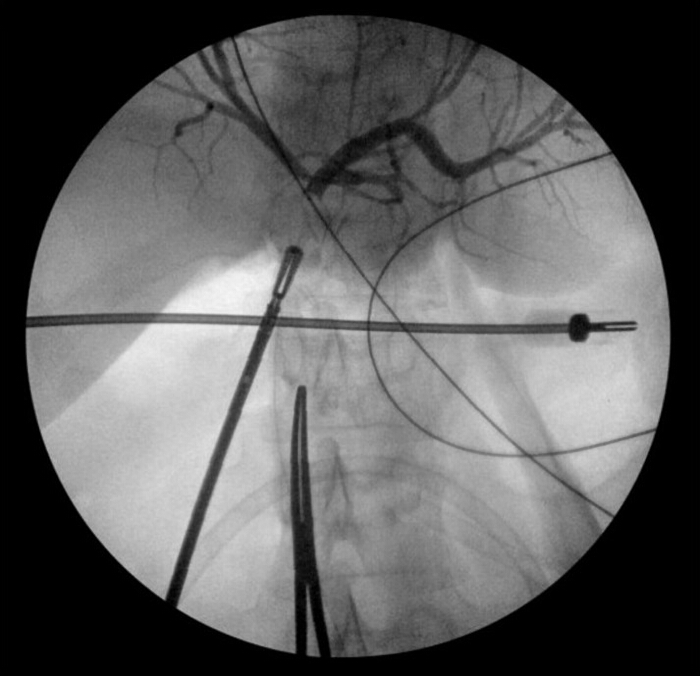
Figure 1: Diagnostic cholangiogram. Cholangiogram taken at the time of intraoperative video consultation. Confirms duct transection just distal to the hepatic bifurcation with abrupt cut-off of injected contrast dye. Please click here to view a larger version of this figure.
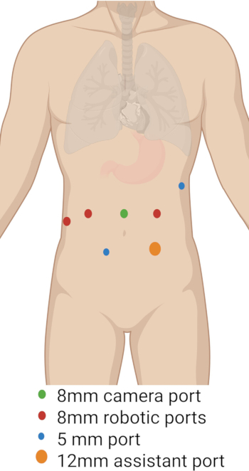
Figure 2: Suggested port layout for robotic-assisted hepaticojejunostomy. Four supraumbilical robotic ports were positioned (green and red dots), with the camera placed at the midline port. Two infraumbilical assistant ports were added, including a larger 12 mm port (orange dot) designated for laparoscopic stapler insertion. Please click here to view a larger version of this figure.

Figure 3: Representative images during robotic-assisted hepaticojejunostomy. Left: Transected common bile duct identified seen after entry into the abdomen. Right: Completed end-to-side, duct-to-mucosa running hepaticojejunostomy anastomosis. Please click here to view a larger version of this figure.
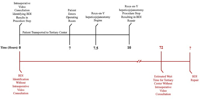
Figure 4: Timeline of BDI repair with and without intraoperative video consultation. The timeline of BDI repair by Roux-en-Y hepaticojejunostomy after intraoperative video consultation (black) compared to the estimated timeline of BDI repair without intraoperative video consultation (red). Please click here to view a larger version of this figure.
| Time from Intra-Opeartive Consultation to Complete Roux-en-Y-hepaticojejunostomy Following Transfer, Hours | 10 |
| Technical Outcomes | Creation of end-to-side, duct-to-mucosa hepaticojejunostomy. Refer to Figure 3 |
| Length of Stay, Days | 6 |
| Duration of JP drain, Days | 6 |
| Perioperative Complications (within 90 days) | Bacteremia on post-operative day 3 requiring 7-day course of antibiotics |
| Need for Revision Surgery | None |
| Need for Advanced Procedural Intervention | *CT-guided drain placement fro 10 x 5 cm peri-hepatic collection (6 months post-operative) *Covered metal stent placement for short segment stricture at hepaticojejunostomy anastomosis (14 months post-operative); patency of anastomosis noted on repeat ERCP (17 months post-operative) |
| Months Post-Operative | 36 |
| Mortality | N/A |
Table 1: Patient outcomes after Roux-en-Y-hepaticojejunostomy.
Discussion
Here we present the case of a patient who experienced an iatrogenic common bile duct transection during a routine laparoscopic cholecystectomy for cholelithiasis at a rural resource-limited institution. If the hospital had hepatobiliary expertise, direct repair or reconstruction with Roux-en-Y hepaticojejunostomy would have been indicated for this major BDI (based on Strasberg classification) discovered intraoperatively. No hepatobiliary surgeons were available at the operating hospital to assist in the repair of the BDI. In this situation, WSES guidelines suggest drain placement and referral to another center with hepatobiliary experience within 0-48 h13. However, given the unique circumstances, as this case occurred during the COVID-19 pandemic, hospital bed availability was scarce. The patient would have either had to wait multiple days for admission to a tertiary institution or be treated with limited resources at the operating facility without advanced gastrointestinal capabilities. Fortunately, telemedicine changed the course of this case.
In terms of reconstructive outcomes, there are many considerations for the creation of the hepaticojejunostomy anastomosis, and the approach should be made on an individualized basis and based on the surgeon's experience. We performed a continuous anastomosis utilizing barbed suture. Although several suture types (e.g., V-loc and polydioxanone [PDS]) have been described, there is a lack of literature directly comparing outcomes. We favor barbed sutures as they allow for a greater amount of tension to be maintained on the suture during the anastomosis creation. In an effort to mitigate the risk of occlusion by inadvertently "backwalling," we elect to place a 4-french plastic stent during suturing, particularly when the duct is small. One study found that when a stent was used, there was no difference in the rate of anastomotic stricture, although further study is warranted to better understand outcomes when an intraductal stent is employed21. Another alternative approach to consider is performing an interrupted suturing technique during anastomosis creation. Multiple recent randomized trials and one meta-analysis have shown no difference in overall biliary complications, including bile leak, biliary stricture, cholangitis, or liver abscess22,23,24. Consistently reported, however, are statistically significant increased material costs and operative time when interrupted suturing is performed.
The incidence of anastomotic stricture in the literature is relatively common, with many citing 20%, but has been reported as high as 69%25. One meta-analysis of 17 studies found concomitant vascular injury (OR 4.96; p = 0.001), post-repair bile leak (OR 8.03; p = 0.003), and repair by a nonspecialist surgeon (OR 11.29; p < 0.0001) are predictors of anastomotic stricture26. Interestingly, in patients undergoing pancreaticoduodenectomy, multiple studies have shown smaller duct size to be predictive of anastomotic stricture. One retrospective cohort study analyzing 241 operative videos found that the duct size of ≤10 mm was 12x more likely to develop stricture and/or cholangitis than larger ducts (p = 0.018)21. Another retrospective study of 103 patients found that duct diameter <6 mm was significantly more likely to stricture than those ≥6.0 mm (25.9% vs 1.3%, p < 0.01)27. Our patient was unfortunately diagnosed with a short-segment stricture without cholangitis in the setting of abdominal pain and elevated liver enzymes, requiring ERCP with covered metal stent placement. The anastomosis was found to be patent on follow-up endoscopic assessment with improved hepatic function studies. He is monitored clinically and biochemically without evidence of recurrence.
Telemedicine has existed in various forms since the inventions of the telephone and radio made it possible for clinicians to provide medical advice to distant colleagues and patients28. Since that time, it has expanded to include a wide array of modalities. Noted benefits of telemedicine for patients include access to clinicians in distant locations, avoidance of spreading communicable disease, and reduced expenses accruing from travel, childcare, or lost work29. Telemedicine is limited in its uses; however, in some cases, due to its lack of in-person care. Despite limitations, telemedicine spiked during the COVID-19 pandemic. This trend was also seen specifically in surgical specialties, as one review concluded that surgical specialties heavily utilized and published articles about the use of telemedicine during this time30. Most commonly, telemedicine is useful for surgical patients who live too far to easily access post-surgical clinic visits.
Another modality of telemedicine, intraoperative consultation, is utilized less commonly in surgery. In the case presented here, intraoperative video consultation uniquely enabled a hepatobiliary surgeon at our tertiary institution to directly visualize the transected bile duct and provide management recommendations in real time. Once BDI was confirmed with a cholangiogram, it became clear that expedited transfer to our academic center was necessary for optimal management. The consultant recommended against converting to a laparotomy and advised the operating surgeon to place an abdominal drain to limit intra-abdominal contamination. This decision was made so that reconstruction with a hepaticojejunostomy could be performed with a robotic approach. While waiting for OR transfer, a CT angiogram was recommended to rule out the possibility of vascular injury. Collectively, these recommendations allowed the patient to be a candidate for direct to operating room (OR) transfer, a process which has long been known to reduce morbidity and mortality in a range of severely injured patients31. A quick referral was crucial, as a late referral to a tertiary center after BDI is an independent prognostic factor for a worse outcome32. Alternatively, had consultation been delayed until after the operation was completed, the initial focus would have been on confirming and characterizing the injury. A several-day wait for a hospital bed would have ensued, and ERCP or percutaneous transhepatic cholangiography with interventionalist colleagues would have further contributed to the delay. This undoubtedly would have placed the patient outside the window in which early reconstruction was favorable due to ongoing intraabdominal contamination and post-surgical inflammation. Overall, this case showcases the value of a relatively novel use of telemedicine via intraoperative consultation. To our knowledge, there has been only one other case reported in which real-time intraoperative, suggesting the unrealized potential to improve patient care in these difficult circumstances33.
Finally, the rural area where this BDI occurred played a significant role in the case. It took place in West Virginia, where the 2020 United States Census reported 55.4% of people live in rural areas, making it the third most rural state34. Fifty-one of West Virginia's 55 counties are considered Health Professions Shortage Areas (HPSAs) or Medically Underserved Areas (MUAs), causing patients to have to travel great distances to receive care35. Thus, it is unsurprising that a facility and surgeon with hepatobiliary expertise were locally unavailable to the patient. This limited access to healthcare is also a likely contributor to West Virginia's status as one of the unhealthiest states. These combined factors place West Virginians in a difficult position in which they are more likely to have a high burden of comorbidities and less likely to access care. Though a multifactorial problem requiring many solutions, telemedicine can ease this burden for patients and local physicians by providing rapid access to care and peer consultation, as occurred in this case. One study in which specialists remotely telementored or telerobotically assisted colorectal surgeries at rural hospitals concluded that this was an effective method for providing community surgeons with advanced training in surgical techniques and providing immediate management advice if surgical complications arose. This further increased surgical confidence and breadth of surgical procedures performed at community hospitals36. Broader implementation of similar strategies would likely benefit all rural areas.
This case provides a unique example of how excellent communication across surgeons in different rural areas led to the rapid discovery and repair of a complete bile duct transection during laparoscopic cholecystectomy. It highlights the benefits of telemedicine, specifically intraoperative video consultation. Here, consultation resulted in an expedited intraoperative patient "workup" that allowed for direct to OR transfer, which would not have been possible otherwise due to the COVID-19 pandemic. This case also emphasizes how intraoperative consultations can be especially beneficial to rural hospitals that may not have access to a wide range of surgical specialists. Together, this unique sequence of events and multi-hospital interprofessional collaboration resulted in efficient repair of the patient's BDI.
Disclosures
The authors have no conflicts of interest to disclose.
Acknowledgements
Figure 2 was created in BioRender. Sestito. (2025) https://BioRender.com/r46u530
Materials
| Name | Company | Catalog Number | Comments |
|---|---|---|---|
| #1 Vicryl Suture | Medtronic | CL-64-M | |
| 0 Vicryl Ties | Ethicon | J646H | |
| 2-0 Silk Suture - Cut to 8" | Ethicon | K833-H | |
| 3-0 V-LOC V-20 6" x 3 | Covidien | VLOCM0604 | |
| 4-0 Monocryl | Ethicon | Y496G | |
| 4-0 Prolene on RB 1 | Ethicon | 8557H | |
| 4-0 V-Loc CV-23 6" x2 | Covidien | VLOCM0024 | |
| 5-0 Polydioxanone RB-2 Cut to 5" | Ethicon | Z148-H | |
| Banded bag 28" x 36" | McKesson | 13628 | Cover non-sterile equipment in sterle field |
| BD ChloraPrep Hi-Lite Orange 26-mL applicator with sterile solution | BD (Becton, Dickinsone and Company) | 930815 | Sterile Chlorhexidine preparation |
| Bladeless optical trocar, 5 mm size, 100 mm length | Covidien | ONB5STF | |
| Bladeless Trocar, 12 mm size, 100 mm length | Covidien | NONB12STF | |
| Bladeless Trocar, 15 mm size, 100 mm length | Covidien | NB15STF | |
| Bladeless Trocar, 5 mm size, 100 mm length | Covidien | NONB5STF | |
| BLAKE Silicone Drain, Size 19 Fr, Round | Ethicon | 2232 | Abdominal drain |
| Cadiere | Intuitive Surgical | 471049 | |
| Clean and protect laparoscope lense | McKesson | 21345 | |
| Coaxial umbilical cable | Medtronic | 203CXC | |
| Da Vinci XI Arm Drape | Intuitive Surgical | 470015 | Sterile drape for robotic arms |
| Da Vinci XI Column Drape | Intuitive Surgical | 470341 | Sterile drape for bedside robot |
| Da Vinci XI Universal Seal 5-12 mm | Intuitive Surgical | 470500 | Robotic Trocar seal |
| Endo Floating Ball Electrodes | Medtronic | 122011 | Monopolar electrocautery device |
| Endo GIA Ultra Universal Stapler | Covidien | EGIAUXL | Laparoscopic stapler |
| Endoscopic surgery swab | Covidien | 173019 | Swab fluid and assist with blunt dissection |
| Exofin Skin Adhesive | McKesson | EX71010 | Skin glue |
| Fenestrated Bipolar | Intuitive Surgical | 471205 | |
| First Entry Access System, 5 x 100 mm | Applied Medical | CTF03 | |
| Garment compress Medium, Calf | Zimmer Biomet | VG501M | Sequential compression device applied to calves for deep venous thrombosis prophylaxis |
| Hook electrocautery | Intuitive Surgical | 470183 | |
| Jackson-Pratt bulb reservoir 100 cc | McKesson | SU1301305 | Drainage suction bulb reservoir |
| Laparoscopic Blade Electrode, 6.5" | Coviden | E14506 | Laparoscopic electrocautery blade |
| Laparoscopic Clip applier 10mm | Covidien | 176657 | Laparoscopic clip applier |
| Laparoscopic Clip applier 5mm | Covidien | 176630 | Laparoscopic clip applier |
| Laparoscopic electrode Flat L-hook | Covidien | E3774-36C | Laparoscopic electrocautery hook |
| Laparoscopic sealer/divider, Maryland, curved Jaw | Covidien | LF1944 | |
| Laparotomy sponge, 4" x 18", X-ray and RF-Detectable | Medtronic | L041804P01C1 | |
| Large needle driver | Intuitive Surgical | 471006 | |
| Large SutureCut needle driver | Intuitive Surgical | 471296 | |
| Maryland Bipolar | Intuitive Surgical | 471172 | |
| Monopolar Curved scissor tip cover accessory | Intuitive Surgical | 400180 | |
| Monopolar curved scissors | Intuitive Surgical | 470179 | |
| Optical Obturator, 8mm, bladeless | Intuitive Surgical | 470359 | Obturator for placement of robotic trocar |
| OR Fluid warming drape | Medline | SDREC44 | |
| Pneumoclear smoke evacuation tube set | STRYKER | 620050350 | Smoke evacuator tubing |
| Prograsp | Intuitive Surgical | 471093 | |
| Skin Stapler | Covidien | 8886803712 | Skin stapler |
| Small grasping forcep | Intuitive Surgical | 471400 | |
| Smoke MGMT EXTD Nozzle for 4in electrode | Medtronic | VSMEN4 | Electrocautery pencil with smoke evacuation |
| Smoke pencil with edge electrode 10 Ft | Medtronic | VSMP10 | Electrode for electrocautery |
| Stapler reload, 60 mm, reinforced | Covidien | SIGTRSB60AXT | |
| Step insufflation/access needle, 100 mm | Covidien | S100000 | Laparoscopic blunt tip access needle |
| Sterile surgical Leggings 31" W X 48" L | McKesson | 89408 | |
| Sterile table Drape, 4' | Grayline Medical | 418HDS | |
| Suction irrigation system, battery operated | McKesson | 250070520 | |
| Suction tube handle, Bulb tip Yankauer | McKesson | K86 | Yankauer Suction handle |
| Suction Tubing 20 FT, 9/32 inch | McKesson | N720A | Suction tubing |
| Surgical Gown | McKesson | 41734 | |
| Surgical utility drape with tape | Medline | DYNJP2405 | Drapes for sterild field |
| SURGICEL Absorbable Hemostat | Ethicon | 1952S | Hemostatic cellulose |
| Tip up Grasper | Intuitive Surgical | 471344 | |
| Vessel Loop Silicone | McKesson | 31145660 | Vessel loop for retraction of vascular, pancreatic neck |
| Vessel sealer extend | Intuitive Surgical | 480422 |
References
- Renz, B. W., Bösch, F., Angele, M. K. Bile duct injury after cholecystectomy: Surgical therapy. Visc Med. 33 (3), 184-190 (2017).
- Connor, S., Garden, O. J. Bile duct injury in the era of laparoscopic cholecystectomy. Br J Surg. 93 (2), 158-168 (2006).
- Mercado, M. A., Chan, C., Orozco, H., Tielve, M., Hinojosa, C. A. Acute bile duct injury. The need for a high repair. Surg Endosc. 17 (9), 1351-1355 (2003).
- Halbert, C., et al. Beyond the learning curve: incidence of bile duct injuries following laparoscopic cholecystectomy normalize to open in the modern era. Surg Endosc. 30 (6), 2239-2243 (2016).
- Olsen, D. Bile duct injuries during laparoscopic cholecystectomy. Surg Endosc. 11 (2), 133-138 (1997).
- Hugh, T. B. New strategies to prevent laparoscopic bile duct injury-surgeons can learn from pilots. Surgery. 132 (5), 826-835 (2002).
- Strasberg, S. M., Hertl, M., Soper, N. J. An analysis of the problem of biliary injury during laparoscopic cholecystectomy. J Am Coll Surg. 180 (1), 101-125 (1995).
- McMahon, A. J., Fullarton, G., Baxter, J. N., O'Dwyer, P. J. Bile duct injury and bile leakage in laparoscopic cholecystectomy. Br J Surg. 82 (3), 307-313 (1995).
- Stewart, L., et al. Right hepatic artery injury associated with laparoscopic bile duct injury: incidence, mechanism, and consequences. Gastrointest Surg. 8 (5), discussion 530-531 523-530 (2004).
- Halbert, C., et al. Long-term outcomes of patients with common bile duct injury following laparoscopic cholecystectomy. Surg Endosc. 30 (10), 4294-4299 (2016).
- Hariharan, D., et al. Quality of Life and Medico-Legal Implications following iatrogenic bile duct injuries. World J Surg. 41 (1), 90-99 (2017).
- Scurr, J. R., Brigstocke, J. R., Shields, D. A., Scurr, J. H. Medicolegal claims following laparoscopic cholecystectomy in the UK and Ireland. Ann R Coll Surg Engl. 92 (4), 286-291 (2010).
- WSES guidelines for the detection and management of bile duct injury during cholecystectomy. World J Emerg Surg. , https://wjes.biomedcentral.com/articles/10.1186/s13017-021-00369-w (2021).
- Stewart, L., Way, L. W. Laparoscopic bile duct injuries: timing of surgical repair does not influence success rate. A multivariate analysis of factors influencing surgical outcomes. HPB (Oxford). 11 (6), 516-522 (2009).
- Johnston, K. J., Wen, H., Joynt Maddox, K. E. Lack of access to specialists associated with mortality and preventable hospitalizations of rural Medicare beneficiaries. Health Aff (Millwood). 38 (12), 1993-2002 (2019).
- Butzner, M., Cuffee, Y. Telehealth interventions and outcomes across rural communities in the United States: narrative review. J Med Internet Res. 23 (8), e29575(2021).
- Broderick, T. J., Harnett, B. M., Doarn, C. R., Rodas, E. B., Merrell, R. C. Real-time Internet connections: implications for surgical decision making in laparoscopy. Ann Surg. 234 (2), 165-171 (2001).
- Glenn, I. C., Bruns, N. E., Hayek, D., Hughes, T., Ponsky, T. A. Rural surgeons would embrace surgical telementoring for help with difficult cases and acquisition of new skills. Surg Endosc. 31 (3), 1264-1268 (2017).
- Rystedt, J. M. L., Montgomery, A. K. Quality-of-life after bile duct injury: intraoperative detection is crucial. A national case-control study. HPB. 18 (12), 1010-1016 (2016).
- Koppatz, H., Sallinen, V., Mäkisalo, H., Nordin, A. Outcomes and quality of life after major bile duct injury in long-term follow-up. Surg Endosc. 35 (6), 2879-2888 (2021).
- Brown, J. A., et al. Video review reveals technical factors predictive of biliary stricture and cholangitis after robotic pancreaticoduodenectomy. HPB (Oxford). 23 (1), 144-153 (2021).
- Hajibandeh, S., et al. Meta-analysis of interrupted versus continuous suturing for Roux-en-Y hepaticojejunostomy and duct-to-duct choledochocholedochostomy. Arch Surg. 407 (5), 1817-1829 (2022).
- Brunner, M., et al. Continuous or interrupted suture for hepaticojejunostomy in pancreaticoduodenectomy (The HEKTIK Trial): findings of a randomized, controlled, single-center superiority trial. Dtsch Arztebl. 121 (21), 696-702 (2024).
- Yadav, T. N., et al. Continuous versus interrupted anastomotic technique for the hepaticojejunostomy: a prospective cohort study. Ann Med Surg (Lond). 86 (4), 1950-1955 (2024).
- Am, S., et al. Long-term impact of iatrogenic bile duct injury. Dig Surg. 37 (1), (2020).
- Halle-Smith, J. M., Hall, L. A., Mirza, D. F., Roberts, K. J. Risk factors for anastomotic stricture after hepaticojejunostomy for bile duct injury: a systematic review and meta-analysis. Surgery. 170 (5), 1310-1316 (2021).
- Nagakawa, Y., et al. Incidence of anastomotic stricture after hepaticojejunostomy with continuous sutures in patients who underwent laparoscopic pancreaticoduodenectomy. Surg Today. 51 (7), 1212-1219 (2021).
- The evolution of telehealth: where have we been and where are we going. Nesbitt, T. S. The Role of Telehealth in an Evolving Health Care Environment: Workshop Summary, , National Academies Press (US). (2012).
- Colbert, G. B., Venegas-Vera, A. V., Lerma, E. V. Utility of telemedicine in the COVID-19 era. RCM. 21 (4), 583-587 (2020).
- Gachabayov, M., Latifi, L. A., Parsikia, A., Latifi, R. The role of telemedicine in surgical specialties during the COVID-19 pandemic: a scoping review. World J Surg. 46 (1), 10-18 (2022).
- Fischer, R. P. Direct transfer to operating room improves care of trauma patients: a simple, economically feasible plan for large hospitals. JAMA. 240 (16), 1731(1978).
- de Reuver, P. R., et al. Referral pattern and timing of repair are risk factors for complications after reconstructive surgery for bile duct injury. Ann Surg. 245 (5), 763-770 (2007).
- Kumar, S., Kumar, P., Chandra, A. Bile duct injury: to err is human; to refer is divine. BMJ Case Rep. 12 (4), e228361(2019).
- State-level 2020 and 2010 Census urban and rural information for the U.S., Puerto Rico, and Island Areas sorted by state FIPS code. , US Census Bureau. https://www2.census.gov/geo/docs/reference/ua/State_Urban_Rural_Pop_2020_2010.xlsx (2023).
- West Virginia State Health Plan on Rural Health. , West Virginia Healthcare Authority. https://hca.wv.gov/policyandplanning/Documents/Background%20Material/shpRurPiper.pdf (1999).
- Sebajang, H., et al. The role of telementoring and telerobotic assistance in the provision of laparoscopic colorectal surgery in rural areas. Surg Endosc. 20 (9), 1389-1393 (2006).
Reprints and Permissions
