We describe a simple yet highly reproducible method to isolate extracellular RNA from the leaf surface and apoplast of Arabidopsis plants.
Method Article
A Stepwise Guide to the Isolation and Analysis of Leaf Surface and Apoplastic RNA Using Arabidopsis Rosettes
In This Article
Summary
Abstract
The surface of plant leaves and intercellular space within leaves (the apoplast) serve as key interfaces for molecular exchange between plants and their interacting microbes. In previous work, we demonstrated the presence of a diverse range of RNA species in the apoplast of Arabidopsis rosettes. More recently, our findings revealed that Arabidopsis plants also actively secrete RNA onto their leaf surfaces. To investigate this, we developed a highly reproducible method to collect both leaf surface wash (LSW) and apoplastic wash fluid (AWF) from the same set of Arabidopsis plants. Additionally, we explored a non-invasive technique involving leaf surface swab (LSS), which enabled us to collect RNA by gently swabbing the leaf surface with sterile cotton-tipped sticks. To minimize the risk of microbial contamination, we filtered LSW, AWF, and LSS fractions before isolating the extracellular RNA (exRNA). Upon analyzing the purified exRNA using denaturing RNA gel electrophoresis, we observed a diverse array of long and small RNA species in LSW, LSS, and AWF RNA samples. Further quantification and comparative analysis between exRNA samples and experimental conditions were performed using the freely accessible software ImageJ. This protocol provides an efficient, cost-effective approach for isolating and analyzing small amounts of exRNA from plant leaves. These RNA preparations can be further characterized through sequencing and other molecular analyses, offering insights into the dynamic roles of exRNA in plant-microbe interactions.
Introduction
Leaves are colonized by a variety of microbes, including bacteria, fungi, oomycetes, and protists, which can help protect plants from biotic and abiotic stressors, ultimately promoting plant growth and development1,2,3,4. The plant leaf has two main compartments to host their interacting microbes: epiphytic (leaf surface) and endophytic (apoplast)2. The leaf surface is protected by a hydrophobic cuticle layer, which prevents water loss and includes components such as wax and cutin. The upper (adaxial) leaf surface often includes specialized hair-like epidermal cells called trichomes, which protect against UV light, regulate temperature, and serve as deterrents to herbivory. In contrast, the apoplast facilitates gas and water exchange for photosynthesis and provides a humid environment conducive to microbial colonization5. Despite these differences, both leaf compartments favor certain microbes, with epiphytic microbial networks being more complex in terms of diversity compared to the endophytic microbial networks5. How the make-up of these microbial communities is regulated in each extracellular compartment remains poorly understood. We speculate that microbial community structure may be determined, in part, by extracellular RNA (exRNA) secreted by the host plant.
In our recent study, we reported the presence of diverse RNA species on the leaf surface that differ from RNA in the apoplast or cell lysates6. While most research on exRNAs in plants has focused on small non-coding RNAs, such as microRNAs (miRNAs) and small interfering RNAs (siRNAs), less attention has been given to other RNA species, such as rRNA- and tRNA-derived fragments, which can potentially regulate cellular processes independent of sequence complementarity, particularly in species lacking RNA interference mechanisms7,8,9.
The method we describe here enables the sequential isolation of RNA from both leaf surface wash (LSW) and apoplastic wash fluid (AWF) using the same set of Arabidopsis plants. Additionally, we describe a non-invasive orthogonal technique involving a leaf surface swab (LSS), which enables the collection of RNA by gently swabbing the sprayed leaf surface with sterile cotton-tipped sticks6. To minimize the risk of microbial contamination, we filter both the LSW and AWF fractions before isolating the extracellular RNA. To assess the quantity and size distribution of exRNA samples, we describe a protocol employing denaturing polyacrylamide gel electrophoresis combined with image analysis using the freely accessible software ImageJ. This protocol provides an efficient, cost-effective approach for isolating and analyzing small amounts of exRNA. These RNA preparations can be further characterized through sequencing and molecular analyses, offering insights into the dynamic roles of extracellular RNA in plant-microbe interactions.
The protocol described below differs from our previously described protocol for isolating RNA from plant extracellular vesicles10. The current protocol does not require any ultracentrifugation steps and enables the isolation of RNA from total extracellular wash fluid, not just from extracellular vesicles. Most significantly, it provides two orthogonal methods for isolating RNA from the leaf surface. To our knowledge, the isolation of RNA from leaf surfaces has not been described prior to our recent publication6.
Protocol
1. Buffer preparation and sterilization
- Prepare vesicle isolation buffer (VIB) comprising 20 mM 2-(N-morpholino) ethanesulfonic acid (MES), pH 6.0, 2 mM CaCl2, and 0.01 M NaCl in 1 L of distilled water.
NOTE: Minor adjustments to the concentration of NaCl in VIB have been made from 0.1 M to 0.01 M to mimic the ionic concentration of the leaf apoplast. - Autoclave the VIB for 20 min at 121 °C, liquid cycle. Alternatively, filter-sterilize the VIB using a 0.45 µm vacuum filter.
NOTE: This step is to ensure that the buffer is sterile, as any microbial contamination could lead to the elicitation of plants and in turn, variability in the results. This buffer can be stored at room temperature (without opening the lid post-sterilization) for short-term storage or at 4 °C for long-term storage.
2. Plant material and growth condition
- Sow Arabidopsis thaliana Col-0 wild type (WT) seeds in 4-inch round plastic pots containing a standard potting mix. After sowing, soak the seeds with a biological fungicide (600 mg/L), cover them with a clear plastic dome, and keep them at 4 °C in darkness for 3 days to induce synchronous germination.
- Transfer the pots with seeds to a temperature-controlled short-day growth chamber set at 24 °C under a 9 h light/15 h dark photoperiod with 150 µmol·m−2·s−1 photosynthetic photon flux density at plant level (50:50 mix of 3,500 and 5,000 K spectrum 32-watt fluorescent bulbs).
- After about 10 days, transfer individual seedlings to 36-cell tray inserts containing potting mix and cover with a clear plastic dome for 1-2 weeks.
NOTE: A typical experiment requires ~12-24 healthy plants for each genotype/treatment. It has been noted that 6-7-week-old plants yield the optimal amount of LSW and AWF RNA with the smallest risk of cellular contamination. - Depending on the potting mix used, water the plants every 10-15 days to avoid drought or flooding stress.
3. Collection of leaf surface RNA
- Centrifugation-based method (Leaf Surface Wash; LSW)
- For each replicate, use 6-7-week-old Arabidopsis plants.
- Add Silwet-L77 (henceforth referred to as wetting agent) to the sterile VIB at a final concentration of 0.001% (1 µL in 100 mL of VIB) before starting the isolation of LSW.
NOTE: Silwet-L77 is a wetting agent, which reduces the surface tension and enables the buffer to coat leaf surfaces. - Transfer 100 mL of VIB to a 500 mL spray bottle.
- Carefully detach Arabidopsis rosettes from their roots using fine scissors.
- Remove excess soil from the petiole using a soft brush.
- Place the rosettes on a flat tray with the abaxial surface facing upwards.
- Spray the abaxial surface with VIB + wetting agent using five pumps of the spray bottle.
- Flip the rosettes carefully and spray the adaxial surface with VIB + wetting agent five times.
- To recover LSW, carefully place the sprayed rosettes inside needleless 60 mL syringes with holes at the bottom (a maximum of two 6-week-old rosettes can fit per syringe) placed inside 250 mL centrifuge bottles. Wrap the syringe with parafilm around the neck to suspend the syringe tip above the bottom of the bottle (See Figure 1 for reference).
NOTE: For details on preparing the 60 mL syringes with holes, refer to the previously published protocol11. - Centrifuge the loaded bottles at 100 g for 10 min at 4 °C (JA-14 rotor).
- Filter the LSW through a 0.22 µm syringe filter and collect the filtrate in a 50 mL centrifugation tube placed on ice.
NOTE: Typically, 12 plants would yield about 30 mL of LSW at the end using this protocol. However, this is just an estimate based on the number of sprays performed at each step. This volume is normally less than the sprayed volume, as part of the sprayed buffer is lost during handling.
- Leaf surface swab (LSS) method
- Spray the detached rosettes with VIB + wetting agent as described in the previous section (steps 3.1.1-3.1.7).
- To recover LSS, swab the adaxial and abaxial surfaces using sterile cotton-tipped sticks and press the soaked tips against the inner wall of a 15 mL centrifugation tube to squeeze out the liquid.
NOTE: The adaxial and abaxial LSS samples can be collected in separate tubes based on the experimental setup and expected outcome. - Filter the LSS samples through a 0.22 µm syringe filter into a 15 mL centrifugation tube placed on ice.
NOTE: Depending on the number of plants swabbed, varying volumes of LSS could be collected per experiment.
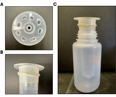
Figure 1: Tools used for leaf surface wash and apoplastic wash fluid isolation. (A) To collect LSW and AWF, two whole Arabidopsis thaliana rosettes are placed in a 60 mL syringe with eight additional holes that are melted into the end of the syringe with heated forceps to create channels for the wash to flow through. (B) This needleless syringe is then wrapped with parafilm at the neck region as depicted. (C) This step allows the syringe to fit into a 250 mL centrifuge bottle without touching the bottom of the bottle. Please click here to view a larger version of this figure.
4. Collection of apoplastic wash fluid (AWF)
- Place the same set of plants used for LSW or LSS isolation in a beaker and gently rinse them twice with distilled water.
- Vacuum infiltrate the rosettes with VIB for 20 s as follows:
NOTE: Although the process of vacuum infiltration is well-described in a previously published protocol11, it has been reproduced here to make the present protocol clear.- Carefully place rosettes in a French press coffee maker with 300-500 mL of VIB. Place the lid on the French press and gently lower the plunger until the plants are fully submerged.
- Place the French press with plants and buffer in a vacuum chamber. Apply a vacuum for 20 s using a vacuum pump.The buffer should just start to boil.
- Release the vacuum and remove the French press from the vacuum chamber, take the lid off, and pour the VIB into a separate 500 mL plastic beaker. Remove the rosettes from the French press.
NOTE:The VIB can be reused multiple times on the same day for one genotype, but once used for vacuum infiltration, the VIB should not be stored for use on another day. - Remove excess VIB from the plants by gently shaking and then brushing the leaves across a paper towel.
- Remove excess buffer from the leaf surfaces by gentle blotting with blotting paper.
- To collect the apoplastic wash fluid (AWF), place the rosettes inside 60 mL needleless syringes placed inside 250 mL centrifuge bottles as described for LSW collection and centrifuge at 4 °C for 30 min at 600 × g with slow acceleration.
- Filter the pooled AWF through a 0.22 µm syringe filter and collect the filtrate in a 15 mL centrifuge tube placed on ice.
NOTE: Typically, 12 plants would yield about 6 mL of AWF at the end using this protocol. Filtered LSW, AWF, and LSS can be used immediately or stored at -80 °C until further use. - Record the fresh weight (FW) of the plants used for each replicate after AWF isolation. Subsequently, use this reading to normalize the amount of RNA in each sample (Figure 2).
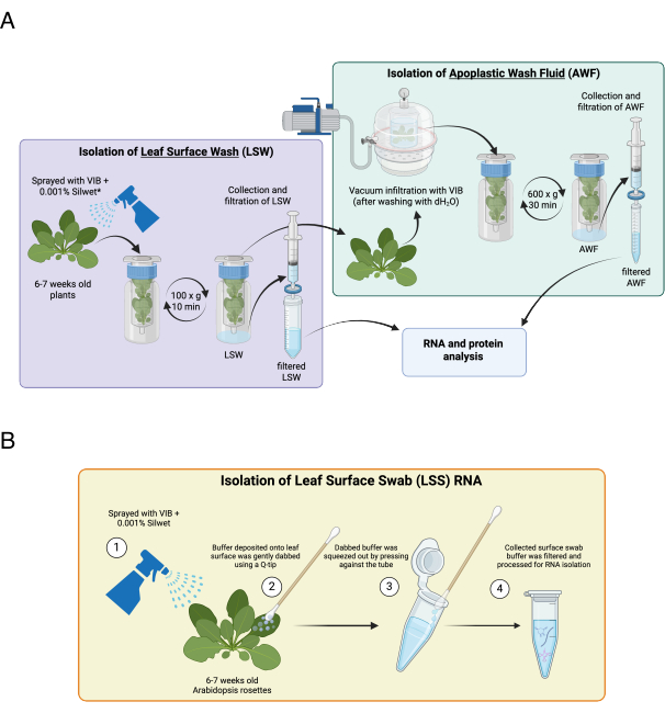
Figure 2: Schematic illustrations of the protocol and the leaf surface swab method. (A) Schematic illustration of the stepwise protocol for the isolation of leaf surface wash and apoplastic wash fluid using Arabidopsis rosettes. (B) Schematic illustration of leaf surface swab (LSS) method for isolating leaf surface RNA. Figures adapted from Figure S1 and Figure S4 of Borniego et al., 20256. Please click here to view a larger version of this figure.
5. Precipitation of RNA
- Precipitate RNA by mixing the required volume of AWF, LSW, or LSS with 0.1 volume of 3 M sodium acetate (pH 5.2) and 1.0 volume of cold isopropanol in a 15 mL centrifugation tube.
- Add glycogen (20 mg/mL stock) to a final concentration of 20 µg/mL.
NOTE: Ensure that the centrifuge tubes used to precipitate RNA can be centrifuged at a high speed of 13,000 × g. Breakage of tubes at this step can lead to loss of RNA samples. - Incubate the mixture at -20 °C for a minimum of 1 h or overnight.
- Centrifuge the tubes at 13,000 × g for 30 min at 4 °C (JA-25.50 rotor).
- Wash the pellet twice with ice-cold 70% EtOH and allow it to air dry for no more than 10 min.
- Resuspend the pellets in 100 µL of ultrapure DNase/RNase-free water or VIB, and transfer the RNA to 1.5 mL centrifuge tubes.
NOTE: After this step, the precipitated RNA can be stored at -80 °C for up to 2 weeks.
6. TRIzol-based RNA isolation
- Add 1 mL of TRIzol Reagent to each tube and mix by shaking on a rotator.
CAUTION: For handling TRIzol, use it in a fume hood, wear gloves, and eye protection. - Place the tubes on a tabletop rotator for 10 min at room temperature (RT).
- Add 200 µL of chloroform to each tube, followed by a brief but vigorous vortexing step.
CAUTION: For handling chloroform, use it in a fume hood, wear gloves, and eye protection. - Allow the tubes to stand at RT for about 3 min, and then centrifuge at 13,000 × g for 15 min at 4 °C.
- Transfer the aqueous phase to labeled 1.5 mL centrifuge tubes and add 10 µg of RNase-free glycogen and one volume of cold isopropanol.
CAUTION: For biohazardous wastes generated (such as phenol waste), collect organic solvents in designated waste containers per institutional guidelines. - Incubate the tubes for no more than 1 h at -20 °C.
NOTE: Precipitating the RNA for more than 1 h at this step reduces the efficiency of RNA isolation and leads to a very low concentration of RNA. - To pellet the RNA, centrifuge the tubes at 13,000 × g for 20 min at 4 °C.
- Wash the RNA pellets twice using ice-cold 70% EtOH and let them air dry before resuspending in 10-20 µL of ultrapure DNase/RNase-free water.
- Store the resuspended RNA at -20 °C or -80 °C for short- or long-term storage, respectively.
7. Removal of phenol or guanidine contamination
- To remove phenol or guanidine contamination from the RNA samples, perform one or two serial precipitations with 96% EtOH and ammonium acetate.
- Add 1 µg of glycogen to 10 µL of RNA samples and mix with 0.5 volume of 7.5 M ammonium acetate prior to the addition of 2.5 volumes of 96-100% EtOH.
NOTE: The final concentration of EtOH should be 70-75% in the entire volume at this step. - Incubate the mixture at -80 °C for 30 min and centrifuge at 13,000 × g for 20 min at 4 °C.
- Wash the RNA pellet twice with 70% EtOH and resuspend in 10 µL of ultrapure DNase/RNase-free water.
8. RNA gel electrophoresis for quantification and comparison
- Prepare mini gel (7.2 cm x 8.6 cm x 0.75 mm) containing 10% or 15% polyacrylamide and 7 M urea in 1x Tris-Boric Acid EDTA (TBE, pH 8.4) using 40% Acrylamide/Bis Solution, 37.5:1. Alternatively, a commercial denaturing mini gel (15% TBE-Urea Gel) can be used to run the RNA samples.
- Mix RNA samples (1:1) with 2x denaturing loading buffer (95% formamide, 10 mM EDTA, 0.02% SDS, 0.02% bromophenol blue, and 0.01% xylene cyanol), denature at 65 °C for 5 min, and place immediately on ice.
- Resolve the RNA samples in 0.5x TBE running buffer at RT at 220 V for approximately 1 h and 15 min.
- For size standards, use a 1:1 mix of low-range ssRNA and 14-30 nt ssRNA ladder markers.
- Stain the gel with 1x SYBR gold nucleic acid gel stain in 0.5x TBE for 10 min, wash twice with distilled water, and image using an imaging system (Figure 3).
- To quantify the RNA resolved in each lane, use the gel analysis method outlined in the ImageJ documentation under the subhead "Gel Analysis": https://imagej.net/ij/docs/menus/analyze.html
- Since 100 ng/µL of CL RNA is loaded every time to compare the banding pattern between CL and exRNA samples, estimate the concentration of exRNA samples relative to CL RNA (Figure 4).
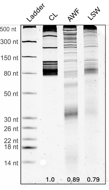
Figure 3: Plant leaves are coated with diverse RNA. 100 ng of RNA from leaf cell lysate (CL), and 2 µL of RNA isolated from AWF and LSW were separated on a 15% denaturing polyacrylamide gel and stained with SYBR Gold nucleic acid stain to visualize and compare the RNA-banding patterns. RNA size standards are shown in the leftmost lane. Densitometry measurements obtained using ImageJ gel analysis are indicated at the bottom of each lane and indicate the amount of RNA relative to the RNA in the total leaf cell lysate lane. Please click here to view a larger version of this figure.
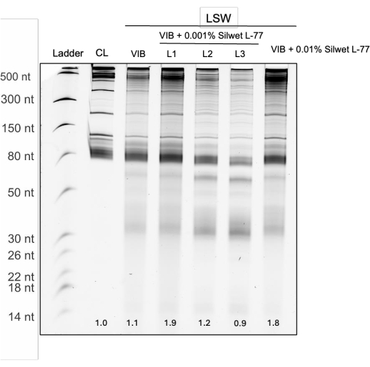
Figure 4: Repeated extraction of LSW RNA. LSW was first isolated using VIB without Silwet L-77 or varying concentrations of Silwet L-77 (0.001% or 0.01%). Repeated isolation of LSW using the same set of Arabidopsis plants can be employed to enrich LSW RNA using VIB + Silwet L-77 (0.001%). L1, L2, and L3 denote RNA samples isolated using LSW collected through three successive rounds of centrifugation post-spraying the same set of plants. Increasing the concentration of the wetting agent from 0.001% to 0.01% did not enhance LSW RNA isolation. Densitometry measurements obtained using ImageJ gel analysis are indicated at the bottom of each lane. Please click here to view a larger version of this figure.
9. Alternate method to quantify RNA using a microtiter plate-based method
- Prepare standard RNA solutions using CL RNA in ultrapure RNase/DNase-free water. Determine the appropriate concentration range for the standards based on the anticipated concentration of the samples. Use the following RNA concentrations: 0, 6.75, 12.5, 25, 50, 100, 200 ng/µL, and prepare a sufficient volume of each standard to allow for two technical replicates.
- Prepare 1.5x SYBR Gold nucleic acid gel stain in 500 mM Tris HCl pH = 8.
NOTE: SYBR Gold is pH dependent and has optimal staining sensitivity between pH = 7.0 and 8.0. - Microplate setup:
- Add 99 µL of dH2O to each well.
- Add 1 µL of RNA to each well. Use 1 µL for both RNA samples and RNA standards.
- Add 50 µL of 1.5x SYBR Gold to each well.
- Set the parameters on the microplate reader as follows:
Shake -Linear or orbital: 0:03 (MM:SS)
Read - Fluorescence Endpoint with excitation wavelength set at 496 nm, and emission wavelength set at 540 nm. - Construct a calibration line using the values of the standards. Using the calibration line equation, calculate the RNA concentrations in the samples.
NOTE: Alternative dyes can be used to quantify RNA in TRIzol-isolated samples. Similar results were obtained with Ribo488 RNA quantification reagent. A detailed manual is provided by the company for this dye. Briefly, dilute the dye 1000x (1 to 1000) using 200 mM Tris pH = 8. Then to make the measurements, add 99 µL of autoclaved dH2O to each well + 1 µL of RNA (sample or standard) + 100 µL of the Ribo488 dilution, incubate 5 min, and quantify fluorescence using a plate reader using 480 nm excitation and 520 nm emission. Standard curves obtained using the two RNA dyes are shown in Figure 5.
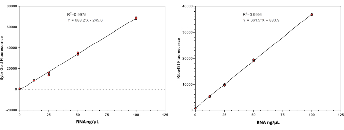
Figure 5: Standard curves obtained using a microtiter plate-based RNA quantification method. Two different RNA dyes, SYBR Gold (left) and Ribo488 (right) yield consistent results with two technical replicates per sample. Either method can be used to perform RNA quantification. However, a standard curve should be obtained every time RNA needs to be quantified. Please click here to view a larger version of this figure.
Results
A schematic overview of the above-described stepwise protocol for isolating leaf surface wash (LSW) and apoplastic wash fractions (AWF) from Arabidopsis plants is shown in Figure 2A. This method allows for the extraction of both LSW and AWF from as few as three to six plants per replicate, while consistently yielding equivalent amounts of RNA from both fractions. To measure RNA concentration in cell lysates, we utilize NanoDrop spectrophotometry. However, for the RNA samples derived from LSW, LSS, and AWF, we rely on densitometric quantification of SYBR Gold-stained polyacrylamide gels. This quantification step is necessary because the extracellular fractions contain an unknown contaminant-likely polysaccharides-that can sometimes copurify and interfere with absorbance-based RNA quantification methods, leading to an overestimation of RNA concentration. To overcome this limitation, we plan to utilize non-phenol-based RNA purification methods in our future work.
Denaturing RNA gel analysis revealed the presence of a broad range of RNA species in LSW and AWF, including both long and small RNAs (Figure 3). Notably, equivalent RNA quantities were successfully isolated from both leaf surface and apoplastic fractions, with the RNA amounts normalized per plant's fresh weight (FW). Surprisingly, the RNA size profiles of the LSW and AWF fractions differed significantly from each other and from the profile of total cell lysate (CL) RNA. These differences suggest that the exRNA fractions are not contaminated with cellular RNA. Among the three fractions, the apoplastic RNA (AWF) exhibited the greatest diversity in size distribution, suggesting that apoplastic RNA is more degraded than leaf surface RNA.
To further examine the presence of RNA on the surface of Arabidopsis leaves, we employed cotton swabs. This method allowed us to separately and sequentially collect RNA from the adaxial (upper) and abaxial (lower) leaf surfaces6. These analyses revealed that both leaf surfaces contained similar amounts of RNA. Moreover, the ability to collect RNA simply by absorbing buffer from the leaf surface without the need for centrifugation further supports the conclusion that RNA is indeed located on the leaf surface, rather than being extracted through the stomata during centrifugation.
One primary adjustment that allowed us to extract an abundant amount of leaf surface RNA was the addition of the wetting agent. We recovered almost two times more RNA upon adding wetting agent (at a very low concentration of 0.001%) to the VIB. This difference in concentration of RNA obtained with and without Silwet L-77 indicates that the use of a wetting agent improves the dissolution of RNA on the leaf surface. However, increasing the concentration of the wetting agent from 0.001% to 0.01% did not enhance LSW RNA isolation (Figure 4). Notably, repeating the isolation of LSW RNA from the same leaves yielded additional RNA (approximately 63% of the recovery obtained from the first wash), which suggests that leaves continually secrete RNA onto the surface, with nearly two-thirds of it being replaced within the approximately 30 min required to process the leaves. To increase total LSW RNA yields when plant material is limiting, it is thus possible to wash the same leaves multiple times in succession. We do not recommend more than three washes, though, as the handling involved may lead to cumulative damage to the leaf material, as assessed by green coloration of the wash fluid. For these experiments, RNA concentration was estimated using RNA gel-based densitometry analysis. However, the above-described microtiter plate-based RNA quantification method can be used for accurate quantification of RNA when assessing multiple samples at once (Figure 5).
Discussion
This protocol details how to isolate leaf surface wash (LSW), leaf surface swab (LSS), and apoplastic wash fluid (AWF) from healthy Arabidopsis rosettes, followed by RNA extraction. Critical steps include the preparation and sterilization of VIB before performing the experiment, growing the plants under controlled conditions, and using Silwet-L77 to ensure proper coating of the leaf surface by the VIB. These procedures should yield equal amounts of RNA from both LSW and AWF normalized by leaf fresh weight. Recovery of RNA from these extracellular fractions requires careful handling, especially with regard to RNA pellets following precipitation steps.
Although the above protocol has been optimized for Arabidopsis leaves, we have also used it successfully on other plant species with only minor modifications. RNA extraction from the leaf surface can be optimized by implementing two primary adjustments: firstly, by conducting multiple rounds of LSW isolation, and secondly, by increasing the concentration of the wetting agent. Plant species that have very hydrophobic leaf surfaces, such as rice12,13, may require higher concentrations of the wetting agent.
Additionally, we described a non-invasive method to collect leaf surface RNA using cotton-tipped swabs. With this leaf surface RNA isolation method, we ensure that the collection tube is placed in an ice bucket. Considering that the RNA on the leaf surface gets secreted and accumulates at room temperature, we assume that it would remain stable even during the collection process. We have also compared the banding patterns of LSW and LSS RNA, and they do not show much difference in terms of degradation.
The above protocol uses TRIzol reagent to purify RNA from either LSW, LSS, or AWF. This reagent employs phenol to remove proteins. We have found, however, that TRIzol-based purification does not remove polysaccharides such as pectin, which are abundant in LSW and AWF6. Such copurifying molecules can cause issues with RNA quantification using spectrophotometry or fluorometry. We therefore rely on RNA gel-based densitometry analysis to assess exRNA concentrations. We are currently investigating column-based purification methods that should provide better separation of RNA from polysaccharides. However, these methods often limit the recovery of small RNAs as the silica membrane is optimized to bind RNA molecules larger than ~200 nucleotides. Small RNAs may pass through the membrane during binding or be lost in wash steps. Therefore, thus far, we rely only on TRIzol-based method for more comprehensive recovery of all RNA sizes, including small RNAs, though it requires careful handling and phase separation.
Additionally, when conducting RNA gel analysis, we do not see fragments smaller than 80 nt for total cellular RNA samples compared to AWF and LSW RNAs, mainly due to low abundance relative to the large amounts of rRNA and tRNA. The presence of smaller bands in the AWF and LSW is due to the degradation of the abundant rRNAs and tRNAs, which occurs naturally after the secretion of RNA in the extracellular compartment of plants, and it is not an artifact introduced during the collection and handling of plants as confirmed by sequencing and northern blot analysis6.
Our leaf surface wash isolation method is a versatile tool with significant potential for a wide range of research applications. It should enable the isolation of biomolecules from the leaf surface, such as RNA, proteins, and other metabolites that may be central to understanding the interactions between plants and microbes. If identification of the microbes that are present in AWF and LSW is desired, this can be accomplished by eliminating filtration through the 0.22 µm filter.
Plants interact with a wide array of microbes, including bacteria, fungi, and viruses, which form complex communities on their surfaces1. These microbial communities can have profound effects on plant growth, disease resistance, and nutrient acquisition2,3,4. By isolating and analyzing leaf surface wash using this method, researchers can isolate exRNA, microbial metabolites, identify microbial communities present on the leaf surface, and investigate how these communities are regulated at a molecular level.
Disclosures
The authors have no conflicts of interest to disclose.
Acknowledgements
This work was supported by two grants from the United States National Science Foundation,
IOS-2243531 and IOS-2141969 to R.W.I.
Materials
| Name | Company | Catalog Number | Comments |
|---|---|---|---|
| 14-30 nt ssRNA ladder marker | Takara | 3416 | |
| 1.5 mL microcentrifuge tubes | VWR | 20170-038 | |
| 10 mL needleless syringe | BD | 303347 | |
| 15 mL falcon tubes | VWR | 89039-668 | |
| 15% Mini-PROTEAN TBE-Urea Gel, 10 well/12 well, 30 µL/20 µL | Bio-Rad | 4566053 / 4566055 | |
| 36-cell tray inserts | True Leaf Market | ||
| 40% acrylamide and bis-acrylamide solution, 37.5:1 | Bio-Rad | 1610148 | |
| 4-inch round pots | True Leaf Market | ||
| 50 mL falcon tubes | VWR | 89039-656 | |
| 50 mL needleless syringe | BD | 309653 | |
| 96 well plate black bottom | Grenier | 655090 | |
| Acrodisc 0.22 μm syringe filter | Pall | 4192 | |
| Acrodisc 0.45 μm syringe filter | Pall | 4454 | |
| Ammonium persulfate (APS) | Sigma-Aldrich | A3678 | |
| Calcium chloride | Sigma-Aldrich | C1016 | |
| Chloroform (RNase-free) | Macron Fine Chemicals | MK444110 | |
| Clear plastic domes | Hummert International | 11 3348 | Any plastic dome can be used to cover the plants and maintain humidity |
| Cotton-tipped stick medical grade | Uline | S-21102 | |
| Ethanol (Rnase-free) | Decon Laboratories, Inc. | 2716GEA | |
| French Press coffee maker, 24 fl oz | Snow Peak | CS-111 | |
| Growing tray without holes 10 x 20 | True Leaf Market | ||
| Ice bucket | Schuett-Biotech, Spongex | 3-680 052 | |
| Isopropanol | Thermo Scientific | 149320050 | |
| JA-14 fixed-angle rotor | Beckman Coulter; model: JA-14 | 339247 | |
| Kimwipes | KCWW, Kimberly-Clark | 34120 | |
| Low range ssRNA lader | NEB | N0364S | |
| MES hydrate | Sigma-Aldrich | M8250 | |
| Microplate reader | BioTek | Synergy H1 | |
| Miracle-Gro | Miracle-Gro | 24-8-16 | It is an all purpose plant food. Depending on soil source used to grow the plants, the application can be adjusted. |
| Nalgene centifuge bottle 250 mL (w/out cap) | Nalgene | 3120 | |
| Paper towels | |||
| ParaFilm M Lab Film | Parafilm | S-25929 | |
| Plastic beaker (500 mL) | Uline | S-21658 | |
| RNase-free glycogen (20 mg/mL) | Thermo Scientific | R0561 | |
| RootShield Plus WP Biological Fungicide | BioWorks | 68539-9 | |
| Scissors | |||
| Silwet L-77 | Phytotech labs | S7777 | Any brand Silwet L-77 can be used. However, Silwet looses its efficacy over the period of time. It is ideal to replace it once a year. |
| Sodium chloride | Sigma-Aldrich | S7653 | |
| Soft nylon hair paint brush | US. Art Supply | No. 6 | |
| Spray bottle | Uline | S-7272 | Any spray bottle can be used but the spray settings should be on shower. |
| Sungro Professional Growing Mix | Sungro | ||
| Sungro Propagation Mix | Sungro | ||
| SYBR Gold Nucleic Acid Gel Stain (10,000x concentrate in DMSO) | Thermo Scientific | S11494 | |
| TEMED | Thermo Scientific | 17919 | |
| TRIzol reagent | Invitrogen | 15596026 | |
| Ultrapure Dnase/Rnase-free water | Invitrogen | 10977015 | |
| Urea | Sigma-Aldrich | U5378 | |
| Vacuum chamber, 0.20 Cu. Ft. | SP Scienceware - Bel-Art Products - H-B Instrument | F42031-0000 | |
| Vacuum pump, 3.5 CFM | Ideal Vacuum Products | P101532 | |
| Weighing scale | Escali | WBB480535 | This is just an example. Any weighing scale can be used. |
References
- Vorholt, J. A. Microbial life in the phyllosphere. Nat Rev Microbiol. 10 (12), 828-840 (2012).
- Ritpitakphong, U., Falquet, L., Vimoltust, A., Berger, A., M'etraux, J., L'Haridon, F. The microbiome of the leaf surface of Arabidopsis protects against a fungal pathogen. New Phytol. 210 (3), 1033-1043 (2016).
- Chaudhry, V., Runge, P., Sengupta, P., Doehlemann, G., Parker, J. E., Kemen, E. Shaping the leaf microbiota: plant-microbe-microbe interactions. J Exp Bot. 72 (1), 36-56 (2021).
- Vacher, C., Hampe, A., Port'e, A. J., Sauer, U., Compant, S., Morris, C. E. The phyllosphere: microbial jungle at the plant-climate interface. Ann Rev Ecol Evol Syst. 47, 1-24 (2016).
- Mahmoudi, M., Almario, J., Lutap, K., Nieselt, K., Kemen, E. Microbial communities living inside plant leaves or on the leaf surface are differently shaped by environmental cues. ISME Commun. 4 (1), ycae103(2024).
- Borniego, M. L., et al. Diverse plant RNAs coat Arabidopsis leaves and are distinct from apoplastic RNAs. Proc Natl Acad Sci U S A. 122 (1), e2409090121(2025).
- Ren, B., Wang, X., Duan, J., Ma, J. Rhizobial tRNA-derived small RNAs are signal molecules regulating plant nodulation. Science. 365 (6456), 919-922 (2019).
- Chery, M., Drouard, L. Plant tRNA functions beyond their major role in translation. J Exp Bot. 74 (7), 2352-2363 (2022).
- Panstruga, R., Spanu, P. Transfer RNA and ribosomal RNA fragments-Emerging players in plant-microbe interactions. New Phytol. 241 (2), 567-577 (2024).
- Baldrich, P., Rutter, B. D., Karimi, H. Z., Podicheti, R., Meyers, B. C., Innes, R. W. Plant extracellular vesicles contain diverse small RNA species and are enriched in 10- to 17-nucleotide "Tiny" RNAs. Plant Cell. 31 (2), 315-324 (2019).
- Rutter, B. D., Rutter, K. L., Innes, R. W. Isolation and quantification of plant extracellular vesicles. Bio Protoc. 7 (17), e2533(2017).
- Zhang, Z., et al. OsHSD1, a hydroxysteroid dehydrogenase, is involved in cuticle formation and lipid homeostasis in rice. Plant Sci. 249, 35-45 (2016).
- Zhou, L., Ni, E., Yang, J., Zhou, H., Liang, H., Li, J., Jiang, D., Wang, Z., Liu, Z., Zhuang, C. Rice OsGL1-6 is involved in leaf cuticular wax accumulation and drought resistance. PLoS One. 8 (5), e65139(2013).
Reprints and Permissions
