This protocol outlines the design of split hybridization probes utilizing fluorescent light-up aptamer DAP-10-42 as a signal reporter and their application for nucleic acid detection and differentiation between targets with single-nucleotide substitutions.
A subscription to JoVE is required to view this content. Sign in or start your free trial.
Method Article
Split Hybridization Probe Utilizing a DNA Fluorescent Light-up Aptamer as a Signal Reporter for Sequence-Specific Nucleic Acid Analysis
In This Article
Summary
Abstract
DNA-based fluorescent light-up aptamers (FLAPs) are promising for bioanalytical assays because they provide a low-cost fluorescent signal readout without the need for labeling of nucleic acid signal reporters with fluorophores and/or quenchers, unlike conventional hybridization probes used for instantaneous nucleic acid detection. Instead, FLAPs non-covalently bind dye ligands , which exhibit intrinsically low fluorescence in aqueous solutions, but become highly emitting upon FLAP binding. This protocol describes an algorithm to design split light-up aptamer sensors (SLASs) utilizing DAP-10-42, the most efficient DNA FLAP reported thus far. When equipped with nucleic acid sequences complementary to a nucleic acid target of interest, SLAS is a promising tool for nucleic acid analysis allowing for the sequence-specific detection of nucleic acid targets with selectivity down to one nucleotide to enable analysis of single-nucleotide substitutions (SNSs). SLASs offer the advantage of a label-free fluorescence-based signal readout, which can be measured with a conventional cuvette-based fluorescent spectrophotometer, a portable fluorometer, or visually observed upon excitation with a handheld light source. The SLAS approach is beneficial for biosensing applications in disease diagnostics, environmental monitoring, and biomolecular research.
Introduction
Nucleic acid analysis using hybridization probes is routinely performed to detect and quantify specific DNA/RNA sequences1. One of the most successful hybridization probes is the Molecular Beacon (MB) probe, which is a hairpin-shaped DNA
oligonucleotide labeled with a fluorophore and a quencher at the opposite termini2,3. It instantaneously (without the need to wash the probe-target complex from the excess of the probe) detects the presence of a nucleic acid sequence complementary to the loop fragment of the probe and is commonly used in quantitative PCR (qPCR). The need to covalently attach two dyes - the fluorophore and the quencher - to the oligonucleotide sequence of the probe increases the cost, particularly when a new MB probe targeting a new nucleic acid sequence is needed. Alternatively, the MB probe can be used as a signal reporter in a multicomponent (split) hybridization probe format, in which two unlabeled oligonucleotides interact with both the target and the MB probe reporter4. The multicomponent probe approach enhances selectivity for nucleic acid recognition and improves the probe's binding to structured targets5,6,7, This approach, nevertheless, does not eradicate the need for covalent labeling of the reporter with a fluorophore8.
Fluorescent Light-up Aptamers (FLAPs)9 are aptamers that have an affinity to low-fluorescent environment-sensitive dyes (fluorogens), where binding by FLAP enhances the fluorescence of the fluorogen manifold. Unlike fluorescently labeled probes and signal reporters, FLAPs do not require sensing elements to be covalently labeled with one or more dyes and thus offer the advantage of label-free fluorescence detection10. This is particularly important during the optimization step when dozens of nucleic acid constructs need to be tested to achieve satisfactory assay performance. Moreover, FLAP assays can be optimized by adjusting the concentration of a fluorogen and/or FLAP sensor rather than changing the sensor's design. These advantages can be significant for low-budget research and teaching laboratories. DNA FLAPs are particularly promising as signal reporters of multicomponent hybridization probes referred to as Split Light-up Aptamer Sensors (SLASs)11. The most efficient DNA FLAP reported so far is DAP-10-42, which was originally selected to enhance the fluorescence of dapoxyl sulfonyl dyes12, with a promiscuous ability to bind and enhance the fluorescence of a variety of aryl methane dyes (e.g., auramine O, AO; crystal violet, CV)13. DAP-10-42 has been converted into SLAS to be used for nucleic acid analysis13,14,15.
The goal of the method described here is to enable sequence-specific analysis of nucleic acid targets of interest using label-free SLAS strands and a fluorogenic dye as a buffer component. This protocol describes an algorithm to design SLAS based on DAP-10-42 and highlights SLAS performance for highly selective detection of nucleic acid sequences differing in as little as a single nucleotide.
The SLAS nucleic acid detection assay requires two custom-synthesized unmodified DNA oligonucleotides used at a final concentration of 0.5-1 µM for maximal performance. The protocol described here utilizes a commercially available Auramine O (AO) dye as a fluorogen. The final AO concentration in the assay ranges from 1-10 µM to ensure high fluorescence enhancement in the presence of a specific nucleic acid target over the blank (absence of the target; Supplementary Figure 1). The necessary components of the assay buffer are potassium and magnesium ions (Supplementary Figure 2). Potassium ions (1.25-40 mM) stabilize the proposed G-quadruplex domain of the FLAP used as a signal reporter. Magnesium ions (2.5-50 mM) reduce repulsion of negatively charged phosphate groups upon SLAS-target interactions. The temperature of the SLAS assay using the reported sequences for the signal reporting domain should not exceed 28 °C to avoid loss of performance16.
SLAS is compatible with single-stranded nucleic acid targets, such as DNA amplicons of asymmetric PCR or an isothermal DNA amplification reaction (e.g., recombinase polymerase amplification, RPA17), RNA products from transcription-mediated amplification (TMA) or nucleic acid sequence-based amplification (NASBA)18, and native RNA molecules such as miRNA, rRNA or ncRNA). Variations in the nucleotide sequence of the target down to a single nucleotide can be detected using the SLAS approach13,14.
Access restricted. Please log in or start a trial to view this content.
Protocol
1. Design of the Split Light-up Aptamer Sensor (SLAS) with DAP-10-42 as a signal reporter
- Design the sequences of two unmodified DNA oligonucleotide strands constituting SLAS. Use a fragment containing nucleotides (nts) 4,6-39 of DAP-10-42 (Table 1, Figure 1A).
- Convert the sequence of stem 1 (nts 1-8 and 36-42) to stem 1' by removing a bulging thymine residue at position nt 5, shortening the stem to 4 bp (nts 4,6-8 and 36-39), and adding a terminal C-G bp (Figure 1A, step 1). Additionally, one of the SLAS strands contains nts 9-29 of the DAP-10-42 core sequence, while another SLAS strand contains nts 30-35 of the sequence.
- Extend the 3'-terminal sequence of the fragment containing nts 9-29 with d(GGTCAT), and the 5'-terminal sequence of the nts 30-35 fragment with d(ATGACC) to form a 6-bp stem 2 (Figure 1A, step 2).
NOTE: The base pairs T4-A39, A6-T38, C7-G37, and G8-C36 of stem 1 in DAP-10-42 are required for the light-up function of the aptamer. The sequence of stem 1' can be modified only at the terminal bp and can be a G-C, A-T, T-A base pair or extended with 1-2 base pairs as needed. The sequence of stem 2 is arbitrary and can be changed. However, it is recommended that the thermodynamic stability of stem 2 is preserved unless the assay temperature is raised above 28 °C. Shortening or otherwise destabilizing stem 2 prevents premature dye binding without a target if a high background of the SLAS-based fluorescent assay is observed. Splitting of the DAP-10-42 nucleotide sequence between nts 28-29, 27-28, or 26-27 instead of nts 29-30 is also allowed (Figure 1B). - To tailor the designed SLAS to a nucleic acid target or interest, extend stem 1' with DNA sequences complementary to the target-target-binding fragments (arms) through d(TT) linkers (Figure 1A, step 3). This yields the sequences of strands SLAS-U and SLAS-S constituting the probe.
- Make the target-binding arm of SLAS-S complementary to a 7-10-nt fragment of the target containing the SNS site, and the target-binding arm of SLAS-U – complementary to a 15-25 nt fragment of the target adjacent to the target-binding arm of SLAS-S (Table 1).
NOTE: It is optional whether to connect the target-binding arm of SLAS-S to nt 4 or nt 39 of the DAP-10-42 sequence (Figure 1, arm 1 or arm 2, respectively). - Assess the melting temperature (Tm) of the duplexes between the target of interest and the target-binding arms of SLAS-S and SLAS-U using the UNAFold Web Server (unafold.org)19.
- Click on the DINAMelt tab on top of the web page; go to Applications and select Two State Melting Hybridization (Supplementary Figure 3A). On the Two-State Melting Hybridization page, enter one of the interacting sequences in the left box and another in the right box (Supplementary Figure 3B). Make sure both sequences are in 5' to 3' order. You can enter more than one pair of interacting sequences by separating them with semicolons.
- At the bottom of the page, indicate the assay temperature (22 °C), monovalent and divalent cation concentrations (20 mM and 25 mM, respectively) to match the assay conditions. Indicate the concentration of the interacting sequences.
- Press Submit (Supplementary Figure 3B). Observe the results that include the Gibbs energy change, enthalpy change, and entropy change of the binding, as well as the Tm values for the corresponding duplexes (Supplementary Figure 3B).
- Make sure the Tm values for the duplexes between the fully matched target (M) and the target-binding arms of SLAS-S and SLAS-U are above the assay temperature, which is 22 °C in the current protocol. Here, the Tm values were 32.2°C and 74.4 °C, respectively (Supplementary Figure 3C).
- Ensure the duplex between SLAS-S and a mismatched target (MM) containing an SNS has a Tm below the assay temperature. This ensures high selectivity of the probe-target recognition, preventing the AO fluorogen from signaling the presence of the targeted nucleic acid analyte if it contains an SNS in the SLAS-binding fragment (Figure 2). Here, the Tm was 21.3 °C (Supplementary Figure 3C), which was sufficient to discriminate the mismatch from the matched complex.
- Adjust the sequences of the target-binding arms by shortening or extending the target-complementary fragments, if needed, to meet the stability requirements indicated.
- Obtain the designed strands SLAS-S and SLAS-U from any commercial vendor supplying custom-made DNA oligonucleotides or synthesize the DNA strands in-house using an automated oligonucleotide synthesizer.
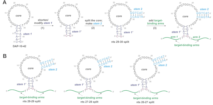
Figure 1: Conversion of a fluorescent light-up aptamer (FLAP) DAP-10-42 into a split light-up aptamer sensor (SLAS). (A)The secondary structure of DAP-10-4212 is predicted by a DNA folding application of the UNAFold Web Server19 at 22 °C, 20 mM Na+, 25 mM Mg2+. The aptamer features a hairpin secondary structure with a core that presumably folds into a G-quadruplex motif stabilized by stem 1. For DAP-10-42 to be converted into a SLAS, (1) stem 1 of the aptamer is shortened to 4 bp and flanked by an additional G-C bp to make stem 1'; (2) the core sequence is split between nts 29 and 30, with the 3'- and 5'-sides of the split site extended with complementary sequences d(GGTCAT) and d(ATGACC), respectively, to make stem 2; (3) stem 1' is extended with the sequences complementary to the adjoining fragments of a target of interest - target-binding arms, with a d(TT)-linker between the target-binding arms and the signal reporting domain of the probe. (B) The core can also be split between nts 28-29, 27-28, or 26-27 without significant loss of the signal and/or fluorescence fold-increase. Please click here to view a larger version of this figure.
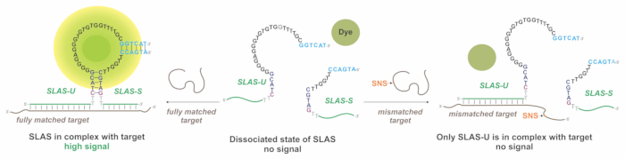
Figure 2: SLAS signal generation mechanism. SLAS is composed of two strands - SLAS-U and SLAS-S - and a fluorogenic dye (e.g., Auramine O; AO). In the absence of a nucleic acid target of interest, both strands are in the dissociated state and unable to bind the dye; no signal is observed (center). In the presence of a fully matched target M, both SLAS-U and SLAS-S hybridize to the adjoining fragment of M to bring the core fragments in proximity. This allows for the formation of the dye-binding site. Binding of AO enhances its fluorescence, which is measured as a fluorescent signal readout (left). In the presence of a mismatched target MM containing a single-nucleotide substitution (SNS) in a fragment interacting with SLAS-S, only SLAS-U can hybridize to the MM target, while SLAS-S is in the dissociated state. This prevents the formation of the dye-binding site, and no signal is observed (right). Please click here to view a larger version of this figure.
2. Amplification of the targeted sequence
- Amplify a fragment of the gene to be analyzed with the SLAS assay. For PCR or RPA amplification, use asymmetric conditions (e.g., with the Forward Primer added at 10-fold excess over the Reverse Primer) to generate a single-stranded amplicon. To amplify a 126 nt fragment from the NANOGP8 gene using Linear After The Exponential (LATE) PCR20, use the primer sequences listed in Table 2. Here, a single-stranded DNA template (Template NANOGP8 or Template G1423C, Table 2) corresponding to the 126-nt fragment of the gene was used. The template was custom-synthesized by Integrated DNA Technologies, Inc.
- Prepare a 100 µL PCR reaction by mixing 10 µL of 10x Standard Taq Reaction Buffer, 2 µL of 10 mM dNTP mix (to have the final concentration of 0.2 mM), 10 µL of 10 µM Forward primer and 10 µL of 1 µM Reverse Primer (for the final concentration of 1 µM and 0.1 µM, respectively), 0.5 µL of 5U/µL Taq DNA polymerase, DNA template and nuclease-free water. For a no-template control, add water instead of the template.
- Use the following thermocycling conditions: initial denaturation at 95 °C for 1 min; 45 cycles of (1) denaturation at 95 °C for 15 s, (2) primer annealing at 60 °C for 30 s, (3) primer extension at 68 °C for 30 s; and final extension at 68 °C for 5 min.
NOTE: Transcription-based amplification techniques such as NASBA18 can be used instead of PCR or RPA, especially if an RNA template is available for amplification. NASBA generates a single-stranded RNA amplicon at a final product concentration in the micromolar range, which is high enough to be used in the downstream SLAS assay without product concentration.
- Precipitate the amplified samples with ethanol by adding 0.1x volume of 3 M sodium acetate (pH 5.5) and 3x volume of ethanol. Incubate at -20 °C for 1 h, centrifuge at 12,000-18,000 x g for 5 min, and discard the supernatant.
- Wash the pellet with 70% ethanol, re-centrifuge, and discard the supernatant again. Air dry the pellet and dissolve in 10 µL of nuclease-free water to be used in the SLAS assay.
3. Fluorescence assay using SLAS
- Prepare stock solutions of AO (0.1 mM in DMSO, 10x), SLAS-S (10 µM in nuclease-free water, 10x), SLAS-U (10 µM in nuclease-free water, 10x), and 4x assay buffer (80 mM Tris-HCl, pH 7.4, 100 mM MgCl2, 80 mM KCl).
NOTE: Potassium ions are necessary to stabilize the presumed G-quadruplex motif of the aptamer, which is part of the dye-binding site. As little as 1 mM KCl (at 25 mM MgCl2) in 1x assay buffer is sufficient to ensure ~14-fold fluorescence enhancement in the presence of the specific target M over the blank (no-target sample; Supplementary Figure 2B). Magnesium ions stabilize the interactions of the nucleic acid strands (between SLAS-S, SLAS-U, and the target). The concentration of Mg2+ in the 1x Assay Buffer should be at least 5 mM (at 20 mM KCl; Supplementary Figure 2C,D). The stock solution of AO (~1 mM) is prepared in DMSO. Stock concentrations of SLAS-S and SLAS-U are arbitrary and can be adjusted if needed as long as the final concentrations of the strands are maintained (see the rationale for the selection of the final SLAS concentration in Discussion).
CAUTION: AO is harmful if swallowed, toxic in contact with skin, and suspected of causing cancer. Always wear gloves when working with AO solutions. The weighing of AO and preparing of its stock solutions should be done under the chemical hood. - Prepare the master mix containing all assay components but the target. Calculate the master mix volume (Vmix) by multiplying the sample volume (60 µL) by the number of samples plus one (n+1):

The number of samples n should include necessary negative and positive controls. - Combine 0.25x Vmix µL of 4x Assay Buffer, 0.1x Vmix µL of 0.1 mM AO, 0.1x Vmix µL of 10 µM SLAS-S, and 0.1x Vmix µL of 10 µM SLAS-S. Add nuclease-free water to the final volume of
 such that it accommodates the target volume added at step 3.4. Vortex and briefly centrifuge the master mix. Aliquot 50 µL of the master mix into n sample tubes.
NOTE: The sample volume can be adjusted depending on the characteristics of the instrument utilized to detect the fluorescent signal. The volume of the master mix that is aliquoted into sample tubes can be adjusted based on the target volume added at step 3.4. The volume of nuclease-free water added to make the master mix should be adjusted correspondingly. For example, if the target-containing sample is added at 10% (V/V) of the final sample volume, water should be added into the master mix up to 0.9x Vmix. - Use one sample as a no-target control (blank), and one sample as a positive control. Add 10 µL of the target-containing sample to the master mix (50 µL) to make the final sample volume of 60 µL containing 10-1000 nM target. For the blank, use nuclease-free water (10 µL) instead of the target. For positive control, use a synthetic DNA oligonucleotide containing the targeted sequence (e.g., target M, Table 1). Add 10 µL of 0.6-3 µM control to make 0.1-0.5 µM final concentration.
NOTE: High fluorescence in this positive control ensures the assay works as expected, even if a negative signal is observed for the tested unknown samples. - Mix the samples and spin down using a microcentrifuge. Incubate the samples at 22 °C for 10-60 min. The signal increases only by <15% between 40 min and 60 min (Figure 3).
- Measure the fluorescence of the samples at 540 nm upon excitation at 475 nm using a fluorescent spectrophotometer.
NOTE: It is possible to use a portable fluorometer that measures fluorescence in thin-walled PCR tubes. The fluorometer needs to have excitation and emission filters suitable for detecting AO fluorescence. An example of a suitable excitation filter is a 495 nm short-pass. An example of a suitable emission filter is 510-580 nm. Alternatively, the signal can be observed visually upon excitation of AO fluorescence in the sample with a UV or Blue light source.
CAUTION: If a UV lamp is used, wear UV-protective goggles.
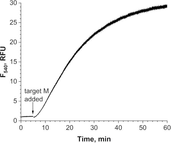
Figure 3: Time-dependence of the signal generation for SLAS-29U/30S. A sample containing AO, SLAS-29U and SLAS-30S was analyzed using the kinetics mode of a fluorescent spectrophotometer (excitation at 475 nm, emission at 540 nm, 5-nm excitation/emission slits) over 60 min. At a 5 min time (indicated with an arrow), target M was added to the sample. The data is averaged from two independent trials. Please click here to view a larger version of this figure.
Access restricted. Please log in or start a trial to view this content.
Results
Performance of SLAS utilizing DAP-10-42 core as a signal reporter was demonstrated using a probe designed to interrogate a fragment of the NANOGP8 gene important for the progression of glioblastoma multiforme (GBM), an aggressive form of brain cancer21. This fragment was reported to contain SNSs in the DNA cargo of exosomes derived from cancer cells22. Therefore, the SLAS was tailored to the SNS site of the gene. Synthetic DNA targets M or MM mimi...
Access restricted. Please log in or start a trial to view this content.
Discussion
In the reported protocol, the critical steps include (i) the SLAS design; (ii) assay temperature; (iii) presence of mono- and divalent metal cations (K+ and Mg2+); (iv) concentrations of SLAS strands.
SLAS design
In designing SLAS strands, the thermodynamic stability of the complexes formed by SLAS-S and SLAS-U strands in the absence of targets, and in the presence of either specific or non-specific target (if SNS-differentiation is needed), is impo...
Access restricted. Please log in or start a trial to view this content.
Disclosures
The authors declare no conflict of interest.
Acknowledgements
Funds from the UCF Office of Undergraduate Research (OUR) for AK are greatly appreciated. Work on optimization of LATE-PCR conditions for the NANOGP8 gene is supported by the Florida Cancer Innovation Fund (grant MOABC).
Access restricted. Please log in or start a trial to view this content.
Materials
| Name | Company | Catalog Number | Comments |
|---|---|---|---|
| Auramine O | MP Biomedicals | 195064 | a fluorogenic dye used for signal reporting |
| Cary Eclipse Fluorometer | Agilent Technologies | G9800A | to measure fluorescent signal of the samples |
| Dimethy Sulfoxide | MP Biomedicals | 196055 | solvent to prepare stock solutions of Auramine O |
| DNA oligonucleotides (primers, SLAS strands, synthetic template for PCR) | IDT | N/A | custome-made and desalted by the vendor, used without further purification |
| dNTP mix, 10 mM | ThermoFisher Scientific | R0191 | for LATE PCR reaction |
| Magnesium Chloride | invitrogen | AM9530G | buffer component; needed for correct association of the probe's strands with the analyzed target |
| Microcentrifuge tubes (1.5-mL ) | Fisherbrand | 02-681-320 | plastic for sample preparation |
| Micropipette (0.1-2.5 µL) | Eppendorph | K56877G | used for sample preparation |
| Micropipette (100-1000 µL) | Eppendorph | K13160F | used for sample preparation |
| Micropipette (20-200 µL) | Eppendorph | J05253M | used for sample preparation |
| Micropipette (2-20 µL) | Eppendorph | G24675G | used for sample preparation |
| Mini Centrifuge | Corning | 6770 | used for spinning down the samples |
| Nuclease-Free Water (not DEPC-Treated) | ThermoFisher Scientific | AM9932 | Use for samples in PCR and SLAS assay |
| PCR tubes (0.5-mL) | invitrogen | AM9932 | to measure fluorescence using Quantus fluorometer |
| Pipett tips (20-200 µL) | VWR | 76322-150 | plastic for sample preparation |
| Pipette tips (1-10 uL) | VWR | 76322-528 | plastic for sample preparation |
| Potassium Chloride | Invitrogen | AM9640G | buffer component; needed for correct folding of the fluorescent light-up aptamer domain of the probe |
| Quantus Fluorometer | Promega | E6150 | portable low-cost fluorometer that can be used for fluorescence measurements |
| Quartz cuvette | Starna | 16.50F-Q-10/Z15 | to be used for fluorescence measurement using Cary Eclipse fluorometer |
| Taq DNA Polymerase with Standard Taq Buffer | New England BioLabs | M0273S | for amplification of a gene fragment to be analyzed with the SLAS assay |
| Tris-HCl buffer (pH 7.4) | Boston Bioproducts | R-314 | buffer component |
| UV lamp | UVP | 95-0016-14 | to excite AO for visual signal observation |
| Vortex Mixer | VWR | 10153-838 | used for mixing the samples |
References
- Wetmur, J. G. DNA Probes: Applications of the principles of nucleic acid hybridization. Crit Rev Biochem Mol Biol. 26 (3-4), 227-259 (1991).
- Tyagi, S., Kramer, F. Molecular Beacons: Probes that Fluoresce upon Hybridization. Nat Biotechnol. 14, 303-308 (1996).
- Kolpashchikov, D. M. An elegant biosensor molecular beacon probe: challenges and recent solutions. Scientifica. 2012, 928783(2012).
- Kolpashchikov, D. M. A binary DNA probe for highly specific nucleic acid recognition. J Am Chem Soc. 128 (32), 10625-10628 (2006).
- Kolpashchikov, D. M. Binary probes for nucleic acid analysis. Chem Rev. 110 (8), 4709-4723 (2010).
- Stancescu, M., Fedotova, T. A., Hooyberghs, J., Balaeff, A., Kolpashchikov, D. M. Nonequilibrium hybridization enables discrimination of a point mutation within 5-40 C. J Am Chem Soc. 138 (41), 13465-13468 (2016).
- Gerasimova, Y. V., Kolpashchikov, D. M. Detection of bacterial 16S rRNA using a molecular beacon-based X sensor. Biosens Bioelectron. 41, 386-390 (2013).
- Gerasimova, Y. V., Hayson, A., Ballantyne, J., Kolpashchikov, D. M. A single molecular beacon probe is sufficient for the analysis of multiple nucleic acid sequences. Chembiochem. 11 (12), 1762-1768 (2010).
- Gao, T., Luo, Y., Cao, Y., Pei, R. Progress in the isolation of aptamers to light-up the dyes and the applications. Analyst. 145, 701-718 (2020).
- Du, Y., Li, B., Wang, E. "Fitting" makes "sensing" simple: Label-free detection strategies based on nucleic acid aptamers. Acc Chem Res. 46 (2), 203-213 (2013).
- Kolpashchikov, D. M., Spelkov, A. A. Binary (split) light-up aptameric sensors. Angew Chem Int Ed Engl. 60 (10), 4988-4999 (2021).
- Kato, T., Shimada, I., Kimura, R., Hyuga, M. Light-up fluorophore--DNA aptamer pair for label-free turn-on aptamer sensors. Chem Commun. 52 (21), 4041-4044 (2016).
- Connelly, R. P., Madalozzo, P. F., Mordeson, J. E., Pratt, A. D., Gerasimova, Y. V. Promiscuous dye binding by a light-up aptamer: Application for label-free multi-wavelength biosensing. Chem Commun. 57 (30), 3672-3675 (2021).
- Kikuchi, N., Reed, A., Gerasimova, Y. V., Kolpashchikov, D. M. Split dapoxyl aptamer for sequence-selective analysis of nucleic acid sequence based amplification amplicons. Anal Chem. 91 (4), 2667-2671 (2019).
- Wang, L., et al. MicroRNA detection in biologically relevant media using a split aptamer platform. Bioorg Med Chem. 69, 116909(2022).
- Gerasimova, Y. V., Nedorezova, D. D., Kolpashchikov, D. M. Split light up aptamers as a probing tool for nucleic acids. Methods. 197, 82-88 (2021).
- Daher, R. K., Stewart, G., Boissinot, M., Bergeron, M. G. Recombinase polymerase amplification for diagnostic applications. Clin Chem. 62, 947-958 (2016).
- Compton, J. Nucleic acid sequence-based amplification. Nature. 350, 91-92 (1991).
- Zuker, M. Mfold web server for nucleic acid folding and hybridization prediction. Nucleic Acids Res. 31, 3406-3415 (2003).
- Pierce, K. E., Sanchez, J. A., Rice, J. E., Wangh, L. J. Linear-after-the-exponential (LATE)-PCR: primer design criteria for high yields of specific single-stranded DNA and improved real-time detection. Proc Natl Acad Sci U S A. 102, 8609-8614 (2005).
- Grochans, S., et al. Epidemiology of glioblastoma multiforme-Literature review. Cancers. 14, 2412(2022).
- Vaidya, M., Bacchus, M., Sugaya, K. Differential sequences of exosomal NANOG DNA as a potential diagnostic cancer marker. PLoS One. 13, e0197782(2018).
- Owczarzy, R., Moreira, B. G., You, Y., Behlke, M. A., Walder, J. A. Predicting stability of DNA duplexes in solutions containing magnesium and monovalent cations. Biochemistry. 47, 5336-5353 (2008).
- Bhattacharyya, D., Arachchilage, G. M., Basu, S. Metal cations in G-quadruplex folding and stability. Front Chem. 4, 2296-2646 (2016).
- Chen, R. F. Fluorescence of free and protein-bound auramine O. Arch Biochem Biophys. 179 (2), 672-681 (1977).
- Klymchenko, A. S. Solvatochromic and fluorogenic dyes as environment-sensitive probes: Design and biological applications. Acc Chem Res. 50 (2), 366-375 (2017).
Access restricted. Please log in or start a trial to view this content.
Reprints and Permissions
