Here, we present a protocol to enable whole-genome sequencing of bacterial sexually transmitted infections from clinical samples using target enrichment. This novel, syndromic panel-based method overcomes challenges of abundant human DNA and low bacterial loads, facilitating genomic surveillance for Chlamydia trachomatis, Neisseria gonorrhoeae, Treponema pallidum, and Mycoplasma genitalium.
Method Article
Generating Whole Bacterial Genomes from Clinical Samples using a Target Enrichment Workflow
In This Article
Summary
Abstract
Obtaining complete genomes of bacterial sexually transmitted infections (STIs) directly from clinical samples is challenging due to the presence of human DNA, microbiota, and very low bacterial pathogen loads. Culture is often not an option, as these species are fastidious, and most samples are taken in transport media that lyse microorganisms, rendering them non-viable for culture. To address these issues, we used a probe panel designed across four species to generate whole-genome sequences of bacterial STIs and performed target enrichment, a two- to three-day hybridization procedure prior to genome sequencing. Our initial results produced excellent genomic data using samples for which direct sequencing was not successful. Target enrichment proved to be highly effective for sequencing bacterial STI genomes, particularly when pathogen load was high (Ct < 30). Tested samples containing Mycoplasma genitalium frequently had Ct values above 30, leading to lower genome recovery success rates; improvements have been observed after optimizing the hybridization temperature from 65 °C to 62.5 °C. In addition to enabling genomic surveillance, the availability of complete genomes can provide valuable insights into antimicrobial resistance, such as identifying acquired resistance determinants associated with Neisseria gonorrhoeae. This method has already led to the identification of a new lineage of Chlamydia trachomatis lymphogranuloma venereum in Buenos Aires, ompA-genotype L4, highlighting the potential of this approach for uncovering novel genomic diversity and improving STI surveillance.
Introduction
The advent of next-generation sequencing (NGS) techniques has facilitated bacterial whole-genome sequencing (WGS), allowing the sequencing of clinical isolates within many species of human bacterial pathogens. The technology has enabled numerous comparative studies through the generation of complete genomes at an unprecedented scale. This has had a large impact on clinical and public health microbiology, as accessing genome data from clinical samples through WGS enables typing and the detection of antimicrobial resistance genes. In addition, metagenomic approaches allow the direct detection of disease-associated pathogens from clinical samples. Such information creates new opportunities for personalized treatment strategies for patients. Additionally, WGS is a powerful tool that provides insights into the evolution of circulating strains of a specific pathogen, as well as their transmission patterns, enabling public health authorities to better understand and respond to pathogen outbreaks1,2,3.
To achieve sufficient sequencing quality for WGS, bacterial isolation through culture is typically needed to obtain the required microbial DNA amounts4,5,6. This represents a limiting factor for those organisms that are difficult to culture, those that cannot be cultivated due to non-viable diagnostic sample processing, or those exposed to antibiotics during patient treatment. Examples of such difficult-to-culture microorganisms include sexually transmitted bacterial pathogens, such as Chlamydia trachomatis, Neisseria gonorrhoeae, Treponema pallidum, and Mycoplasma genitalium7,8,9. These species are challenging to culture due to several factors, including their intracellular nature, fragility, fastidious growth requirements, and slow replication rates. For these reasons, developing and implementing techniques that enable WGS despite low DNA content -- as is often the case in patient samples -- is crucial for performing genomic analyses in clinical settings.
Target enrichment is the most promising method for genome sequencing when microbial DNA is scarce10. For instance, in the context of pathogens causing sexually transmitted infections (STIs), only a small fraction of the DNA present in a patient sample originates from the pathogen. In the case of C. trachomatis infections, pathogen DNA has been found to comprise up to a maximum of 0.6% of the total DNA, but values are typically much lower11. The target enrichment method consists of selectively capturing the microbial DNA of interest prior to NGS. This is achieved using specifically designed biotin-tagged RNA probes that are complementary to the target microbial DNA. Adapter ligation is performed to generate a genomic library, prior to hybridization, during which these probes bind to the complementary microbial DNA. Subsequently, streptavidin-coated magnetic beads are used to capture the biotin-labeled nucleic acid complexes, exploiting the strong biotin-streptavidin interaction. Finally, a polymerase chain reaction (PCR) amplifies the selected DNA fragments. These key conceptual steps are necessary for understanding the target enrichment strategy. However, additional procedural steps, such as washing and quality control, are also involved in the full protocol10,12.
Before initiating the sequencing protocol, it is important to evaluate the quantity of input DNA and ensure that conditions throughout the workflow preserve its integrity. According to the manufacturer's recommendations, the standard protocol requires 10-200 ng of DNA in a volume of 7 µL, which corresponds to a concentration range of approximately 1.4 to 28.6 ng/µL. When the DNA concentration is lower, a modified version of the protocol using 17 µL is advised, allowing for concentrations between 0.6 and 11.8 ng/µL while maintaining the required input amount12. Given the low concentrations often obtained from clinical specimens, we routinely implemented the 17 µL protocol variant. Hybridization and sequencing performance are associated with DNA quality, underscoring the importance of appropriate sample storage and transport conditions to preserve nucleic acid integrity. Therefore, proper handling practices such as stabilizing collection media, rapid processing, maintaining cold-chain conditions, and minimizing freeze-thaw cycles are important measures to optimize DNA quality. As well as required DNA concentrations, DNA fragment length is an indicator of integrity. We recommend minimizing DNA degradation during sample handling and storage to obtain longer fragments and optimize sequencing outcomes. This is particularly relevant in clinical settings, where variability in collection methods, medium, and handling is common. We successfully applied this protocol to DNA extracted from both 2-SP medium and NAAT transport media, with mean fragment lengths down to 1kb, as detailed in the representative results. Finally, successful sequencing was defined as achieving more than 95% genome coverage with a mean read depth exceeding 10x10.
To date, target enrichment has been successfully applied by various research groups to a wide range of microorganisms and viruses, including pathogens causing STIs, noroviruses, livestock viruses, and plant pathogens10,13,14,15,16. However, so far, probe design has generally been restricted to single pathogens rather than a panel targeting complete genomes of multiple syndromically linked pathogens. Ideal targets are organisms with small, conserved genomes, which indeed include C. trachomatis, T. pallidum, and M. genitalium. While N.gonorrhoeae has a more diverse genome and larger pan-genome, the probes to cover it can still be rationalized in a design with the other three bacteria. There is some divergence in %G+C across these bacteria, and others that may be syndromically linked, which means that the conditions may need to be optimized.
The goal of this protocol is to obtain complete genomes directly from clinical samples to enable phylogenetic and epidemiological analyses. We demonstrate a novel and effective strategy using a probe panel tailored to C. trachomatis, N. gonorrhoeae, T. pallidum, and M. genitalium to recover complete genomes using extracted DNA from clinical samples such as urogenital and anorectal swabs. This approach has been shown to be successful and was applied in the characterization of the lymphogranuloma venereum (LGV) outbreak in Buenos Aires, enabling the description of a new lineage of C. trachomatis LGV10,13.
The target enrichment procedure will be described, including the use of a commercially available DNA capture method with post-capture pooling for next-generation sequencing (NGS) employed to obtain complete genomes directly from clinical samples from patients diagnosed with C. trachomatis, N. gonorrhoeae, T. pallidum, and/or M. genitalium infections. For optimal success, it is recommended to select samples with a Ct value below 30, as higher Ct values reflect lower pathogen loads and are less likely to yield complete genomes. DNA extract for the method can be obtained from clinical samples of various sample types and collection media. DNA extraction procedures can be performed either manually or with automated systems, using kits designed for the isolation of pathogen or viral DNA from human swab samples (see Table of Materials).
The probe set used in the target enrichment procedure (see Table of Materials) was specifically designed to capture the genomic diversity of C. trachomatis, N. gonorrhoeae, T. pallidum, and M. genitalium. Custom probes were generated using multiple complete genomes for each species, optimizing coverage of their known pan-genomes10. The design criteria included a minimum sequence homology of 90% to the reference sequences and a tiling frequency of 1x-2x to enhance target capture efficiency. To optimize cost-effectiveness, the algorithm used in probe design minimized the number of probes while maintaining comprehensive genomic representation. The final probe set consisted of 242,000 probes for the multiplex designs targeting C. trachomatis, N. gonorrhoeae, M. genitalium, and T. pallidum. The entire target enrichment protocol can be performed in 2 or 3 days. Clear indications of stopping points are provided for both the two-day and three-day workflow options.
ETHICS STATEMENT
This study proposes methodological improvements based on previously published protocols10,13. The procedures described involve only de-identified samples and do not include patient data; therefore, no specific ethical approval was required for the methodological aspects. The representative results were obtained from two sets of previously studied samples: Argentinian samples collected under the approved protocol Detección de C. trachomatis en pacientes con rectitis infecciosa: prevalencia y tipificación (Gobierno de la Ciudad de Buenos Aires, approval no. 201723), with written informed consent obtained from all participants; and anonymized Finnish samples, for which no specific ethical approval was required. Prior to database submission of any resulting sequencing data, human reads need to be removed.
Protocol
NOTE: The working space should be divided into a pre-PCR zone (amplicon-free workspace) and a post-PCR zone (amplicon-handling workspace) to prevent contamination. Unless explicitly stated otherwise, all steps must be performed in one of the two controlled workspaces. All master mixes and PCR reaction mixes should be prepared in the amplicon-free workspace, while all subsequent steps involving amplified DNA should be conducted in the amplicon-handling workspace. All quantities in this protocol are calculated to process eight samples.
1. DNA fragmentation
- Measure the DNA concentration of the DNA extracts from clinical samples containing target pathogen DNA (DNA) using a sensitive, accurate, fluorescence-based method (double-stranded DNA [dsDNA] assay, using 1 µL of sample, with a detection range up to 120 ng/µL; see Table of Materials). This quantification method is used consistently throughout the protocol whenever DNA concentration is measured.
- Dilute each DNA sample to a concentration of 10 - 200 ng using nuclease-free water, ensuring a final volume of 17 µL in each PCR tube strip. To achieve optimal sequencing results, use the highest possible amount of input DNA within the recommended range.
NOTE: In cases where the total DNA concentration is below the recommended minimum of 10 ng in 17 µL, supplementation with background DNA (e.g., human genomic DNA) may be used to meet the input requirements, as suggested by the manufacturer. This can help stabilize the library preparation reaction and improve sequencing performance in low-input clinical samples. - Thaw the fragmentation buffer (5x), vortex, and place on ice.
- Program the thermal cycler according to Table 1. Start the program and pause immediately.
NOTE: The duration of the 37 °C incubation step may vary depending on the initial DNA fragment size and the desired NGS read length. For high-quality DNA samples, an incubation time of 15 min is recommended to obtain fragments of 150 to 200 bp suitable for 2 100x reads, and 10 min to obtain fragments of 180 to 250 bp for 2 150x reads. - Retrieve the required vials for the fragmentation master mix from -20°C storage. Prepare the master mix based on Table 2. Seal the tube and vortex at high speed for 5-10 s. Briefly spin to remove bubbles and keep the master mix on ice.
- Add 3 µL of fragmentation master mix to each well containing 17 µL of input DNA. Mix by pipetting up and down 20x, seal the PCR tube strip, vortex at high speed for 5-10 s, and spin briefly.
- Place the PCR tube strip in the thermal cycler (see Table of Materials) and resume the program illustrated in Table 1 at the beginning of step 1.
- Once the 4 °C hold step is reached, remove the PCR tube strip from the thermal cycler and add 30 µL of nuclease-free water. Place the PCR tube strip on ice.
Table 1: Thermal cycler program for enzymatic fragmentation. Please click here to download this Table.
Table 2: Preparation of Fragmentation Master Mix Please click here to download this Table.
2. Library preparation
- Preparation of ligation master mix
NOTE: Ligation master mix should be prepared at this point because it needs 30-45 min to equilibrate to room temperature.- Retrieve ligation buffer from -20°C storage and thaw on ice. Retrieve the T4 DNA ligase from -20°C storage, mix by inverting the vial, and keep on ice.
- Prepare the ligation master mix according to Table 3. Seal the tube and vortex at high speed for 10-20 s, spin briefly, and keep at room temperature for 30-45 min before use.
- End repair and dA-tail fragments
- Program the thermal cycler according to Table 4.
- Retrieve the end repair tailing buffer from -20 °C storage and thaw on ice. Once thawed, vortex at high speed, spin briefly. Retrieve the end repair-A tailing enzyme mix, mix by inverting the vial, and keep on ice.
- Prepare the end repair/dA-tailing master mix according to Table 5 (see Table of Materials). Mix by securely sealing the tube and vortex at high speed for 10-20 s, spin briefly, and keep on ice.
- Pipette 20 µL of end repair/dA-tailing master mix into each well containing 50 µL of DNA fragments (from steps 1.1-1.8). Seal the PCR tube strip, mix thoroughly by vortexing at high speed for 5-10 s. Briefly spin the PCR tube strip and place it in the thermocycler. Resume the program of Table 4.
- Ligation of MBC-tagged adaptors
- Once the program in Table 4 completes its final step, place the PCR tube strip on ice. Set up the thermal cycler according to the parameters in Table 6. Start the program and immediately pause it.
- Add 25 µL of room temperature ligation master mix to each sample prepared in step 2.1. Seal the PCR tube strip, vortex for 5-10 s, and spin briefly.
- Retrieve the adapter oligo mix for MBC-tagged libraries (see Table of Materials) from -20 °C storage and thaw on ice. Once thawed, vortex at high speed for 5-10 s. Briefly spin and keep on ice.
- Add 5 µL of adapter oligo mix for MBC-tagged libraries to each sample. Seal the PCR tube strip and vortex at high speed for 5-10 s, spin briefly. Immediately place the PCR tube strip into the thermal cycler and continue running the program as specified in Table 6.
- Clean-up of ligation
- Perform DNA purification using magnetic beads. Retrieve the beads from 4 °C storage and allow them to reach room temperature before use.
- Prepare 4 mL of 70% ethanol using nuclease-free water. Keep at room temperature.
NOTE: It is strongly recommended to freshly prepare a 70% ethanol solution for use in the clean-up steps to ensure optimal performance. For 70% ethanol preparation, use nuclease-free water immediately before use. - Vortex the magnetic bead suspension thoroughly until the mixture is uniform, and the color appears consistent.
- When the program in Table 6 reaches the 4 °C hold step, transfer the PCR tube strip from the thermal cycler to room temperature. Add 80 µL of bead suspension to each well. Seal the PCR tube strip and vortex for 5-10 s, briefly spin.
- Incubate the bead suspensions for 5 min at room temperature. Transfer the PCR tube strip to a magnetic stand (see Table of Materials) and wait 5-10 min until the solution becomes clear.
- While keeping the PCR tube strip in the magnetic stand, carefully remove and discard the cleared solution from each well, avoiding contact with the beads.
- With the PCR tube strip in the magnetic stand, perform two washes with 70% ethanol in the following way: add 200 µL of 70% ethanol to each sample. Wait 1 min and remove 70% ethanol. Avoid touching the beads while removing the solution.
- After the second wash, seal the PCR tube strip and briefly spin the samples to collect residual ethanol. Place the PCR tube strip in the magnetic stand for 30 s.
- Remove the collected ethanol while avoiding touching the beads. To remove the residual ethanol, remove the PCR tube strip from the magnetic stand and keep unsealed (ideally within a hood) for up to 5 min. Ensure the beads are fully dried but not cracked in appearance; this would reduce the efficiency of the elution process.
- Elute the DNA by adding 35 µL of nuclease-free water to each sample well. Mix thoroughly by pipetting up and down 10x-15x, seal the tubes, and vortex for 5-10 s. Confirm the absence of visible clumps. In case there are any, vortex until dissolved.
- Incubate the samples at room temperature for 5 min. Place the PCR tube strip in a magnetic stand and wait approximately 5 min until the solution clears.
- Carefully remove the cleared supernatant (~34 µL) from each well and transfer to a corresponding well in a fresh PCR tube strip. Keep the samples on ice and discard the beads.
NOTE: For a 3-day protocol, this marks the end of Day 1. Store the samples at 4 °C overnight.
- Amplification and dual index
NOTE: Select the appropriate index primer pair for each sample to ensure proper identification during sequencing. Each index sequence consists of an 8 bp tag incorporated within the primers used for the amplification of the pre-capture libraries.- Retrieve the high-fidelity DNA polymerase buffer with dNTPs (5x) from -20 °C storage and thaw on ice. Retrieve the selected indexed sequencing primers from -20 °C storage and keep on ice.
NOTE: Ensure to record the specific barcode assigned to each sample for accurate identification and traceability throughout the sequencing process. - Program the thermal cycler according to Table 7. Start the program and pause immediately.
NOTE: The manufacturer recommends performing 8 to 14 pre-capture PCR cycles depending on the quality and quantity of input DNA. In this protocol, 11 cycles were implemented following the manufacturer's recommendations, resulting in high-quality sequencing data10,12. - Retrieve the high-fidelity fusion enzyme for PCR amplification from -20 °C storage. Pipette the enzyme up and down to mix while avoiding excessive agitation, and vortex the high-fidelity DNA polymerase buffer to ensure homogeneity. Prepare the pre-capture PCR reaction mix (as described in Table 8). Vortex the final mix and briefly spin down, keep on ice.
- Dispense 11 µL of the pre-capture PCR reaction mix (prepared as described in Table 8) into each sample well containing the purified DNA library. Add 5 µL of the selected index primer pair to each reaction. Seal the PCR tube strip and vortex for 5 s, and then quickly spin down.
- Resume the thermal cycler program as specified in Table 7. Once the temperature reaches 98 °C, place the PCR tube strip in the thermal cycler and securely close the lid.
- Retrieve the high-fidelity DNA polymerase buffer with dNTPs (5x) from -20 °C storage and thaw on ice. Retrieve the selected indexed sequencing primers from -20 °C storage and keep on ice.
- Clean-up of the library
- Allow magnetic beads to equilibrate to room temperature for at least 30 min before use. Prepare 4 mL of 70% ethanol using nuclease-free water. Keep at room temperature.
- Vortex the magnetic bead suspension thoroughly until the mixture is uniform, and the color appears consistent.
- When the program in Table 7 reaches the 4 °C hold step, transfer the PCR tube strip from the thermal cycler to room temperature. Add 50 µL of bead suspension to each sample. Seal the PCR tube strip and vortex for 5-10 s, briefly spin (without pelleting the magnetic beads).
- Incubate the bead suspensions for 5 min at room temperature. Transfer the PCR tube strip to a magnetic stand and wait 5-10 min until the solution becomes clear.
- While keeping the PCR tube strip in the magnetic stand, carefully remove and discard the clear solution from each well. Avoid touching the beads while removing the solution.
- With the PCR tube strip in the magnetic stand, perform two washes with 70% ethanol in the following way. Add 200 µL of 70% ethanol to each sample. Wait 1 min and remove 70% ethanol. Avoid touching the beads while removing the solution.
- After the second wash, seal the PCR tube strip and briefly spin it to collect residual ethanol. Place the PCR tube strip in the magnetic stand for 30 s. Remove the residual ethanol while avoiding touching the beads.
- To remove the residual ethanol, remove the PCR tube strip from the magnetic stand and keep unsealed (ideally within a hood) for up to 5 min. Ensure the beads are fully dried but not cracked in appearance, as this would reduce the efficiency of the elution process.
- Elute the library DNA by adding 15 µL of nuclease-free water to each sample tube. Mix thoroughly by pipetting up and down 10x-15x, seal the PCR tube strip, and vortex for 5-10 s. Confirm the absence of visible clumps. In case there are any, vortex until dissolved.
- Incubate the samples at room temperature for 5 min. Place the PCR tube strip in a magnetic stand and wait approximately 5 min until the solution clears.
- Carefully remove the cleared supernatant (~15 µL) from each well and transfer to a corresponding well in a fresh PCR tube strip. Keep the samples on ice and discard the beads.
- Quality control
- Measure the DNA concentration of the purified library DNA. Due to the higher sensitivity, use a fluorescence-based method to quantify the DNA in 1 µL.
Table 3: Preparation of ligation master mix. Please click here to download this Table.
Table 4: Thermal cycler program for end repair/dA-tailing. Please click here to download this Table.
Table 5: Preparation of end repair/dA tailing master mix. Please click here to download this Table.
Table 6 Thermal cycler program for ligation. Please click here to download this Table.
Table 7: Pre-capture PCR thermal cycler program. Please click here to download this Table.
Table 8: Preparation of pre-capture PCR reaction mix. Please click here to download this Table.
3. Sample hybridization/capture
- Hybridization libraries to probe
- Program the thermal cycler according to Table 9. Start the program and pause immediately.
NOTE: The manufacturer recommends a hybridization temperature of 65°C. This temperature was reduced to 62.5 °C, resulting in improved outcomes. - Retrieve the hybridization buffer from -20 °C storage, thaw on ice, and keep at room temperature until use. Retrieve both the RNase blocker and the oligonucleotide blocker mix from -20 °C storage and thaw on ice. Keep these vials on ice until use.
- Calculate the volume required for each sample to contain 500-1000 ng of DNA library, based on the DNA concentration measured in step 2.7. Transfer the calculated volume into a new PCR tube strip and adjust the total volume to 12 µL with nuclease-free water.
- Vortex and quickly spin the oligonucleotide blocker mix. Add 5 µL to each sample tube. Seal the PCR tube strip and transfer to the thermal cycler. Resume the thermal cycling program described in Table 9 and run until Step 3, where additional reagents need to be added directly to the sample wells in the thermal cycler.
- Prepare the 25% RNase block solution according to Table 10. Seal the tube and vortex at high speed for 10-20 s, spin briefly, and keep on ice until use.
NOTE: Avoid prolonged exposure of probe-containing solutions to room temperature. Keep at room temperature only during the preparation steps. - Retrieve the probe vial from -80 °C storage and keep the vial in a cooling rack while not in use. Transfer to ice for thawing. Probes are recommended to be used as provided, but can be diluted if tested10,13.
- Once all reagents listed in Table 11 have reached room temperature, combine to prepare the probe hybridization mix. Mix the solution by vortexing at high speed for 5 s, spin down briefly, and proceed immediately to the next step.
- When the thermal cycler reaches Step 3 of the program described in Table 9, add 13 µL of the room temperature probe hybridization mix to each sample well, keeping the tubes in the thermal cycler throughout the process. Mix thoroughly by pipetting up and down slowly 8x-10x.
- Securely seal the PCR tube strip, briefly vortex, and spin down the tubes. Ensure no bubbles are present, then promptly return the strip tubes to the thermal cycler. Double check that tubes are appropriately sealed.
- Resume the thermal cycling program in order to perform hybridization of the prepared library DNA samples with the probes. Hybridization proceeds overnight
NOTE: For a 2-day protocol, this marks the end of Day 1. For a 3-day protocol, this marks the end of Day 2.
- Program the thermal cycler according to Table 9. Start the program and pause immediately.
- Preparation of streptavidin beads
- Retrieve the streptavidin-coated magnetic beads (streptavidin beads) from 4 °C storage. Vortex the vial thoroughly to ensure the beads are fully resuspended. The suspension must be homogeneous.
- For each hybridization sample, transfer 50 µL of the fully resuspended beads into individual tubes of a new PCR tube strip.
- Wash the streptavidin beads in the following way: add 200 µL of binding buffer to each tube, mix by pipetting up and down. Seal the tubes, vortex at high speed for 5-10 s, and spin down briefly. Place the PCR tube strip into a magnetic stand. Allow the solution to clear for 5 min, then carefully remove and discard the supernatant. Repeat steps 2x, completing a total of three washes.
- Resuspend the beads in 200 µL of binding buffer.
- Capture of hybridized libraries
- Once the hybridization thermal cycling program indicated in Table 9 has completed its final step, transfer the PCR tube strip to room temperature.
- Transfer the entire volume (~30 µL) from each tube to the corresponding tubes containing 200 µL of pre-washed streptavidin beads. Mix thoroughly by pipetting up and down 5x-8x, then securely seal the strip tube.
- Place the strip tube on a 96-well plate mixer and mix vigorously at 1800 rpm for 30 min at room temperature. Ensure proper mixing of the liquid within the wells.
NOTE: If a 96-well plate mixer is unavailable or the samples are not mixing effectively, consider alternative methods such as securing the strip tubes horizontally on the upper surface of the mixer or using a temperature-controlled shaker (with heating option off) to ensure thorough sample agitation. - During this incubation time, prepare six aliquots of 200 µL of high-temperature wash buffer for each sample (i.e., in this case, 6 x 8 strip tubes each with 200 µL). Seal the tubes and incubate in the thermal cycler held at 70 °C until needed.
- Once the 30 min capture incubation is complete, briefly spin the PCR tube strip to collect the liquid. Place the PCR tube strip in a magnetic stand to collect the beads. Wait until the solution becomes clear (approximately 2 min), then carefully remove and discard the supernatant while avoiding touching the beads.
- After removing the PCR tube strip from the magnetic stand, add 200 µL of room-temperature wash buffer. Pipette up and down until streptavidin beads are fully resuspended (15x-20x).
- Place the PCR tube strip in the magnetic stand and wait for the suspension to clear (approximately 1 min). Then, remove and discard the supernatant.
NOTE: To ensure capture specificity, it is crucial to maintain the bead suspension at 70 °C throughout the washing procedure described below. Therefore, verify that the high-temperature wash buffer is pre-warmed to 70 °C before use. - Wash the streptavidin beads 6x with aliquoted and pre-warmed to 70 °C high-temperature wash buffer as described below.
- Remove the PCR strip tube containing the beads from the magnetic stand. Add 200 µL of 70 °C pre-warmed high-temperature wash buffer to each tube containing the beads. Pipette up and down until beads are fully resuspended (15x-20x). Carefully seal the PCR tube strip, vortex at high speed for 8 s, and quickly spin without pelleting the beads.
- Place the sample strip tube containing the resuspended beads in the thermal cycler block at 70 °C for 5 min. Transfer the strip tube from the thermal cycler to the magnetic stand at room temperature.
- Wait 1 min for the solution to clear. Then, remove and discard the supernatant. Repeat 5x for a total of six washes.
- Ensure that all wash buffer has been completely removed, then add 25 µL of nuclease-free water to each sample tube. Slowly resuspend the beads by pipetting up and down 8x, trying to avoid bubble formation.
- Keep the PCR strip containing the samples on ice until they are ready to be used in the PCR reactions described below.
- Amplification-enriched libraries
- Program the thermal cycler according to Table 12. Start the program and pause immediately.
NOTE: The manufacturer recommends performing 10 to 16 post-capture PCR cycles depending on the size of the probe design. In this protocol, 22 cycles were implemented, resulting in successful pathogen genome recovery10,12. - Retrieve both the high-fidelity DNA polymerase buffer with dNTPs (5x) and the post-capture primer mix (see Table of Materials) from -20 °C storage, thaw, and keep on ice. Vortex at high speed and quickly spin down before use.
- Retrieve the high-fidelity fusion enzyme for PCR amplification (see Table of Materials) from -20 °C storage. To properly mix the enzyme solution, pipette up and down the liquid to avoid excessive agitation. Then, prepare the post-capture PCR reaction mix by combining all reagents according to Table 13. Vortex the final mix and briefly spin down, keep on ice.
- Add 25 µL of the post-capture PCR reaction mix to each sample tube (each containing 25 µL of bead-bound target-enriched DNA). Pipette up and down to mix the reactions until the bead suspension is uniform. Securely seal the PCR tube strip. Do not spin down the PCR tube strip at this stage.
- Place the PCR tube strip in the thermal cycler and resume the thermal cycler program as specified in Table 12. Once the program reaches its final step, spin the PCR tube strip briefly. Place the PCR tube strip in the magnetic stand, wait approximately 2 min for the solution to clear.
- Transfer approximately 50 µL of supernatant from each PCR tube strip into the corresponding well of a new PCR tube strip. The strip tube containing the streptavidin beads can be discarded.
- Program the thermal cycler according to Table 12. Start the program and pause immediately.
- Clean-up of final libraries
- Allow magnetic beads to equilibrate to room temperature for at least 30 min before use. Prepare 4 mL of 70% ethanol using nuclease-free water. Keep at room temperature.
- Vortex the magnetic bead suspension thoroughly until the mixture is uniform, and the color appears consistent.
- Add 50 µL of bead suspension to each sample tube. Seal the tubes and vortex for 5-10 s, briefly spin (without pelleting the magnetic beads).
- Incubate the bead suspensions for 5 min at room temperature. Transfer the PCR tube strip to a magnetic stand and wait 5-10 min until the solution becomes clear.
- While keeping the PCR tube strip in the magnetic stand, carefully remove and discard the clear solution from each well. Avoid touching the beads while removing the solution.
- With the PCR tube strip in the magnetic stand, perform two washes with 70% ethanol in the following way: add 200 µL of 70% ethanol to each sample. Wait 1 min to allow disturbed beads to settle and remove the 70% ethanol. Avoid touching the beads while removing the solution.
- After the second wash, seal the tubes and briefly spin the PCR tube strip to collect residual ethanol. Place the PCR tube strip on the magnetic stand for 30 s.
- To remove the residual ethanol, remove the PCR tube strip from the magnetic stand and keep unsealed (ideally within a hood) for a maximum of 5 min. Ensure the beads are fully dried but not cracked in appearance, this would reduce the efficiency of the elution process.
- Elute the library DNA by adding 25 µL of nuclease-free water to each sample tube. Mix thoroughly by pipetting up and down 10x-15x, seal the PCR tube strip, and vortex for 5-10 s. Confirm the absence of visible clumps.
- Incubate the samples at room temperature for 5 min. Place the PCR tube strip in a magnetic stand and wait approximately 5 min until the solution clears.
- Carefully remove the cleared supernatant (approximately 25 µL) from each well and transfer to a corresponding well in a fresh PCR tube strip. Keep the samples on ice and discard the beads.
- Quality control
- Measure the DNA concentration of the purified library DNA. To achieve better sensitivity, use a fluorescence-based method to quantify the DNA in 1 µL.
- Next generation sequencing
NOTE: Depending on the capacity of the platform used and on the amount of sequencing data required per sample, indexed libraries from several batches can be multiplexed in a single sequencing run.- Before pooling the samples, adjust the DNA concentration of each sample to 5 ng/µL. Pool the libraries in equimolar amounts to maintain a balanced representation.
- Sequence the prepared libraries using a high-throughput short-read sequencing system with, for example, 150 bp paired-end read configuration (see Table of Materials).
Table 9: Thermal cycler program for hybridization. Please click here to download this Table.
Table 10: Preparation of RNase block solution. Please click here to download this Table.
Table 11: Preparation of Probe Hybridization Mix. Please click here to download this Table.
Table 12: Post-capture PCR thermal cycler program. Please click here to download this Table.
Table 13: Preparation of Post-capture PCR Reaction Mix. Please click here to download this Table.
Results
A study on the C. trachomatis LGV outbreak in Buenos Aires, Argentina, showed some novel features in the samples, namely, 10 nucleotide substitutions in the ompA gene compared to ompA-genotype L1, and an unusual Multi-Locus Sequence Typing (MLST) sequence type 217,18,19. We aimed to investigate the genomes from these clinical samples, placing them within the context of global data. To achieve this, we compared the performance of a target enrichment approach with direct metagenomic sequencing for WGS of bacterial STIs from clinical samples, in an attempt to overcome the challenges posed by low bacterial loads and abundant human and microbial DNA.
DNA used was either from rectal swab samples collected between 2017 and 2023 in Buenos Aires, Argentina, placed in 2-SP medium (sucrose-phosphate medium) supplemented with 2% fetal calf serum (FCS) and antibiotics (gentamicin 25 µg/mL, vancomycin 0.5 mg/mL, and amphotericin B 2.5 µg/mL) and stored at −80 °C, or pooled urethral, anorectal, and pharyngeal swabs collected at the University Hospital Zurich (USZ) in NAATs (Nucleic Acid Amplification Tests) buffer. DNA from the Buenos Aires samples was extracted using a silica-based manual extraction kit, while DNA from USZ samples was extracted using an automated magnetic bead-based system.
All clinical samples included in this study were confirmed positive by an in-house developed multiplex real-time PCR assay (see Table of Materials) with qPCR cycle threshold (Ct) values ranging as follows: C. trachomatis: 17-34, N. gonorrhoeae: 24-31, T. pallidum: 27-36, and M. genitalium: 30-37. DNA concentration was quantified using a high-sensitivity fluorometric assay prior to sequencing to characterize the input material of the clinical samples (see Table of Materials).
To compare direct metagenomic sequencing to target enrichment, we used 32 samples, of which 29 were positive for C. trachomatis, nine for N. gonorrhoeae, five for T. pallidum, and three for M. genitalium. Direct metagenomic sequencing did not result in any complete genomes and indeed barely achieved target data above the baseline seen in negative samples. Target enrichment with hybridization at 65 °C resulted in vastly better results for each pathogen, and successful genomes were obtained in many cases from this method (Figure 1).
To assess the optimum sample selection for target enrichment, we evaluated the relationship between diagnostic qPCR Ct values and the efficiency of genome recovery, as measured by the percentage of on-target reads (%OTR). This metric reflects the proportion of sequencing reads that map to the target organism's genome and is commonly used to assess enrichment performance. A significant correlation was found between pathogen load and sequencing success for C. trachomatis, with a Ct cut-off for success around Ct30. Although data for the other target pathogens were limited, a similar trend was observed, where genome sequencing success occurred predominantly in samples with pathogen load below Ct30 (Figure 2).
This experiment with a hybridization temperature of 65 °C gave genome sequencing success rates with target enrichment of 75%-80% for C. trachomatis, N. gonorrhoeae, and T. pallidum, based on samples with Ct values below 30. The three M. genitalium positive samples had Ct values of 30-37 and achieved lower genome coverage and mean read depth. In order to improve these results, we experimented with reducing the hybridization temperature, which improves hybridization due to the low %G+C content in the M. genitalium genome. Using lower hybridization temperatures, such as the now-recommended temperature of 62.5 °C, we can improve recovery of M. genitalium genomes without compromising recovery of other genomes (Figure 3). These results demonstrate that target enrichment is an effective approach for obtaining valuable genomic information from clinical samples and can be adapted to a wide range of genomes, also across a panel-based design.
Phylogenetic analysis of the successfully enriched C. trachomatis genomes showed that the majority of samples from Buenos Aires cluster within the ompA-genotype L2b clade, suggesting a successful lineage with global circulation. The analysis also revealed the presence of a novel, distinct lineage in Argentina, causing LGV. The samples from this clade differ by approximately 600 SNPs from the other LGV lineages, and this lineage is proposed as ompA-genotype L4 (Figure 4). A detailed description of the samples used in Figure 1, Figure 2, and Figure 4 is provided in Supplementary Table 1.
These findings underscore the value of target enrichment in studying pathogen diversity and demonstrate a high degree of genome sequencing success from clinical samples.

Figure 1: Enrichment of genomes using target enrichment compared to direct metagenomic sequencing. Percentage of on-target reads (%OTR), defined as the percentage of sequencing reads mapping to the genome of the target organism, shows the degree of enrichment achieved with target enrichment. The line represents the median of all data points, and the box indicates the 50% interquartile range (IQR). Data point colors correspond to the sample source: pale blue for Argentina and red for Switzerland. Genome sequencing success, defined as coverage >95% and mean read depth >10, is represented by large points, while failure is indicated by small points. Note the different scales on the y-axis. Abbreviations: CT = C. trachomatis, NG = N. gonorrhoeae, TP = T. pallidum, MG = M. genitalium. This figure has been modified from10. Please click here to view a larger version of this figure.
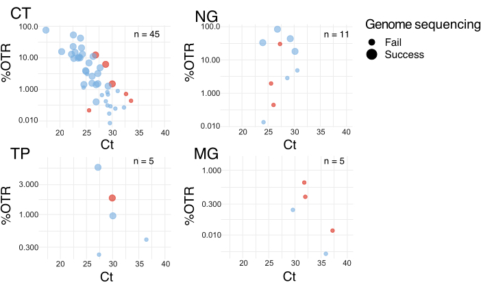
Figure 2: Correlation of %OTR from target enrichment with diagnostic qPCR Ct values. Data point colors represent the sample source: pale blue for Argentina and red for Switzerland. The genome sequencing success, defined as coverage >95% and mean read depth >10, is indicated by large points, while failure is represented by small points. Note the different scales on the y-axis. Abbreviations: CT = C. trachomatis, NG = N. gonorrhoeae, TP = T. pallidum, MG = M. genitalium. This figure has been modified from10. Please click here to view a larger version of this figure.
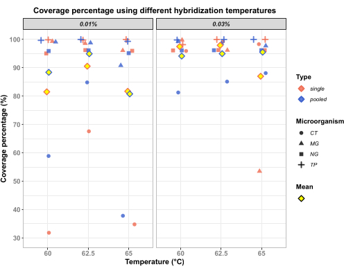
Figure 3: Effect of hybridization temperature on recovery of complete genomes across all STI bacterial pathogens in ten samples. Human DNA in the range of that in clinical samples was spiked with known concentrations of target bacterial DNA separately (single) and also altogether to represent a sample with multiple infections (pooled). Two experiments, each with the five pathogens/combinations, were performed: 0.01% and 0.03% to represent the approximate percentage of DNA from each microorganism relative to the total DNA in the sample (the remaining DNA corresponds to human DNA). Recovery of M. genitalium is improved at lower temperatures. C. trachomatis DNA was spiked at a low concentration; further experiments show that the temperatures did not affect the recovery of C. trachomatis genomes (data not shown). Abbreviations: CT = C. trachomatis, MG = M. genitalium, NG = N. gonorrhoeae, TP = T. pallidum. Please click here to view a larger version of this figure.
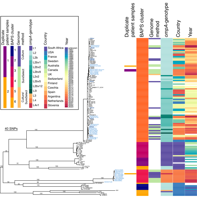
Figure 4: Recombination-adjusted phylogeny of LGV strains from Argentina. The analysis includes all available LGV genomes with published data. Metadata displayed to the right of the phylogeny includes duplicate patient samples, Bayesian Analysis of Population Structure (BAPS; cluster = clade, with the polyphyletic BAPS cluster 6 representing the genetic diversity of the population), genome sequencing method (data from this study), ompA genotype, country, and year, as indicated in the key at the top left. Missing data is shown in white. Genome names from this study are highlighted in light blue for Argentina and dark blue for Finland (not obtained by target enrichment). These colors are used solely to distinguish the genomes generated in this study. Bootstrap values from 1000 ultrafast bootstraps are displayed as percentages on major branches. This figure has been modified from13. Please click here to view a larger version of this figure.
Supplementary Table 1: Description of materials, reagents, instruments, and equipment used in the study. The table includes detailed information on the commercial name, manufacturer, and catalogue number of the components used for DNA extraction, library preparation, target enrichment, and sequencing. It also includes items used for in-house qPCR assays and general molecular biology procedures. Equivalent alternatives may be used for items marked with an asterisk (*). Please click here to download this Table.
Discussion
Generating complete bacterial genome data from clinical samples is particularly challenging. Yet we need this data to gain information on circulating clones and to investigate antimicrobial resistance profiles, particularly in fastidious and difficult-to-culture bacteria. Using alternative methods such as deep sequencing or adaptive sequencing with Oxford Nanopore Technologies, sufficient data for whole-genome sequence analysis cannot be obtained. In cases such as STI samples, target enrichment is the optimum technique to access the full genome, and this method can be used across a panel of bacterial STI pathogens. While it is not a cheap procedure, the results it can offer cannot be obtained in any other way10.
Appropriate samples should be carefully selected, ideally to answer relevant and important questions in diagnostics and research. The medium used for collection does not necessarily play a critical role, with successful sequences obtained from both 2-SP and NAAT media. Sample storage is important, with the aim of maintaining DNA integrity. The most important factor was found to be diagnostic load, aiming for approximate Ct values under Ct30. For samples with Ct values over 30, full genome sequencing success is less likely, but partial data may provide relevant information in some cases.
The procedure is not trivial, involving many steps including PCR cycles, hybridization, and many bead washes. Careful attention to the procedure is required for optimal results. The critical steps of the protocol -- DNA fragmentation, hybridization with capture probes, and magnetic bead-based capture with washing -- require careful execution and proper handling. The protocol can be learned by researchers and technicians after a couple of attempts. In the first rounds, while learning, the 3-day protocol can be an advantage; with more experienced users, the 2-day protocol can speed up the results. As the reagents are expensive, it can be worth running a test run without the expensive components, for example, the probes. For alternative bait sets, the hybridization temperature can be adjusted, which may improve the sensitivity or specificity of the enrichment. If sufficient data is generated, and a considerable proportion of PCR duplicates are observed, the number of pre-capture and post-capture PCR cycles can also be adjusted.
Data obtained from these experiments can be used for typing, with MLST and other relevant targets extracted. It can further be analyzed in an epidemiological context, globally or locally, compared with relevant genomes for contemporary insights. Recombination and mobile element analysis across the genome also infers changes over time and under selective pressure, and possible co-infections. With sufficient data and associated metadata, temporal calculations can be made, and the inference of the ancestors of specific characteristics and lineages, as well as possible transmission routes and mechanisms. All of these advances in our knowledge of an important group of pathogens can inform public health strategies. Our protocol and representative results show the use of syndromic panel target probe sets, which are effective in bacterial STI samples and could be extended to additional sample types.
Disclosures
The authors declare that there are no conflicts of interest.
Acknowledgements
We sincerely thank all those who contributed to this project. From the University of Zürich, we acknowledge Dr Fanny Wegner, Srinithi Purushothaman, Dr Frank Imkamp, Dr Tim Roloff, Dr Dominique Braun, and Prof Dr Adrian Egli for their involvement in the development and optimization of the technique. We extend our gratitude to Daniel Gander, Valéria Pires, Arianita Asani, and Stefan Antener for their outstanding technical assistance with sequencing. From the University of Buenos Aires, we thank Dr Andrea Carolina Entrocassi, Dr María Lucía Gallo Vaulet, Dr Deysi López Aquino, Dr Laura Svidler López, and Dr Luciana La Rosa for their valuable contributions. Finally, we are grateful to Dr Ibrahim Kisakesen and Dr Andreas Schreiber from Agilent for their support. This work was supported by the STIDirect Grant from the Gottfried und Julia Bangerter-Rhyner-Stiftung and by the STISeq Grant STWF-24-009 from the Stiftung für wissenschaftliche Forschung an der Universität Zürich. Additional unrestricted funds from the University of Zurich were provided.
Materials
| Name | Company | Catalog Number | Comments |
|---|---|---|---|
| 8-channel pipette 0.5-10 µL | Eppendorf | 3125000010 | Eppendorf Research plus, 8-channel, 0.5-10 µL, medium gray, variable volume. |
| 8-channel pipette 10-100 µL | Eppendorf | 3125000036 | Eppendorf Research plus, 8-channel, 10-100 µL, yellow, variable volume. |
| Adapter Oligo Mix for MBC-tagged libraries | Agilent Technologies | 5500-0147 | Part of SureSelect XT HS2 Library Preparation Kit for ILM (Pre PCR). |
| Automated nucleic acid extraction system using a viral/pathogen DNA extraction kit | Qiagen | 937036 (Kit) / 9001297 (Instrument) | QIAsymphony SP/AS instrument used with QIAamp DSP Virus/Pathogen Kit for automated DNA extraction. |
| Binding Buffer | Agilent Technologies | 5190-9687 | Part of SureSelect Target Enrichment Kit ILM Hyb Module Box 1 (Post PCR). |
| DNA extraction kit and automated instrument | Zymo Research Corporation | D3024/D3025 | Quick-DNA Miniprep Kit used for total DNA extraction from clinical specimens. Manual or automated workflow compatible. |
| End Repair- A Tailing Enzyme Mix | Agilent Technologies | 5500-0147 | Part of SureSelect XT HS2 Library Preparation Kit for ILM (Pre PCR). |
| End Repair Tailing Buffer | Agilent Technologies | 5500-0147 | Part of SureSelect XT HS2 Library Preparation Kit for ILM (Pre PCR). |
| Ethanol | VWR Chemicals BDH | 20816.298 | Ethanol absolute ≥99.5% Ph. Eur., USP |
| Fluorometer with High Sensitivity dsDNA Assay / Fluorescence-based method to quantify DNA | Thermo Fisher Scientific (Invitrogen) | Q33238, Q33231 | Qubit 4 Fluorometer with 1X dsDNA High Sensitivity (HS) Assay Kit. |
| Fragmentation Buffer | Agilent Technologies | 5191-4080 | Part of SureSelect Enzymetic Fragmentation Kit with the name ''5X Fragmentation Buffer''. |
| Fragmentation Enzyme | Agilent Technologies | 5191-4080 | Part of SureSelect Enzymetic Fragmentation Kit. |
| High-fidelity DNA polymerase buffer | Agilent Technologies | 5500-0147 | Part of SureSelect XT HS2 Library Preparation Kit for ILM (Pre PCR) with the name ''Herculase II Buffer''. |
| High-fidelity DNA polymerase buffer with dNTPs (5X) | Agilent Technologies | 5190-9687 | Part of SureSelect Target Enrichment Kit ILM Hyb Module Box 1 (Post PCR) with the name ''Herculase II Buffer''. |
| High-fidelity fusion enzyme for PCR amplification | Agilent Technologies | 5191-5688 | Part of SureSelect Target Enrichment Kit ILM Hyb Module Box 1 (Post PCR) with the name ''Herculase II''. |
| High-temperature Wash Buffer | Agilent Technologies | 5190-9687 | Part of SureSelect Target Enrichment Kit ILM Hyb Module Box 1 (Post PCR) with the name ''Wash buffer 2''. |
| Hybridization Buffer | Agilent Technologies | 5500-0147 | Part of SureSelect XT HS2 Library Preparation Kit for ILM (Pre PCR). |
| Indexed sequencing primers | Agilent Technologies | 5500-0147 | Part of SureSelect XT HS2 Index Primer Pairs 1-96 for ILM (Pre PCR). |
| In-house multiplex real-time PCR assay | Tib Molbiol | N/A | In-house developed and accredited assay based on Lightmix Kits for STI detection |
| Ligation Buffer | Agilent Technologies | 5500-0147 | Part of SureSelect XT HS2 Library Preparation Kit for ILM (Pre PCR). |
| Magnetic beads for clean up | Agilent Technologies | 5191-5740 | SureSelect DNA AMPure XP Beads. |
| Magnetic stand | Permagen Labware | MSRLV08 | 0.2mL PCR 8 Strip Magnetic Separator 5 µL ~0.2mL Volume. |
| Next-generation sequencing (NGS): High-throughput short-read sequencing system | Illumina, Inc. | N/A | Paired-end 150 bp sequencing (PE150) performed on Illumina NextSeq1000 and MiSeq platforms. |
| Nuclease-free water | BioConcept | 3-07F04-H | Water for Molecular Biology, DNA/DNAse/RNase free, sterile, PCR Inhibitor free. |
| Oligonucleotide Blocker Mix | Agilent Technologies | 5500-0147 | Part of SureSelect XT HS2 Library Preparation Kit for ILM (Pre PCR). |
| PCR tube strip | avantor, delivered by VWR | 732-3608 | PCR tube strips with attached Flat Caps. |
| Post-Capture Primer Mix | Agilent Technologies | 5190-9687 | Part of SureSelect Target Enrichment Kit ILM Hyb Module Box 1 (Post PCR). |
| RNA Probe | Agilent Technologies | 5191-6916 | SureSelect Custom Tier4 6Mb-11.9Mb Probe, eg Design ID S3465164). |
| RNase Blocker | Agilent Technologies | 5500-0147 | Part of SureSelect XT HS2 Library Preparation Kit for ILM (Pre PCR). |
| Room-temperature Wash Buffer | Agilent Technologies | 5190-9687 | SureSelect Target Enrichment Kit ILM Hyb Module Box 1 (Post PCR) with the name ''Wash buffer 1''. |
| Streptavidin beads | Agilent Technologies | 5191-5742 | SureSelect Streptavidin Beads. |
| T4 DNA Ligase | Agilent Technologies | 5500-0147 | Part of SureSelect XT HS2 Library Preparation Kit for ILM (Pre PCR) . |
| Target enrichment method / DNA capture method | Agilent Technologies | G9983-90000 | SureSelect XT HS2 DNA Kit with Library Prep, Fast-Hyb Target Enrichment, and Post-capture Pooling Workflow. Protocol Version F0, May 2023. For Illumina Platform NGS |
| Target enrichment probe set for C. trachomatis, N. gonorrhoeae, T. pallidum, and M. genitalium | Agilent Technologies / Enriching Microbiology Laboratory (IMM, UZH) | Design ID S3465164 | Custom-designed probe set (~242,000 probes) for multiplex capture of four STI pathogens. Design based on multiple reference genomes. |
| Thermal cycler | Thermofischer | 4375305 | Veriti 96-Well Thermal Cycler. |
References
- Lefterova, M. I., Suarez, C. J., Banaei, N., Pinsky, B. A. Next-generation sequencing for infectious disease diagnosis and management: a report of the Association for Molecular Pathology. J Mol Diagn. 17 (6), 623-634 (2015).
- Gwinn, M., Maccannell, D., Armstrong, G. L. Next generation sequencing of infectious pathogens. JAMA. 321 (9), 893-894 (2019).
- Hasman, H., et al. Rapid whole-genome sequencing for detection and characterization of microorganisms directly from clinical samples. J Clin Microbiol. 52 (1), 139-146 (2014).
- Gautam, S., KC, R., Leong, K. W. C., Mac Aogáin, M., O'Toole, R. F. A step-by-step beginner's protocol for whole genome sequencing of human bacterial pathogens. J Biol Methods. 6 (1), e110(2019).
- Baert, L., et al. Guidance document on the use of whole genome sequencing (WGS) for source tracking from a food industry perspective. Food Control. 130, 108148(2021).
- Didelot, X., et al. Transforming clinical microbiology with bacterial genome sequencing. Nat Rev Genet. 13 (9), 601-612 (2012).
- Scidmore, M. A. Cultivation and laboratory maintenance of Chlamydia trachomatis. Curr Protoc Microbiol. 11, (2006).
- Edmondson, D. G., Norris, S. J. In vitro cultivation of the syphilis spirochete Treponema pallidum. Curr Protoc. 1 (2), e44(2021).
- Pitt, R., et al. Challenges of in vitro propagation and antimicrobial susceptibility testing of Mycoplasma genitalium. J Antimicrob Chemother. 77 (11), 2901-2907 (2022).
- Büttner, K. A., et al. Evaluating methods for genome sequencing of Chlamydia trachomatis and other sexually transmitted bacteria directly from clinical swabs. Microb Genom. 11 (2), 001353(2025).
- Seth-Smith, H. M. B., et al. Whole-genome sequences of Chlamydia trachomatis directly from clinical samples without culture. Genome Res. 23 (5), 855-866 (2013).
- SureSelect XT HS2 DNA kits: library preparation (+/-MBC) / fast-Hyb target enrichment / post-capture pooling workflow for Illumina platform NGS. , Agilent Technologies. https://www.agilent.com/cs/library/usermanuals/public/G9983-90000.pdf (2023).
- Büttner, K. A., et al. Chlamydia trachomatis genomes from rectal samples: description of a new clade comprising ompA-genotype L4 from Argentina. Microb Genom. 11 (2), 001350(2025).
- Brown, J. R., et al. Norovirus whole-genome sequencing by SureSelect target enrichment: a robust and sensitive method. J Clin Microbiol. 54 (10), 2530-2537 (2016).
- Dao, T. D., et al. Application of the SureSelect target enrichment system for next-generation sequencing to obtain the complete genome sequence of bovine leukemia virus. Arch Virol. 163 (11), 3155-3159 (2018).
- Cai, W., et al. SureSelect targeted enrichment, a new cost effective method for the whole genome sequencing of Candidatus Liberibacterasiaticus. Sci Rep. 9 (1), 1-8 (2019).
- Büttner, K. A., et al. ompA sequencing and multilocus sequence typing of lymphogranuloma venereum cases in Buenos Aires reveal new Chlamydia trachomatis genotypes. Microorganisms. 12 (3), 587(2024).
- La Rosa, L., et al. Chlamydia trachomatis anorectal infections by LGV (L1, L2 and L2b) and non-LGV serotypes in symptomatic patients in Buenos Aires Argentina. Int J STD AIDS. 32 (14), 1270-1278 (2021).
- Rodríguez Fermepin, M., et al. More than L2b in rectal lymphogranuloma venereum. Lancet Microbe. 2 (2), e55(2021).
Reprints and Permissions
