This article presents a co-culture model of bovine theca and granulosa cells. This method has the potential to serve as a reliable foundation for analyses focused on the study of paracrine communication and substrate transport between the somatic cells within the follicle.
Method Article
A Modified Co-Culture System for Understanding Granulosa-Theca Cell Interactions in the Bovine Ovary
In This Article
Summary
Abstract
A reliable co-culture of granulosa (GCs) and theca cells (TCs) has the potential to facilitate a more comprehensive understanding of the interactions, signaling pathways, and substrate exchange between both cell compartments in the bovine ovarian follicle. Utilizing commercially available cell culture inserts, a reproducible co-culture model of bovine GCs and TCs was established. It was observed that primary theca and granulosa cells require considerably more time to attach to the insert membrane compared to cell lines. Initially, the TCs were seeded onto the inverted insert membrane, with a truncated tube serving as an inoculation container to prevent medium leakage. Following a 72 h incubation period, the insert was inverted, allowing the GCs to be cultured on the opposite side of the membrane. This co-culture was then subjected to an additional 6 days of incubation at 37 °C and 5% CO2, with media exchange every other day. The expression of marker genes indicated cell-type-specific patterns, with CYP17A1 being highly abundant in TCs and not expressed in GCs. Conversely, CYP19A1 showed high levels in GCs and only low levels in TCs. The hormone analysis supported the presence of a physiological co-culture system, as evidenced by the synthesis of estradiol. This modified co-culture model supports reproducible studies of paracrine signaling and substrate transport between the somatic cells of the follicle.
Introduction
During folliculogenesis, a surge in luteinizing hormone (LH) triggers a cascade of physiological changes in the ovary, ultimately leading to the release of a mature oocyte during ovulation1,2. This process is tightly regulated by the follicular somatic cells, which provide essential support for oocyte maturation and ensure proper ovulatory function3. The follicle is composed of two types of steroidogenic somatic cells, an inner layer of granulosa cells (GCs) and an outer layer of theca cells (TCs). Among these, GCs undergo the most pronounced morphological and functional changes following the LH surge, highlighting their critical role in orchestrating the final stages of follicular development4. Consequently, GCs are of considerable interest in the analysis of factors that might influence folliculogenesis, ovulation, and luteinization.
A cell culture model of bovine GCs mimicking an estradiol-active status was previously established5,6,7,8,9,10,11. This serum-free culture model allows a reliable analysis of GC physiology and possible effector analyses in signaling pathways, gene regulation, and hormone synthesis12,13,14. However, the follicle structure is much more complex, as highlighted by the 2-cell-2-gonadotropin hypothesis15. This underscores the dependence and interconnected nature of the cell types in the context of steroid synthesis being compartmentalized between the two layers.
A number of studies have described TC culture models that are similar to previously established GC mono-culture systems16,17,18. However, a co-culture system of porcine theca and granulosa cells was reported, in which both cell populations are cultivated in a single dish at a ratio of 4:1 of GCs to TCs19,20,21. In this model, it could be shown that there is a functional system and interaction between granulosa and theca cells, revealing a higher amount of estradiol in the co-culture system than in the mono-culture. However, it should be noted that this model is not able to adequately analyze the interaction and substrate exchange between granulosa and theca cells, as both cell types might be differently affected. Therefore, the analysis on molecular levels of GCs and TCs independently from each other is nearly not possible without an elaborate sorting procedure, which itself might affect cell physiology. Furthermore, it is important to consider that this model may not accurately replicate the situation prior to the LH surge, when GCs and TCs are separated by a basement membrane3. In the bovine, a membrane-based cell culture system was proposed, in which both cell types are cultivated on either side of a collagen membrane, thereby reflecting the in vivo situation more precisely22,23,24,25,26.
Adapting the initial concept of a membrane-based system, this method aimed to establish a co-culture model for bovine GCs and TCs with cell culture inserts to enable future studies of paracrine signaling between both cell compartments. The presented method utilizes commercially available hanging cell culture inserts instead of a custom-based membrane system, offering advantages such as higher reproducibility, standardized quality, and improved product stability. This approach ensures consistency across experiments, making it a valuable tool for researchers studying follicular cell interactions. The culture procedure includes an initial seeding of 1.7-1.8 x 10⁵ theca cells on the underside of the insert, followed by approximately 1.0 x 10⁵ granulosa cells on the upper surface after inversion and a 72 h pre-incubation. Cultures are maintained in defined medium under standard conditions (37 °C, 5% CO2) with media exchanged every other day. To enable paracrine exchange across compartments, 0.4 µm cell culture inserts made of polyester, such as polyethylene terephthalate (PET), were used (commercially available). Membranes of this pore size permit diffusion of soluble macromolecules, such as hormones, growth factors, and proteins, while preventing cell migration27,28.
By enabling spatial separation while maintaining paracrine communication, this co-culture system provides a physiologically relevant environment for investigating granulosa-theca cell interactions, signaling pathways, and substrate exchange. The model is particularly suited for studies requiring controlled conditions, such as hormone regulation, gene expression analysis, or drug testing. Researchers interested in follicular dynamics, reproductive physiology, or ovarian disorders may find this method applicable to their studies. Additionally, the use of standardized inserts allows for easy adaptation to different experimental needs, including modifications in culture duration, cell types, or treatment conditions.
Protocol
Bovine ovaries were obtained from a local commercial abattoir. In this experimental setup, the preparation of the ovaries is performed within 2 h after receiving. The collection of slaughterhouse byproducts does not require ethical approval according to German Law.
NOTE: The isolation and cryo-preservation of bovine GCs has already been described and shown in a previous method report6 and can be performed accordingly.
1. Preparation of reagents and setup for ovarian follicle dissection
- Prepare 1x PBS (pH 7.4) supplemented with 100 IU penicillin, 0.1 mg/mL of streptomycin, and 0.5 µg/mL of amphotericin for transport of ovaries. The solution can be stored at 4 °C for up to 4 weeks.
- Prepare the digestion solution containing 10x Hanks' salt buffer supplemented with 25 mM HEPES, 100 IU penicillin, 0.1 mg/mL of streptomycin, and 0.5 µg/mL of amphotericin. The digestion solution can be stored at 4 °C for 2-3 weeks.
- Fill 1 mL of digestion solution in 1.5 mL tubes for each planned dissected follicle and pre-warm the solution to 37 °C.
- Add 10 µL of Collagenase NB8 to achieve a 0.1% concentration of the digestion solution shortly before preparation.
- Prepare the following dishes for preparation, isolation, and washing procedures.
- Glass dishes with a minimum diameter of 3 cm to place the ovaries for dissection.
- A 6-well plate with 1 mL 1x PBS (supplemented with antibiotics) for washing.
- A 12-well dish with approximately 10 µL of digestion solution.
- For cryo-preservation, prepare a freezing solution consisting of 80% fetal calf serum (FCS) and 20% DMSO.
NOTE: The final concentration of DMSO for cryo-preservation will be 10%.
2. Follicle dissection with subsequent digestion of bovine theca cells
- Perform the cell digestion procedures as well as the cell culture work under a laminar flow bench.
- Wash the ovaries 2-3x in 1x PBS (supplemented with antibiotics, see step 1.1) to remove blood and other residues from the surface. Place the ovaries in a tumbler, fill it with 1x PBS so that the ovaries are covered, and discard the solution afterwards. Repeat this step 3-4x until the ovaries are clean.
- Place one ovary in a glass dish and select follicles of 5-11 mm, measured with a ruler, for dissection.
- Aspirate the follicular fluid with a 3 mL syringe and an 18 G needle by puncturing one selected follicle. Discard the follicular fluid.
- Cut the follicle open with scissors, starting at the point where the needle was inserted during aspiration. Use a pair of binoculars for precise cutting.
- Use tweezers at the cutting edge of the follicle to grasp the innermost layer, the theca interna.
- Try to peel off the theca interna from the inner side of the open follicle wall.
NOTE: Depending on the age or stage of the follicle, the theca cell layer can be more easily peeled off, resulting in an intact theca layer.
- Place the theca cell layer in a dish filled with 1x PBS (supplemented with antibiotics, see step 1.1.) to remove the remaining granulosa cells.
- Gently scrape the granulosa cells from the inner side of the membrane with a scalpel. The cells should detach easily in large clusters.
- Place the theca cell layer in a new dish filled with 1x PBS to wash it.
- Carefully rinse the membrane by swirling in the fresh 1x PBS to remove any loose cells.
NOTE: From the next step onwards, work under the laminar flow box.
- Transfer the theca interna into the prepared digestion solution of the 12-well plate (see step 1.2 and 1.3.3) and cut it into small pieces, presumably 1-3 mm, using a scalpel.
NOTE: It is recommended to digest theca cells from different follicles independently to prevent the clogging of tissues from different origins. - Transfer the theca snippets into a prepared 1.5 mL reaction tube (see step 1.2.1 and 1.2.2). Incubate the theca in a thermos-shaking incubator at 800 rpm at 37 °C for 45-50 min.
- After shaking for 30 min, begin vortexing the reaction tubes for 3-5 s and place them back in the shaking incubator.
- Repeat this step 3-4x in the remaining incubation time.
- After incubation, resuspend the digested cells with a pipette.
- Eliminate the undigested tissue by filtration with the 100 µm cell strainer placed on a 50 mL tube, and proceed immediately with counting and cryo-preservation of the cells to remove the digestion media.
3. Counting and cryo-preservation of TCs
- To count the number of viable cells, prepare a tube with 1.5 µL of a 0.25% trypan blue solution. Living cells will remain unstained, while dead cells appear blue due to the access of trypan blue through broken cell barriers.
- Add 15 µL of the digested cell suspension to the trypan blue solution and mix gently.
- Place 10 µL of the mixture in both chambers of a hemocytometer. Count the number of living (= colorless) cells in one big square per chamber.
- Determine the number of living cells with the mean of both chamber squares following standard guidelines. Note that the proportion of living cells may differ between ovaries.
- Freeze undigested portions of the theca cell suspension (see step 2.9) for an intended comparison as a non-cultured control and to perform an assessment of potential cross-contamination with GCs (as shown in Figure 2).
- Transfer the undigested tissue of each dissected theca interna that remains in the 100 µm cell strainer (see step 2.10) into a labeled tube.
- Shock-freeze the tube in liquid nitrogen before storing at -80 °C.
- Perform cryo-preservation of the isolated theca cells in order to facilitate improved planning of cell cultures, with particular regard to the number of cells required.
- Centrifuge the digested cell suspension at 500 x g for 5 min at room temperature (in average 20 °C). Discard the supernatant.
- Carefully dissolve the cell pellet in 1 mL of 100% FCS. Add 1 mL of freezing media (see step 1.4) to the cell solution. The final concentration of DMSO for cryo-preservation is 10%. Transfer the mixture into the specified 2 mL cryo vials.
- Place the vials in a cooler device for a controlled decrease in temperature. In this setup, a specific freezing container was used, which guarantees a freezing rate of -1 °C/min when placed for at least 4 h in -80 °C. Afterwards, the vials can be stored in liquid nitrogen storage systems.
4. Preparations for cell culture work
- Cut off the lid from a 1.5 mL reaction tube. Then, cut the reaction tube approximately 1 cm from the opening with the help of a scalpel (preferably warmed) or an appropriate hose cutter. Afterwards, autoclave the cut tubes and lids. The lids might be required for subsequent procedures of the cell culture (see step 5.5).
NOTE: Ensure that the 1.5 mL tubes tightly fit onto the insert chosen. - Ensure to coat the inserts with 0.02% collagen R for GC's and TC's attachment in the culture.
NOTE: Perform the procedure of coating under a sterile laminar flow hood.- Use purified water (suitable for cell culture) to prepare a 0.02% collagen R solution. Prepare 80 µL of this solution for each cell culture insert required.
- Place the insert upside down in a lid of a 24-well plate. Gently add 40 µL of collagen R solution to the membrane of the insert, ensuring that it remains in place. Leave the collagen R solution to dry for 4-5 h under the laminar flow hood.
- Turn the insert and hang it in the wells of the 24-well plate to coat the second side again with 40 µL of collagen R solution. Let the solution dry again for 4-5 h.
- Irradiate the inserts with UV light for 10-15 min to diminish contamination.
- The inserts should be stored within the 24-well plate (preferably in the sterile delivery bag) and in a fridge at 4 °C and can be used up to 4 weeks.
- Prepare the media8,10 as follows:
NOTE: Perform the procedure of media preparation under a sterile laminar flow hood.- Supplement the α-Minimial Essential Medium (α-MEM) with 2 mM L-glutamine, 0.084% sodium bicarbonate, 0.1% BSA, 20 mM HEPES, 4 ng/mL sodium selenite, 5 µg/mL transferrin, 10 ng/mL insulin, and 1 mM non-essential amino acids.
NOTE: To ensure consistency, it is recommended to prepare enough medium for the entire experiment in advance. The prepared medium can be stored at 4 °C. For the described setup, use 750 µL per cell culture well. - Further, add 100 IU penicillin, 0.1 mg/mL of streptomycin, and 0.5 µg/mL of amphotericin to the prepared media.
- Pre-warm the media at 37 °C in a water bath.
NOTE: After the pre-warming of media, the antibiotics are stable for approximately 48 h. Therefore, aliquot only the desired amount of media for warming. - Additionally, supplement the α-MEM with 20 ng/mL of follicle stimulating hormone (FSH), 50 ng/mL of R3 IGF-1, and 2 µM androstenedione for a physiological cell environment7,8,10,11.
NOTE: Add these components shortly prior to the onset of culture or media exchange. In order to prevent a compromised activity, it is imperative to avoid repeated freeze-thaw cycles of the FSH and IGF-1 stock solutions.
- Supplement the α-Minimial Essential Medium (α-MEM) with 2 mM L-glutamine, 0.084% sodium bicarbonate, 0.1% BSA, 20 mM HEPES, 4 ng/mL sodium selenite, 5 µg/mL transferrin, 10 ng/mL insulin, and 1 mM non-essential amino acids.
5. Co-culture of TCs and GCs
- Prepare the inoculation chamber for the culture of theca cells (Figure 1A).
- Place the coated insert upside down into a larger plate (e.g., 12-well plate).
- Place the cut and autoclaved tube on an inverted insert (see step 4.1). Ensure that the tube is firmly seated on the insert to prevent media leakage.
- Thaw the cryo-preserved theca cells quickly in a water bath of 37 °C for 3-5 min and transfer the cell suspension immediately after thawing into the pre-warmed media.
- Centrifuge the theca cell suspension at 500 x g for 3 min at room temperature (in average 20 °C) and discard the supernatant. Add the supplemented α-MEM and carefully resuspend the cells.
- Seed 1.7-1.8 x 10⁵ cells per insert in the inoculation chamber in a final volume of 200 µL media. Include at least three technical replicates per condition.
- To close the cell culture dish with a lid, it might be necessary to increase the distance between the dish and the lid when using the inoculation chamber.
- Use the cut and autoclaved caps from the 1.5 mL reaction tubes (see step 4.1) and place them in each corner of the cell culture plates. The caps will act as supports to lift the lid of the plate slightly, providing enough space for the inoculation chamber while still covering the culture dish adequately.
- Close the well plate carefully, ensuring it remains stable and does not touch the membrane or chamber.
- Gently transfer the plate into a humidified cell culture incubator.
- Incubate the theca cells for 3 days at 37 °C with 5% CO2 to allow cell attachment on the membrane.
- Turn the insert after 3 days of culture to prepare for seeding with GCs (Figure 1B).
- Prepare a 24-well plate and fill with 500 µL of media in each of the desired wells.
- With a pipette, remove all media from the inoculation chamber. Carefully remove the inoculation chamber from the insert with tweezers.
- Place the insert with the attached theca cells into the prepared 24-well plate, ensuring that the TCs face down (see Figure 1B for more details).
- To seed granulosa cells from cryo-preservation, perform steps 5.2 and 5.3.
- Seed approximately 1.0 x 10⁵ cells per insert in a final volume of 250 µL of media that is placed inside the insert.
- Subsequently, incubate the co-culture at 37 °C with 5% CO2 for six more days.
- Perform a media exchange every 48 h after GC seeding when only two-thirds of the culture media is replaced. This should reduce the stress of the procedure and help to adapt to the newly added media.
6. Subsequent analysis of cultured theca and granulosa cells
- Perform morphological assessment of theca and granulosa cells throughout the cell culture with a phase-contrast microscope to monitor attachment and spreading of both cell types (see Figure 3).
- For the steroid hormone analysis, collect the media and freeze at -20 °C. Use an appropriate method to analyze the concentrations of the desired steroid hormones (e.g., in this workflow, estradiol was analyzed via RIA5,29).
NOTE: A steroid hormone analysis helps to evaluate the physiological status of the culture7. - To analyze the gene expression, firstly isolate the RNA with an appropriate technique (e.g. kit procedures).
NOTE: Be aware that during the isolation procedure, both cell types are prepared independently from each other.- Dissociate the cells from both sides of the membrane with an appropriate reagent (e.g., trypsin or accutase). Place the dissociation medium in both the insert (e.g., 200 µL of accutase) and wells of the plate (e.g., 500 µL of accutase) so that the membrane is completely surrounded by the dissociation medium.
- Collect the granulosa cells from the insert, whereas the detached theca cells can be collected from the well. Transfer every cell type into a 1.5 mL tube.
- Centrifuge the cells at 500 x g for 3 min and discard the supernatant.
- Lyse the cells, perform RNA isolation and afterwards measure RNA concentration.
NOTE: The protocol can be stopped after lysis of cells for most isolation procedures so that samples can be stored at -20 °C for short term. For a longer storage, the lysed cells can be stored at -80 °C. - Subsequently, synthesize cDNA to measure the abundance of transcripts of interest by qPCR. The cDNA synthesis utilizes 100 ng of RNA with random hexamer and oligo-dT primers. For qPCR, gene-specific primers were used as previously described5,30. In brief, cycle conditions were as follows: pre-incubation at 95 °C for 5 min, 40 cycles denaturation at 95 °C for 20 s, annealing at 60 °C for 15 s, extension at 72 °C for 15 s, and single-point fluorescent acquisition for 10 s. An external standard of five different concentrations (5x10-12- 5x10-16 g DNA/reaction) was co-amplified. For normalization of transcript abundance, the geometric mean of RPLP0 and TBP was calculated.
- To eliminate potential cross-contamination, evaluate the purity of theca and granulosa cell preparations by analyzing the gene expression of the cell-specific markers CYP17A1 and CYP19A1 from the freshly isolated cell preparations of TCs and GCs (Figure 2).
NOTE: A statistical evaluation should include a minimum of three replicates of different cell cultures. In this experimental setup, a t-test or the one-way ANOVA was performed after checking for normality and variance.
Results
Although TCs and GCs were isolated using well-established mechanical separation techniques based on follicular compartmentalization, the expression of cell-type-specific genes CYP17A1 and CYP19A1 was evaluated to eliminate the risk of cross-contamination. CYP17A1 was exclusively expressed in freshly isolated TCs, while CYP19A1 was predominantly expressed in freshly isolated GCs (Figure 2).
The co-culture system enables the compartmentalization of bovine theca and granulosa cells on opposing sides of a membrane insert, thereby mimicking the in vivo spatial organization. After three days in culture, the TCs exhibited a flattened and elongated morphology as they spread across the surface of the collagen-coated insert membrane, indicating successful attachment (Figure 3A). When cultured alone for the entire 9-day period, TCs continued to proliferate and reached confluence by day 9 (Figure 3B). In contrast, GCs cultured alone for 6 days on the collagen-coated insert membrane developed a fibroblast-like phenotype, which is characteristic of GCs in vitro (Figure 3C). Additionally, as previously described, GCs tended to form clusters, a typical feature of these cells in culture5,7,31,32. In the co-culture system, no apparent morphological differences were observed in either TCs or GCs when compared to their respective time-matched mono-cultures (Figure 3D), indicating that the insert-based environment maintains normal cellular behavior. However, due to technical limitations, microscopic evaluation of the co-culture remains challenging, particularly in distinguishing and assessing TC morphology independently from the other cells. To overcome these limitations, an optimized protocol for cell fixation is currently being addressed in accordance with exploring alternative strategies for cell identification in the absence of cell-specific antibodies for immunostaining.
The functionality of the co-culture system was assessed by analyzing hormone production and the expression of key marker genes for TCs and GCs33,34, confirming the cell-type-specific steroidogenic roles. A comparison between the GC mono-culture and the co-culture model revealed similar estradiol (E2) levels produced in vitro. However, as expected, the TC mono-culture produced only negligible amounts of E2, consistent with the 2-cell-2-gonadotropin hypothesis (Figure 4A)15, which states that theca cells lack the ability to synthesize E2 independently.
To further evaluate the physiological relevance of the culture conditions, the expression of key marker genes33 was examined. In accordance with the 2-cell-2-gonadotropin hypothesis, CYP19A1 was predominantly expressed in GCs, whereas CYP17A1 was mainly detected in TCs, a pattern observed in both mono- and co-culture conditions (Figure 4B,C). Notably, the transcript abundance of CYP19A1 was higher in GCs within the co-culture system compared to the GC mono-culture, despite no observed differences in estradiol concentrations.
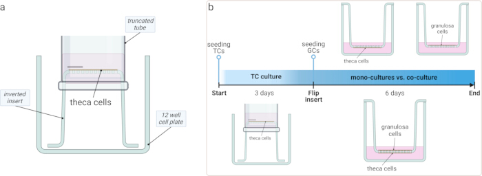
Figure 1: Schematic representation of cell culture setup. (A) A diagram illustrating the custom-built inoculation chamber, which utilizes a truncated tube on the inverted insert to prevent media leakage throughout the initial culture period. (B) Timeline of the experimental procedure: TCs were cultured on the inverted insert for three days to allow attachment. The insert was flipped, and GCs were seeded on the opposite side, followed by an additional six days of culture. The study compared TCs and GCs cultured separately to the TC-GC co-culture system. Please click here to view a larger version of this figure.
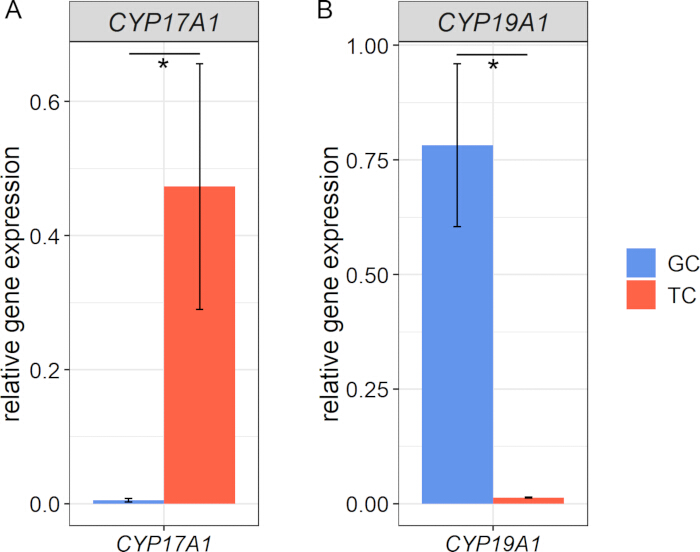
Figure 2: Gene expression of cell-type-specific markers in freshly isolated granulosa and theca cells. (A) CYP17A1 was exclusively expressed in TCs, while (B) CYP19A1 was predominantly detected in GCs, confirming the specificity and initial purity of the isolated cell populations prior to culture. Data are presented as relative expression normalized to the geometric mean of the reference genes RPLP0 and TBP. Means and standard error of means are shown, data were tested for normality and variance, asterisks indicate significant differences (n=3, t-test). Please click here to view a larger version of this figure.
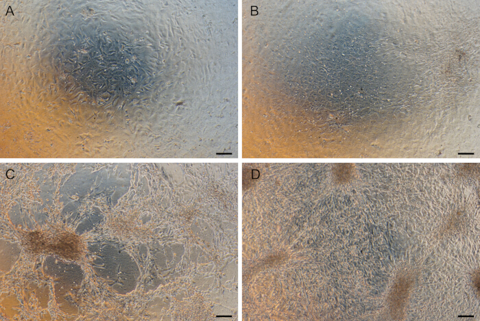
Figure 3: Morphology of theca and granulosa cells in culture. (A) TCs cultured for 3 days were attached to the collagen-coated insert membrane. (B) TCs grew to confluence after the entire 9-day period in the mono-culture. (C) GCs display the typical fibroblast-like phenotype after 6 days in culture. (D) Co-culture of TCs and GCs shows no apparent morphological difference compared to respective time-matched mono-cultures. This image was taken after 6 days of GC culture and 9 days of TC culture (at the endpoint of the co-culture experiment). Scale bars 100 µm. Please click here to view a larger version of this figure.
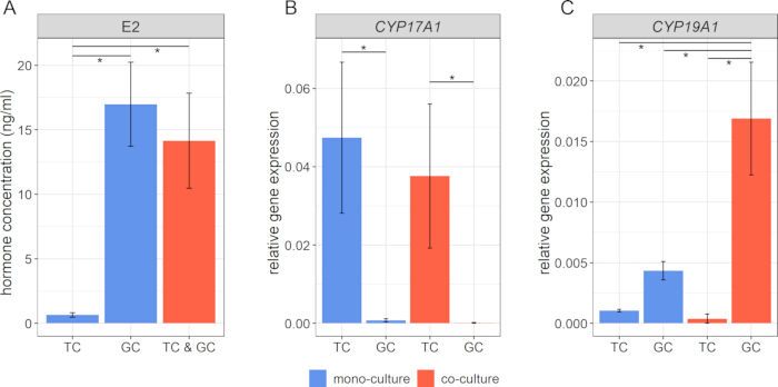
Figure 4: Analysis of functional markers in the co-culture system. (A) Hormone concentrations of estradiol (E2) confirm physiological functionality of the cell culture system. (B, C) Gene expression analysis of the TC marker CYP17A1 and the GC marker CYP19A1 demonstrates the identity and functional state of both cell populations. The geometric mean of the reference genes RPLP0 and TBP was used to calculate the relative expression. Means and standard error of means are shown, data were tested for normality and variance, asterisks indicate significant differences (n=3, one way ANOVA). Please click here to view a larger version of this figure.
Discussion
The prerequisite to perform this co-culture model is a well-performed isolation of both cell types to prevent any cross-contamination. The purity of both cell preparations was evaluated by the gene expression of the TC-specific marker CYP17A1 and the GC-specific marker CYP19A1. As shown before, the technique of GC and TC isolation showed no relevant cross-contamination30.
This co-culture model allows for the simultaneous culture of theca and granulosa cells on opposite sides of a membrane, mimicking their spatial organization within the follicle and supporting the physiological state of both cell populations3. The model's validity is reinforced by its hormone production profile and gene expression patterns that align with the 2-cell-2-gonadotropin hypothesis15. As expected, the low estradiol levels in the TC mono-culture confirm that theca cells alone are unable to synthesize estradiol, supporting established knowledge of follicular steroidogenesis35. Despite the higher CYP19A1 expression in co-cultured GCs, estradiol levels remained comparable to the GC mono-culture, suggesting the influence of regulatory mechanisms such as substrate availability, feedback inhibition, or enzyme activity differences. Further exploration into the signaling pathways and underlying mechanisms is warranted, as a subject for future studies. Nevertheless, the establishment of a functional co-culture system of bovine TCs and GCs is imperative, as provided in this study. The present study focused on estradiol production and mRNA expression levels of CYP17A1 and CYP19A1 as key indicators of the co-culture model's functionality. Both represent sensitive markers of granulosa and theca cell function4,8,36 and are directly aligned with the 2-cell-2-gonadotropin hypothesis15. While recognizing the importance of protein expression, the lack of reliable antibodies precluded its accurate assessment; therefore, a comprehensive analysis of protein levels was beyond the scope of this initial investigation.
In general, this model is a further development of the traditional 2D culture model, which partly mimics the structure of the ovarian follicle3. The results support a physiological status of both cell compartments revealed by the expression of sensitive markers for GC and TC physiology.
An advantage over traditional 2D culture models is facilitating paracrine interactions between theca and granulosa cells while maintaining their spatial separation, better mimicking the in vivo follicular structure. In contrast, on-dish co-culture systems, where TCs and GCs are mixed in a single well, lack this structural organization. Studies using such systems have commonly applied a GC:TC ratio of 4:119,20,21, but this approach does not reflect the compartmentalization found in vivo and might, therefore, not be feasible for analyzing substrate exchange. Furthermore, alterations found on the cell level cannot be easily detected, when this GC-TC mixture has to be sorted in a complex manner.
Three-dimensional co-culture models for granulosa and theca cells remain scarce, but early attempts have shown promising results. The group of Kotsuji22,23,24,37 previously described the interaction of granulosa and theca cells in culture using a collagen membrane as a physical separator. However, this system relied on a custom-built collagen membrane, making reproducibility and standardization challenging. In contrast, the presented co-culture model benefits from commercially available hanging culture inserts, which enhance reproducibility, standardization, and product quality, ensuring broader applicability across laboratories. Although the herein presented model resulted in higher estradiol production than the previous models, the comparison of these models is difficult, as it lacks unity of cell culture supplements, especially the concentrations used26 or the additional usage of IGF-122,23,24,37.
One of the most crucial steps in the procedure is the culture of TCs within the inoculation chamber. Ensuring that the chamber is securely placed on the inverted insert is essential to prevent media leakage, which could otherwise compromise TC attachment and survival. Therefore, it is advised to firmly press the inoculation chamber on the inverted insert with the help of tweezers and visually inspect the placement of the inoculation chamber. Additionally, removing the inoculation chamber and flipping the insert after three days requires technical precision. Some skill training may be necessary to perform this step without disrupting and/or damaging the attached cells. Another important consideration is the seeding density of GCs. Higher cell density exceeding optimal levels of 1.0 x 10⁵ cells per insert can lead to differentiation of GCs, potentially affecting steroidogenic output. Moreover, the presented cryo-preservation method yielded in viability after thawing of 80-90% for both cell types.
Although the described model offers a physiological and reproducible system to study granulosa and theca cell interactions, some limitations should be considered. The method is adaptable to other species as well; however, species-specific differences might require optimization of cell culture parameters, like cell densities, timing, or media composition. The scalability of this model is limited to the availability of the cell culture inserts, but it might be sufficient for most effector studies. Finally, this co-culture model is a further step towards mimicking the in vivo situation of the follicle, but still lacks the full complexity of the ovarian follicle.
In conclusion, the presented model can be easily modified to accommodate different experimental needs, such as extending the culture period, adjusting cell numbers or ratios, or incorporating additional factors. With further development, this system represents a valuable tool for studying follicular function, steroidogenesis and cell signaling between both cell compartments. Additionally, it holds potential for applications in reproductive toxicology, providing a controlled platform to assess the impact of different compounds on follicular cells.
Disclosures
All authors declare that they have no conflicts of interest.
Acknowledgements
We would like to express our appreciation to Veronica Schreiter, Maren Anders and Olivier Seidel for their excellent technical assistance. Furthermore, we would like to acknowledge the commercial abattoir "DANISH CROWN Teterower Fleisch GmbH" (Germany), from whom we obtained the ovaries for this study.
Materials
| Name | Company | Catalog Number | Comments |
|---|---|---|---|
| 10x Hank's Balanced Salt Solution HBSS (w Mg/Ca, Phenol Red) | Bio&Sell | BS.L201-10 | dissolve in ultrapure water; adjust pH to 7.4 of the digestion solution (HBSS and HEPES) and perform sterile filtration before use |
| 18 G needle, 40 mm, diameter 1.2 mm | Roth | 281T.1 | disposable needle, short cut |
| 3 ml syringe (Omnifix Luer) | Roth | LY22.1 | |
| accutase | Merck | A6964 | company branch: Sigma-Aldrich |
| amphotericin B (250 µg/ml) | Bio&Sell | BS.AB17.01050 | |
| androstenedione | Merck | 46033 | we use a stock solution of 10 mM, diluted in 100 % ethanol; company branch: Supelco |
| binocular Carl Zeiss Technival2 | Carl Zeiss Jena | not applicable | used together with incident light Schott KL 1600 LED |
| BSA (5%) | Merck | A3311 | company branch: Sigma-Aldrich |
| cell strainer (100 µm pore size) | Sarstedt | 83.3945.100 | fitting to 50 ml centrifugation tubes |
| collagen R (0.2 %) | Serva | 47254 | |
| Collagenase NB8 (10%) | Nordmark | S1745601 | |
| CoolCell LX cell freezing container | Merck | CLS432002 | product from Corning |
| cryo-preservation vials (2 ml) | TPP Faust | TPP89020 | |
| culture dish, 12-well, flat bottom | TPP Faust | TPP92112 | |
| culture dish, 24-well, flat bottom | TPP Faust | TPP92424 | |
| culture dish, 6-well, flat bottom | TPP Faust | TPP92006 | |
| DMSO | Roth | 4720.1 | |
| DPBS w/o Ca2+, Mg2+ | Bio&Sell | BS.L182-10 | dissolve in ultrapure water and autoclave before use |
| estradiol, 17b-[2,4,6,7-3H] | Hartmann Analytik | ART1555 | |
| fetal calf serum (FBS Superior stabil) | Bio&Sell | FBS.S0615 | |
| FSH (~7,000 IU/mg; from human pituitary) | Merck | F4021 | we prefer to use a stock solution of 2 µg/ml, diluted in 0.9 % NaCl; company branch: Sigma-Aldrich |
| hanging cell culture inserts (0.4 µm pore size) | cellQART | 9320412 | while ordering be aware of the different properties of inserts; we prefer a clear PET membrane for phase contrast microscopy |
| HEPES (1 M) | Roth | 9105.2 | adjust pH to 7.4 of the digestion solution (HBSS and HEPES) and perform sterile filtration before use |
| holo-transferrin (cattle) | Merck | T1283 | dissolve in sterile water; company branch: Sigma-Aldrich |
| innuPREP RNA Mini Kit 2.0 | IST innuscreen | 845-KS-2040250 | |
| insulin (10 mg/ml; from bovine pancreas) | Merck | I0516 | dissolve in 1x PBS; company branch: Sigma-Aldrich |
| L-Glutamin (200 mM) | Bio&Sell | BS.K0283 | |
| LR3-IGF-1 | Merck | I1146 | we use a stock solution of 200 µg/ml, dissolved in 10 mM HCl and 1x PBS with 1 mg/ml BSA; company branch: Sigma-Aldrich |
| NanoDrop 1000 Spectrophotometer | ThermoScientific | not applicable | RNA concentration measurement |
| NEA (100x) | Bio&Sell | BS.K0293 | |
| penicillin/streptomycin (10,000 IU/10 mg/ml) | Bio&Sell | BS.A2213 | |
| purified water, suitable for cell culture | Merck | W3500 | company branch: Sigma-Aldrich |
| SensiFAST cDNA Synthesis Kit | Bioline | BIO-65054 | supplied through distribution partner BioCat |
| sodium bicarbonate (7.5%) | Bio&Sell | BS.L1713 | |
| sodium selenite | Merck | S9133 | dissolve in sterile water; company branch: Sigma-Aldrich |
| thermoshaker TS-100C | Fischer Scientific | 12357627 | used together with HC18 block (Cat.-No. 12337627) |
| α-MEM | Bio&Sell | BS.F0915 |
References
- Adams, G. P., Jaiswal, R., Singh, J., Malhi, P. Progress in understanding ovarian follicular dynamics in cattle. Theriogenology. 69 (1), 72-80 (2008).
- Forde, N., et al. Oestrous cycles in Bos taurus cattle. Anim Reprod Sci. 124 (3-4), 163-169 (2011).
- Hennet, M. L., Combelles, C. M. The antral follicle: A microenvironment for oocyte differentiation. Int J Dev Biol. 56 (10-12), 819-831 (2012).
- Christenson, L. K., et al. Research resource: Preovulatory LH surge effects on follicular theca and granulosa transcriptomes. Mol Endocrinol. 27 (7), 1153-1171 (2013).
- Baufeld, A., Vanselow, J. Increasing cell plating density mimics an early post-LH stage in cultured bovine granulosa cells. Cell Tissue Res. 354, 869-880 (2013).
- Baufeld, A., Vanselow, J. A Tissue culture model of estrogen-producing primary bovine granulosa cells. J Vis Exp. (139), e58208(2018).
- Gutierrez, C. G., Campbell, B. K., Webb, R. Development of a long-term bovine granulosa cell culture system: Induction and maintenance of estradiol production, response to follicle- stimulating hormone, and morphological characteristics. Biol Reprod. 56 (3), 608-616 (1997).
- Hamel, M., Vanselow, J., Nicola, E. S., Price, C. A. Androstenedione increases cytochrome P450 aromatase messenger ribonucleic acid transcripts in non-luteinizing bovine granulosa cells. Mol Reprod Dev. 70, 175-183 (2005).
- Chandrasekher, Y. A., Fortune, J. E. Effects of oxytocin on steroidogenesis by bovine theca and granulosa cells. Endocrinology. 127 (2), 926-933 (1990).
- Silva, J. M., Price, C. A. Effect of follicle-stimulating hormone on steroid secretion and messenger ribonucleic acids encoding cytochromes P450 aromatase and cholesterol side-chain cleavage in bovine granulosa cells in vitro. Biol Reprod. 62 (1), 186-191 (2000).
- Gong, J. G., McBride, D., Bramley, T. A., Webb, R. Effects of recombinant bovine somatotrophin, insulin-like growth factor-I and insulin on bovine granulosa cell steroidogenesis in vitro. J Endocrinol. 143 (1), 157-164 (1994).
- Baufeld, A., Koczan, D., Vanselow, J. L-lactate induces specific genome-wide alterations of gene expression in cultured bovine granulosa cells. BMC Genomics. 20 (1), 273(2019).
- Baddela, V. S., Sharma, A., Plinski, C., Vanselow, J. Palmitic acid protects granulosa cells from oleic acid induced steatosis and rescues progesterone production via cAMP dependent mechanism. Biochim Biophys Acta Mol Cell Biol Lipids. 1867 (8), 159159(2022).
- Sharma, A., et al. Elevated free fatty acids affect bovine granulosa cell function: a molecular cue for compromised reproduction during negative energy balance. Endocr Connect. 8 (5), 493-505 (2019).
- Hillier, S. G., Whitelaw, P. F., Smyth, C. D. Follicular oestrogen synthesis - The two-cell, two- gonadotrophin model revisited. Mol Cell Endocrinol. 100, 51-54 (1994).
- Campen, K. A., Lavallee, M., Combelles, C. M. H. The impact of bisphenol S on bovine granulosa and theca cells. Reprod Domest Anim. 53 (2), 450-457 (2018).
- Glister, C., Richards, S. L., Knight, P. G. Bone morphogenetic proteins (BMP) -4, -6, and -7 potently suppress basal and luteinizing hormone-induced androgen production by bovine theca interna cells in primary culture: could ovarian hyperandrogenic dysfunction be caused by a defect in thecal BMP signaling. Endocrinology. 146 (4), 1883-1892 (2005).
- Wrathall, J. H., Knight, P. G. Effects of inhibin-related peptides and oestradiol on androstenedione and progesterone secretion by bovine theca cells in vitro. J Endocrinol. 145 (3), 491-500 (1995).
- Shores, E. M., Picton, H. M., Hunter, M. G. Differential regulation of pig theca cell steroidogenesis by LH, insulin-like growth factor I and granulosa cells in serum-free culture. J Reprod Fertil. 118 (2), 211-219 (2000).
- Gregoraszczuk, E. L., Ptak, A., Karniewska, M., Ropstad, E. Action of defined mixtures of PCBs, p,p'-DDT and its metabolite p,p'-DDE, on co-culture of porcine theca and granulosa cells: steroid secretion, cell proliferation and apoptosis. Reprod Toxicol. 26 (2), 170-174 (2008).
- Rak, A., Drwal, E., Wróbel, A., Gregoraszczuk, E. Resistin is a survival factor for porcine ovarian follicular cells. Reproduction. 150 (4), 343-355 (2015).
- Kotsuji, F., Tominaga, T. The role of granulosa and theca cell interactions in ovarian structure and function. Microsc Res Tech. 27 (2), 97-107 (1994).
- Orisaka, M., et al. Effects of ovarian theca cells on granulosa cell differentiation during gonadotropin-independent follicular growth in cattle. Mol Reprod Dev. 73 (6), 737-744 (2006).
- Tajima, K., et al. Role of granulosa and theca cell interactions in ovarian follicular maturation. Microsc Res Tech. 69 (6), 450-458 (2006).
- Tajima, K., et al. Effects of ovarian theca cells on apoptosis and proliferation of granulosa cells: changes during bovine follicular maturation. Biol Reprod. 66 (6), 1635-1639 (2002).
- Allegrucci, C., Hunter, M. G., Webb, R., Luck, M. R. Interaction of bovine granulosa and theca cells in a novel serum-free co-culture system. Reproduction. 126 (4), 527-538 (2003).
- Santaguida, S., et al. Side by side comparison between dynamic versus static models of blood-brain barrier in vitro: A permeability study. Brain Res. 1109 (1), 1-13 (2006).
- Pardridge, W. M. The Isolated Brain Microvessel: A Versatile Experimental Model of the Blood-Brain Barrier. Front Physiol. 11, 398(2020).
- Schneider, F., Brüssow, K. P. Effects of a preovulatory administered depot gonadotrophin-releasing hormone agonist on reproductive hormone levels and pregnancy outcome in gilts. Reprod Fertil Dev. 18 (8), 857-866 (2006).
- Nimz, M., Spitschak, M., Schneider, F., Fürbass, R., Vanselow, J. Down-regulation of genes encoding steroidogenic enzymes and hormone receptors in late preovulatory follicles of the cow coincides with an accumulation of intrafollicular steroids. Domest Anim Endocrinol. 37, 45-54 (2009).
- Amsterdam, A., May, J. V., Schomberg, D. W. Synergistic effect of insulin and follicle-stimulating hormone on biochemical and morphological differentiation of porcine granulosa cells in vitro. Biol Reprod. 39 (2), 379-390 (1988).
- Monga, R., Sharma, I., Datta, T. K., Singh, D. Characterization of serum-free buffalo granulosa cell culture and analysis of genes involved in terminal differentiation from FSH- to LH-responsive phenotype. Domest Anim Endocrinol. 41 (4), 195-206 (2011).
- Bao, B., Garverick, H. A. Expression of steroidogenic enzyme and gonadotropin receptor genes in bovine follicles during ovarian follicular waves: A review. J Anim Sci. 76 (7), 1903-1921 (1998).
- Bao, B., et al. Changes in messenger ribonucleic acid encoding luteinizing hormone receptor, cytochrome P450 side chain cleavage, and aromatase are associated with recruitment and selection of bovine ovarian follicles. Biol Reprod. 56 (5), 1158-1168 (1997).
- Orisaka, M., Tajima, K., Tsang, B. K., Kotsuji, F. Oocyte-granulosa-theca cell interactions during preantral follicular development. J. Ovarian. Res. 2 (1), 9(2009).
- Gilbert, I., Robert, C., Dieleman, S., Blondin, P., Sirard, M. A. Transcriptional effect of the LH surge in bovine granulosa cells during the peri-ovulation period. Reproduction. 141 (2), 193-205 (2011).
- Kotsuji, F., Kamitani, N., Goto, K., Tominaga, T. Bovine theca and granulosa cell interactions modulate their growth, morphology, and function. Biol Reprod. 43 (5), 726-732 (1990).
Reprints and Permissions
