Neurofibromatosis type 1 (NF1) leads to benign peripheral nerve sheath tumors (neurofibromas) that require a specific nerve microenvironment for growth. An optimized orthotopic xenograft model was developed, using intraneural injection of human Schwann cells into NSG mice. This model recapitulates human neurofibromas and provides a valuable platform to evaluate therapeutic strategies.
Method Article
An Orthotopic Sciatic Nerve Xenograft for Neurofibromatosis Type 1 Neurofibromas
* These authors contributed equally
In This Article
Summary
Abstract
Neurofibromatosis type 1 (NF1) is a genetic disorder caused by pathogenic variants in the NF1 gene, leading to the loss of neurofibromin function and the development of benign peripheral nerve sheath tumors known as neurofibromas. While genetically engineered mouse models (GEMMs) have advanced our understanding of NF1 pathophysiology, they often fail to replicate the spontaneous tumorigenesis and microenvironmental complexity of patient-derived neurofibromas. A key challenge in modeling benign neurofibromas is their dependency on the nerve microenvironment, which conventional xenograft approaches do not recapitulate. Here, an optimized orthotopic xenograft model was developed for NF1-associated neurofibromas. Using intraneural injection of immortalized neurofibroma-derived human Schwann cells into the sciatic nerve of NSG immunodeficient mice, this model recapitulates key features of human neurofibroma biology, including nerve-dependent growth and tumor microenvironment interactions. This protocol allows dynamic, real-time monitoring of tumor progression via bioluminescence imaging, providing a reproducible and quantitative method to assess neurofibroma growth. Unlike GEMMs, this model allows for controlled tumor initiation and precise evaluation of preclinical therapeutic strategies. Additionally, it serves as a bridge between GEMMs and patient-derived xenograft models, enhancing the translational relevance of neurofibroma research. By offering a physiologically relevant setting for studying tumor-nerve interactions, this model provides a valuable platform for investigating neurofibroma biology and evaluating potential therapeutic interventions. Future refinements, including the integration of immune components or advanced imaging techniques, may further enhance its application to NF1 research.
Introduction
Neurofibromatosis type 1 (NF1) is a genetic disorder caused by pathogenic variants in the NF1 gene, leading to the loss-of-function of neurofibromin, a key tumor suppressor that downregulates the RAS-mitogen-activated protein kinase (MAPK) signaling pathway1. NF1 is a tumor predisposition syndrome2 with major clinical manifestations involving the nervous system, skin, and skeletal system, primarily affecting neural crest-derived tissues. A hallmark of NF1 is the development of benign peripheral nerve tumors known as neurofibromas. These tumors are composed primarily of Schwann cells harboring biallelic NF1 inactivation (NF1-/-)3, along with other stromal elements including NF1+/- fibroblasts, perineurial cells, immune cells, and extracellular matrix components4.
Animal models are essential for understanding neurofibroma pathophysiology and for testing therapeutic strategies. Mice homozygous for an Nf1 knockout (KO) mutation (Nf1-/-) died in utero; Nf1+/− mice are viable, fertile, and cancer-prone, like their human counter-parts5. However, these animals do not develop hallmark features of the human NF1 disease: Nf1+/− mice manifested neither pigmentation abnormalities nor neurofibromas. A large variety of genetically engineered mouse models (GEMMs) have been established in the past decade to independently model some of the symptoms, through conditional-KO approaches. Some GEMMs have been developed to induce neurofibroma formation by conditionally inactivating Nf1 in Schwann cell precursors6,7,8.
While these GEMMs have significantly advanced our understanding of NF19, their phenotypic complexity and heterogeneity require alternative and complementary mouse models for therapeutic assays. Orthotopic xenografts provide a valuable alternative by allowing human cells to be implanted directly into their native anatomical niche, thus offering a more physiologically relevant model. However, most xenograft-based studies have focused on malignant peripheral nerve sheath tumors (MPNSTs)10, probably since the neurofibromas do not graft ectopically, thus leaving a gap in modeling benign tumors.
This study describes a novel orthotopic NF1 neurofibroma model, based on the intraneural injection of immortalized neurofibroma-derived human Schwann cells (hTERT NF1 ipNF04.4 CVCL_UI78 cell line) into the sciatic nerve of NOD SCID gamma (NSG) immunodeficient mice. This approach takes full advantage of the native nerve microenvironment, allowing benign tumor formation, while allowing dynamic monitoring through bioluminescence imaging (BLI).
This protocol aims to: (1) Establish a reproducible orthotopic neurofibroma model for NF1, (2) Provide a robust preclinical platform to study tumor-nerve interactions and evaluate novel therapies, and (3) Bridge the gap between GEMMs11,12 and patient-derived xenografts (PDX)13 by offering an intermediate, controlled experimental system. This model enables rapid and cost-effective tumor development -typically within 5 days post-injection -without requiring the generation of transgenic mouse lines, thereby combining the accessibility of xenografts with the anatomical relevance of orthotopic placement.
This neurofibroma orthotopic cell line-derived tumor xenograft (CDX) mouse model offers several advantages over alternative methods. Unlike GEMMs, which rely on genetic predispositions to drive tumorigenesis, this model allows for direct control over tumor initiation and progression.
The protocol of orthotopic engraftment of malignant peripheral nerve sheath tumors (MPNSTs)10 was adapted to the models of benign neurofibromas. This represents a significant step forward in translational NF1 research, with potential applications in identifying new therapeutic targets and testing innovative treatment strategies.
Protocol
All procedures were reviewed and approved by the ethics committee of the Institut Curie CEEA-IC #118 (Authorization #50186 given by the National Authority) in compliance with the international guidelines and were conducted by trained personnel following ethical guidelines for animal research. NOD SCID gamma (NSG) mice (NOD.Cg-Prkdc<scid> Il2rg<tm1Wjl>/SzJ) were selected for this study due to their severe immunodeficiency, which allows for successful human cell engraftment. The reagents and the equipment used are listed in the Table of Materials.
1. Animal model and cell line preparation
- Perform hair removal at the surgical site using an electric razor the day before grafting to streamline the procedure for large cohorts of NSG mice.
- To prevent bilateral hindlimb impairment, inject only one sciatic nerve (left side) per mouse in all experiments. Monitor mice for signs of distress, weight loss, and motor deficits.
- After the procedure, allow mice to fully recover on a heating pad in a cage without bedding to prevent accidental inhalation of bedding material. Once the mice are fully mobile and show no signs of grogginess, reintroduce them into the animal facility.
- For xenografting, use the NF1 ipNF04.4 (CVCL_UI78) cell line, an hTERT-immortalized human Schwann cell line derived from an NF1 patient14. These cells were stably transduced with a CMV-luciferase construct, pLenti CMV Puro LUC (w168-1), allowing for longitudinal tracking via BLI using a commercial luciferase assay system.
- Maintain cells in DMEM supplemented with 5% fetal bovine serum (FBS) and 1% Penicillin-Streptomycin at 37 °C with 5% CO2. Expand cells to 80% confluency to maintain optimal growth conditions.
- On the day of grafting, trypsinize cells (≤4 min at 37 °C); count and resuspend them at 5 × 10³ cells in 4 µL per mouse.
2. Surgical procedure for orthotopic xenografting
- Pre-surgical preparation
- Prepare the surgical station prior to anesthesia induction by thoroughly disinfecting the working area. Cover all contact surfaces of the surgical microscope and lighting system with sterile transparent film, and ensure that all surgical instruments have been previously sterilized by autoclaving.
- Pretreat mice with pre-operative analgesia with subcutaneous buprenorphine (0.1 mg/kg) 30 min before surgery to ensure adequate initial analgesia.
- Induce and maintain anesthesia via intraperitoneal injection of ketamine (80 mg/kg) and xylazine (15 mg/kg), prepared freshly in sterile saline and adjusted based on body weight (following institutionally approved protocols). Confirm the depth of anesthesia by a toe pinch reflex before proceeding.
- Maintain body temperature using a heating pad throughout the entire procedure, including anesthesia, grafting, and recovery.
- Apply lubricant drops to the eyes to prevent corneal desiccation.
- Injection procedure
- Place the mouse on its right side, with hind legs gently spread apart. Cover the surgical area with a sterile drape before starting the incision.
- Prepare a sterile surgical field by applying povidone-iodine in several passes in a centrifugal circular motion.
- Incision and dissection: make a small skin incision (~5 mm) parallel to the femur using fine scissors. Carefully separate the underlying muscle to expose the sciatic nerve: use fine forceps and micro scissors to gently separate the muscle layers, taking care not to damage blood vessels. Identify the sciatic nerve as a white, cord-like structure located medial to the femur.
- Store the cell suspension in a 1.5 mL tube and gently flick to resuspend any settled cells. Load 4 µL of the cell suspension into a 10 µL Hamilton syringe with a 33 G needle (which was chosen to minimize nerve trauma while maintaining high cell viability).
- Insert the needle parallel to the sciatic nerve axis (to prevent perforation) to a depth of ~2 mm. Slowly start injecting to create pressure and a gap between the nerve and the peripheral sheath. Slowly advance the needle for another 4 to 5 mm. Then slowly inject the suspension while withdrawing the needle from the nerve. Avoid any tension or movement of the nerve.
- Perform the procedure without stretching the sciatic nerve to prevent any damage, and don't use clamps. Wait 10-15 s before slowly withdrawing the needle to minimize reflux.
- After injection, ensure that the sciatic nerve appears slightly swollen at the injection site, with no leakage or discoloration. Successful injection is indicated by a stable nerve appearance without extravasation.
- Close the surgical incision using 3 to 4 simple interrupted sutures with non-absorbable material (3-0, 30 mm, 3/8 c) to avoid inflammation at the surgical site. If the surgery lasts longer than 10 min, administer 1 mL of Ringer lactate (s.c.). Administer Meloxicam (5 mg/kg, subcutaneously) at the end of the day.
- Tag each mouse for unique identification using an ear punch or tag immediately after surgery and before recovery from anesthesia.
- Allow mice to recover in a temperature-controlled environment and monitor for post-surgical complications.
3. Bioluminescence imaging for engraftment and tumor growth monitoring
- To assess successful engraftment and tumor progression, perform BLI at multiple time points. For in vivo monitoring, use an in vivo imaging system to capture light emitted from the enzymatic reaction between the firefly luciferase, expressed by the tumor cells, and its substrate, D-Luciferin. This approach provided an accurate method for visualizing and quantifying tumor activity, offering valuable insights into tumor dynamics over time.
- Quantify bioluminescence signals at specific time points post-grafting, including days 5, 12, 19, 26, 30, and 33 post-grafting (Figure 1). Typically, image five mice simultaneously to streamline the process and maintain consistency across measurements.
4. Imaging setup
- Anesthetize mice with 2% isoflurane during image acquisition and use a heating pad integrated into the IVIS imager.
- Position mice consistently in the same orientation and image them 10 min following intraperitoneal injection of D-Luciferin at a dose of 10 µL/g of body weight (stock concentration: 15 mg/mL in PBS, freshly prepared and filtered).
- Acquire image during 1 min, using the data acquisition software, ensuring that no pixels become saturated during the process.
- Measure bioluminescent signal intensity as relative photon counts/s. Use the imaging system with the following settings: Exposure time: Auto; Binning: Medium; F/Stop: 1.2; Field of view: 12.5 cm; Software: Living Image Software v4.7.3.
- Measure light emission from tumor regions, reported as relative photon counts per second, using proprietary software. Visualize bioluminescence intensity using pseudocolor scaling which is overlaid on black-and-white images captured simultaneously.
5. Criteria for engraftment success
- Exclude mice if they exhibit no detectable bioluminescence or if the signal decreases over time.
- Include only mice with a progressive increase in luminescence between days 5 and 12.
- Proceed to tumor monitoring using bioluminescence imaging as described in the Representative Results section.
Results
Using the procedure described above, an orthotopic sciatic nerve tumor model was successfully established in NSG mice with immortalized neurofibroma-derived cells (ipNF04.4). Bioluminescence imaging with D-luciferin revealed a clear localization of the tumor in the sciatic nerve as early as day 5, with evidence of exponential growth over time. Engraftment was successful in 90% (37/41) of the injected mice. Tumor progression was monitored on days 5, 12, 19, 26, 30, and 33 (Figure 1A), and no non-specific signals were observed throughout the monitoring period. Mice were euthanized on day 33, in accordance with institutional guidelines, at the first sign of distress or excessive tumor burden. Upon dissection, the tumor appeared well-developed and integrated within the sciatic nerve, with no signs of abscess formation (Figure 1B). Notably, mice exhibited normal ambulation immediately after recovery from anesthesia.
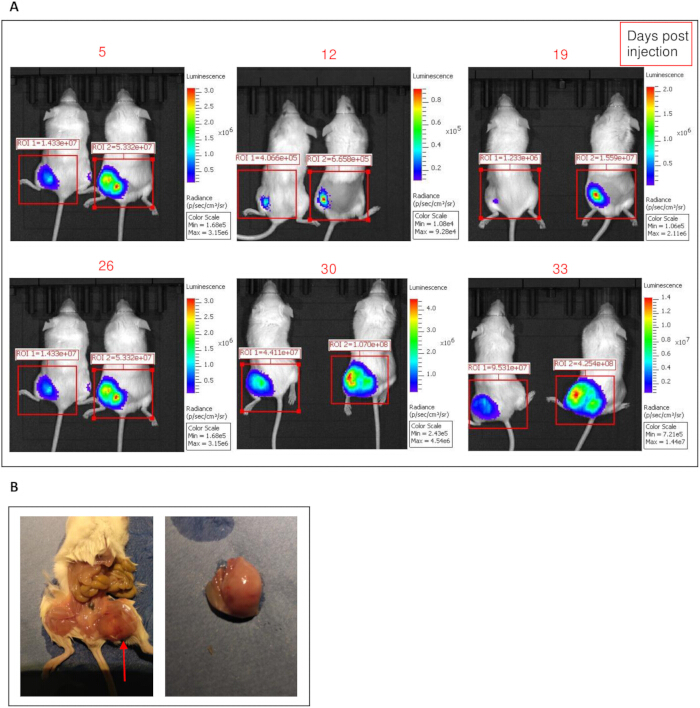
Figure 1: Bioluminescence imaging and macroscopic analysis of orthotopic sciatic nerve xenografts in NSG mice. (A) Longitudinal bioluminescence imaging (BLI) of two NSG mice injected intraneurally with ipNF04.4 cells into the left sciatic nerve. Images were captured at days 5, 12, 19, 26, 30, and 33 post-injection, illustrating progressive tumor growth over time in the two same mice. The increase in bioluminescent signal intensity reflects successful engraftment and tumor expansion within the nerve microenvironment. (B) Macroscopic view of the sciatic nerve at day 33 post-grafting, showing a well-developed, integrated tumor without signs of abscess formation. Please click here to view a larger version of this figure.
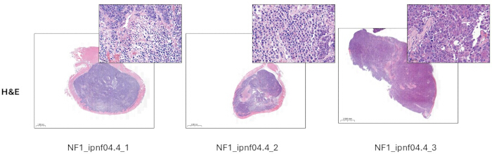
Figure 2: Photomicrograph of the graft site from this mouse demonstrating tumor growth and focal invasion. Paraffin-embedded tissue sections stained with hematoxylin and eosin (H&E). Examples of typical tumors from three mice are shown. Scale bars: 2000 µm. Please click here to view a larger version of this figure.
Supplementary Figure 1: Representative body weight evolution of NSG mice (n=15) over the course of the xenograft experiment. No significant weight loss was observed. Please click here to download this figure.
Discussion
The successful establishment of an orthotopic neurofibromas cell line-derived xenograft (CDX) model in NSG mice represents a significant advancement in NF1 research, addressing key limitations of existing models. The protocol confirms that human immortalized NF1-associated Schwann cells (ipNF04.4)14,15,16 can be efficiently engrafted into the sciatic nerve of immunodeficient mice, leading to reproducible tumor development within a physiologically relevant microenvironment. Orthotopic xenografts of Schwann cell lines were mentioned in the original studies establishing human Schwann cell lines14,15, and a detailed protocol is provided here.
Key to the success of this protocol was the meticulous preparation and monitoring of both the cellular components and the host animals. During the procedure, anesthesia was carefully managed, ensuring that the mice were kept in an appropriate state of anesthesia without significant stress. To minimize any potential damage, the sciatic nerve was not stretched with clamps or forceps, and great care was taken to avoid mechanical stress on the tissues. The use of luciferase-based BLI allowed for non-invasive, longitudinal monitoring of tumor growth, providing quantitative and reproducible data. This imaging strategy not only confirmed graft viability but also allowed for the identification of early deviations in tumor dynamics. The absence of significant weight loss (Supplementary Figure 1), motor dysfunction, or other signs of distress in the mice highlights the compatibility of this protocol with the ethical principles of animal welfare and the 3Rs (Replacement, Reduction, Refinement).
Compared to GEMMs, which rely on conditional Nf1 inactivation6,7,9, this approach offers several distinct advantages. GEMMs require specific genetic modifications, but they may not fully replicate the complex tumor-stroma interactions observed in human neurofibromas1,17. This model allows for the direct study of human Schwann cells in their native nerve niche, preserving cell-extrinsic factors4. Unlike GEMMs, where tumors develop unpredictably over time, the xenograft model allows precise spatiotemporal control over tumor formation. This model provides a rapid and reproducible platform for testing candidate therapies, with BLI enabling real-time monitoring of tumor progression and treatment response.
Furthermore, these findings align with previous studies on MPNSTs, where orthotopic xenografting has been shown to enhance the fidelity of tumor models5. However, benign neurofibromas pose unique challenges, as their growth is highly dependent on specific nerve interactions, which conventional subcutaneous grafting methods fail to recapitulate6. The results confirm that intraneural injection provides an optimal environment for sustained neurofibroma development.
Thus, this in vivo model can be considered a relevant complementary alternative to GEMMs, for preclinical therapeutic testing. However, there are limitations to the model. Human Schwann cells derived from neurofibromas have been immortalized and exhibit a tumor phenotype that could be considered more aggressive than that of a plexiform neurofibroma, with dense cell proliferation (Figure 2). No evidence of distant dissemination has been observed in animals, and complete malignant transformation has not been demonstrated. Further studies are needed to show whether the cell phenotype could correspond to the "intermediate" phenotype reminiscent of atypical neurofibroma, like atypical neurofibromatous neoplasms with unknown biological potential (ANNUBP)18. Another critical limitation of the model lies in its dependence on immunodeficient mice. The absence of an adaptive immune system in NSG mice limits the study of immune-tumor interactions, which are increasingly recognized as key contributors to neurofibroma biology7,19,20,21. Future refinements could incorporate humanized mouse models or alternative strategies to better capture the immunological aspects of the disease. Additionally, the reliance on luciferase expression for tumor monitoring, while highly effective, may limit the applicability of this protocol to experimental setups lacking access to BLI equipment. While BLI is a sensitive and non-invasive method, it does not provide direct insights into histopathological changes. Complementary analyses, such as MRI or advanced histology, could further improve the characterization of neurofibroma progression.
Several procedural steps may require optimization to ensure successful tumor engraftment. Injection failure can result from improper needle positioning or excessive leakage of the cell suspension. To mitigate this, the needle must be carefully inserted parallel to the sciatic nerve and withdrawn slowly after injection. Variability in bioluminescence signals may arise from inconsistent luciferin dosing or misalignment during imaging. It is recommended that the timing of the post-luciferin injection (10 min) be standardized and that identical imaging settings be maintained across all animals. If variability in tumor growth remains high, confirm cell viability before injection and minimize animal stress during handling.
In conclusion, this study establishes a novel and reproducible orthotopic neurofibroma xenograft model, offering an effective preclinical platform for studying NF1-associated benign tumors. By leveraging human Schwann cells in their natural nerve microenvironment, this model closely mimics human neurofibroma biology while enabling real-time tumor monitoring and controlled therapeutic interventions. This model represents an essential step between GEMMs and patient-derived models (PDXs), paving the way for more clinically relevant approaches to neurofibroma research and treatment.
Future refinements should aim to integrate immune components, such as humanized mouse models, to study the role of the tumor microenvironment6. Expanding imaging modalities, including MRI or PET scans, could complement BLI with structural and metabolic insights22. Extending experimental timelines will be essential to investigate long-term neurofibroma development and potential malignant transformation risks. By bridging the gap between GEMMs and PDX models, this platform enhances translational research in NF1, offering new opportunities for therapeutic innovation in benign peripheral nerve sheath tumors.
Disclosures
The authors declare no competing financial interests
Acknowledgements
This work was supported by the Institut national du cancer (Inca_18828). We would like to thank the HistIM platform (directed by Maryline Favier) and Pierre Sohier, at the Institut Cochin.
Materials
| Name | Company | Catalog Number | Comments |
|---|---|---|---|
| Buprenorphine | Buprecare | For pre-operative analgesia | |
| Heating mats | ANIPHY | VI-653246 | For maintaining normothermia during mouse surgery |
| hTERT NF1 ipNF04.4 (CVCL_UI78) cell line | Applied Biological Materials | T0194 | Immortalized human NF1-null Schwann cell line |
| Isoflurane | Iso-Vet | For mouse anesthesia during bioluminescence imaging | |
| IVIS Lumina Series III | Revvity | For bioluminescence imaging | |
| Ketamine | Ketamine 1000 Virbac | For surgical anesthesia in mice | |
| Living Image Software | Revvity | Included with IVIS | For image acquisition and quantification |
| Luciferase Assay System | Promega | E1500 | For BLI detection |
| Meloxicam | METACAM | Non-steroidal anti-inflammatory drug (NSAID) | |
| Monofilament non-absorbable suture | ETHILON | F2419H | For suturing the incision |
| Needle for syringe | Hamilton | Custom | ga33/12,7mm/pst4-12° |
| NOD SCID gamma (NSG) mice | Charles River | 614NSG | |
| pLenti CMV Puro LUC (w168-1) | Addgene | 17477 | For expression of firefly luciferase |
| Povidone iodée dermique 10% | Viatris | 330200775 | Topical antiseptic for skin disinfection |
| Scalpel blade | FST | 10015-00 | For skin incision during surgery |
| Stereo Microscope L125 | Leica | For visualizing cell injection site in the sciatic nerve | |
| Syringe 10 µl | Hamilton | 1701RN | For cell injection into sciatic nerve |
| Xilazine | Rompun 2% | For surgical anesthesia in mice |
References
- Brems, H., Beert, E., De Ravel, T., Legius, E. Mechanisms in the pathogenesis of malignant tumours in neurofibromatosis type 1. Lancet Oncol. 10 (5), 508-515 (2009).
- Rasmussen, S. A., Friedman, J. M. NF1 gene and neurofibromatosis 1. Am J Epidemiol. 151 (1), 33-40 (2000).
- Gutmann, D. H., et al. Neurofibromatosis type 1. Nat Rev Dis Primers. 3 (1), 17004(2017).
- Mazuelas, H., et al. Modeling iPSC-derived human neurofibroma-like tumors in mice uncovers the heterogeneity of Schwann cells within plexiform neurofibromas. Cell Rep. 38 (7), 110385(2022).
- Jacks, T., et al. Tumour predisposition in mice heterozygous for a targeted mutation in Nf1. Nat Genet. 7 (3), 353-361 (1994).
- Radomska, K. J., et al. Cellular origin, tumor progression, and pathogenic mechanisms of cutaneous neurofibromas revealed by mice with Nf1 knockout in boundary cap cells. Cancer Discov. 9 (1), 130-147 (2019).
- Chen, Z., et al. Spatiotemporal loss of NF1 in Schwann cell lineage leads to different types of cutaneous neurofibroma susceptible to modification by the Hippo pathway. Cancer Discov. 9 (1), 114-129 (2019).
- Wu, J., et al. Plexiform and dermal neurofibromas and pigmentation are caused by Nf1 loss in desert hedgehog-expressing cells. Cancer Cell. 13 (2), 105-116 (2008).
- Li, S., Chen, Z., Le, L. Q. New insights into the neurofibroma tumor cells of origin. Neurooncol Adv. 2 (Suppl 1), i13-i22 (2020).
- Turk, A. N., Byer, S. J., Zinn, K. R., Carroll, S. L. Orthotopic xenografting of human luciferase-tagged malignant peripheral nerve sheath tumor cells for in vivo testing of candidate therapeutic agents. J Vis Exp. (49), e2558(2011).
- Jessen, W. J., et al. MEK inhibition exhibits efficacy in human and mouse neurofibromatosis tumors. J Clin Invest. 123 (1), 340-347 (2013).
- Jousma, E., et al. Preclinical assessments of the MEK inhibitor PD-0325901 in a mouse model of neurofibromatosis type 1: MEK inhibition in neurofibroma. Pediatr Blood Cancer. 62 (10), 1709-1716 (2015).
- Blanchard, Z., Brown, E. A., Ghazaryan, A., Welm, A. L. PDX models for functional precision oncology and discovery science. Nat Rev Cancer. 25, 153-166 (2024).
- Li, H., Chang, L. J., Neubauer, D. R., Muir, D. F., Wallace, M. R. Immortalization of human normal and NF1 neurofibroma Schwann cells. Lab Invest. 96 (10), 1105-1115 (2016).
- Muir, D., Neubauer, D., Lim, I. T., Yachnis, A. T., Wallace, M. R. Tumorigenic properties of neurofibromin-deficient neurofibroma Schwann cells. Am J Pathol. 158 (2), 501-513 (2001).
- Wu, M., Wallace, M. R., Muir, D. Tumorigenic properties of neurofibromin-deficient Schwann cells in culture and as syngrafts in Nf1 knockout mice. J Neurosci Res. 82 (3), 357-367 (2005).
- Rasmussen, S. A., Friedman, J. M. NF1 gene and neurofibromatosis 1. Am J Epidemiol. 151 (1), 33-40 (2000).
- Miettinen, M. M., et al. Histopathologic evaluation of atypical neurofibromatous tumors and their transformation into malignant peripheral nerve sheath tumor in patients with neurofibromatosis 1: A consensus overview. Hum Pathol. 67, 1-10 (2017).
- Pundavela, J., et al. Stimulator of interferon gene facilitates recruitment of effector CD8 T cells that drive neurofibromatosis type 1 nerve tumor initiation and maintenance. Sci Adv. 10 (42), eado6342(2024).
- Fletcher, J. S., Pundavela, J., Ratner, N. After Nf1 loss in Schwann cells, inflammation drives neurofibroma formation. Neurooncol Adv. 2 (Suppl 1), i23-i32 (2020).
- Le, L. Q., Parada, L. F. Tumor microenvironment and neurofibromatosis type I: Connecting the GAPs. Oncogene. 26 (32), 4609-4616 (2007).
- Wu, J. C., Sundaresan, G., Iyer, M., Gambhir, S. S. Non-invasive optical imaging of firefly luciferase reporter gene expression in skeletal muscles of living mice. Mol Ther. 4 (4), 297-306 (2001).
Reprints and Permissions
