This protocol describes a sciatic nerve transection and resuture model in mice to study peripheral nerve regeneration using adult dorsal root ganglion neurons. It enables in vivo analysis of axon regrowth, muscle reinnervation, and functional recovery following nerve injury.
Method Article
Mouse Sciatic Nerve Transection/Resuture Procedure for Studying Nerve Repair and Functional Recovery
In This Article
Summary
Abstract
Nerve repair is crucial for restoring function following axonal injury. However, mature neurons in the central nervous system (CNS) have a limited ability to regenerate their axons after injury due to diminished intrinsic growth capacity and a hostile environment. In contrast, neurons in the peripheral nervous system (PNS) exhibit robust regenerative potential. Among them, adult dorsal root ganglion (DRG) neurons are particularly well-known for their ability to regenerate effectively following peripheral nerve injuries, making them an ideal model for studying the cellular and molecular mechanisms underlying nerve repair. Here, a modified microsuture technique, termed "loop-before-transection," is described for generating a sciatic nerve transection and resuture model in mice, followed by analysis of axon regeneration. Behavioral assessments and muscle reinnervation are also demonstrated to evaluate functional recovery. After sciatic nerve transection and resuture, animals initially exhibit behavioral deficits, but full recovery is typically observed by 30 days post-injury. Applying drugs that promote axonal fusion at the injury site significantly accelerates functional recovery. This model system offers a powerful tool for investigating the mechanisms governing mammalian nerve repair in vivo.
Introduction
Axonal injury is a common and severe consequence of brain and spinal cord damage, often caused by traumatic injuries, neurotoxins, or neurological disorders that disrupt neural connectivity and impair axonal regeneration1,2. Not allneurons possess an intrinsic capacity for regeneration, which is crucial for the repair of severed axons.The rate of axonal regeneration is influenced by multiple factors, including the neuron's intrinsic growth potential, protein synthesis efficiency, cytoskeletal dynamics, clearance of myelin debris, and the availability of growth factors3,4.
In the adult mammalian central nervous system (CNS), axonal injury leads to a diminished regenerative capacity, primarily due to a lack of intrinsic growth-promoting factors and the presence of inhibitory extrinsic cues1,5. In contrast, axons in the peripheral nervous system (PNS) possess a robust ability to regenerate following injury, owing to the intrinsic self-repair mechanisms of peripheral neurons and the rapid reactivation of pro-regenerative growth programs. Upon injury, the distal segment of the axon becomes disconnected from the neuronal soma and undergoes Wallerian degeneration, characterized by axonal disintegration and fragmentation6,7. Efficient clearance of axonal and myelin debris by glial cells is essential for successful regeneration, as it prevents scar formation that could impede axonal growth3,8. This process, together with cytoskeletal remodeling of injured axons, supports the regrowth of the proximal stump and facilitates target reinnervation. These events occur in partially overlapping phases, during which injury-induced activation of transcription factors such as ATF3 and c-Jun promotes pro-regenerative gene expression programs4,9,10.
Regenerative axonal fusion represents an efficient mechanism for restoring neuronal connectivity following axonal injury11. This process was first described in the crayfish Procambarus clarkii in 196712 and has since been documented across multiple species13,14,15,16, including Caenorhabditis elegans17. Although direct evidence for axonal fusion in mammals remains limited, fusion-inducing strategies have been successfully employed to enhance nerve repair11. Notably, the chemical fusogen polyethylene glycol (PEG) promotes membrane resealing and has been shown to facilitate functional recovery following both peripheral nerve and spinal cord injuries in mammalian models18,19,20,21,22,23,24. Recent findings indicate that glutathione peroxidase (GPX) depletion is sufficient to promote axonal fusion in C. elegans25. Similarly, Gpx4 conditional knockout mice or those treated with the GPX inhibitor ML162 displayed accelerated functional recovery after sciatic nerve transection, potentially through enhanced axonal fusion25.
The sciatic nerve is a mixed nerve composed of sensory, somatic motor, and autonomic motor axons26. It arises from the L4 and L5 spinal segments in rats27, and from the L3 and L4 segments in mice28. Due to its accessibility and anatomical consistency, sciatic nerve injury has become one of the most widely used models for studying peripheral nerve regeneration29,30. These models enable the investigation of both intrinsic neuronal mechanisms and extrinsic environmental factors that influence axonal growth and functional recovery3,4,9. The use of sciatic nerve crush as an experimental paradigm in neuroscience dates back to the late 19th century29. In addition to nerve crush models, nerve transection is widely used to study axon regeneration and functional recovery. However, the classical sciatic nerve transection followed by suture in mice is often less reproducible and reliable than in rats, due to the nerve's small size and fragile texture31.
Using a modified sciatic nerve transection and resuture model, termed "loop-before-transection", reproducible sciatic nerve transection and resuture models were successfully generated25. This approach eliminates the need for post-transection nerve handling by pre-inserting the suture prior to cutting, thereby reducing mechanical stress and minimizing variability due to surgical technique. Moderate levels of GPX modulation were found to contribute to functional recovery after complete transection of the mouse sciatic nerve25. Co-treatment of the low concentrations ML162 (0.25 µM)32, GPX inhibitor, and polyethylene glycol (PEG, 500 mM)18,19,20,21,22,23,24 on the sciatic nerve transection (SNT) / resuture lesion successfully enhanced the behavior score assessed by pinprick, foot fault asymmetry, and toe spreading test25,33,34. Amputated distal axon fragments of mammalian dorsal root ganglion (DRG) neurons degenerate within 3 days, resulting in disruption of neuromuscular junctions (NMJ)6,35, and regenerative DRG neurons will reinnervate muscles to recover their functions. SNT/resuture-ML162/PEG co-treatment group showed innervated NMJs and enhanced behavior scores at the early time point (3 days post-operation), indicating early recovery of neuromuscular function25.
Here, the "loop-before-transection" microsuture method and drug administration that enabled the above observations in mice are described. A previously established PEG-fusion protocol, which enables rapid repair of severed rat nerves, was modified18. Polyethylene glycol (PEG) was initially reported to promote exogenous reconnection of cut crayfish medial giant axons (MGA) in vitro36. PEG-fusion protocols improved behavioral outcomes in 25%-30% of trials involving crush-severance injuries of the rat sciatic nerve, though functional recovery was not observed at early time points34. Subsequent refinements incorporated the concept of endogenous membrane sealing37. After nerve injury, rapid plasmalemmal repair is neuroprotective and prevents neurodegeneration. Ca2+-induced accumulation of membrane-bound vesicles facilitates this sealing process38. Complete nerve transection disrupts the axolemma as well as the endoneurial, perineurial, and epineurial sheaths at the lesion site. Microsuturing of the epineurial sheath aligns the proximal and distal nerve stumps. During the suture step, the use of Ca2+-free hypotonic saline reduces the formation of membrane-bound vesicles, keeping the endoneurial and perineurial sheaths open and allowing them to be closely approximated by the epineurial sutures. Following this, the application of Ca2+-containing saline induces vesicle accumulation to seal remaining plasmalemmal disruption18.
In this protocol, Adult FVB mice (8-10 weeks old) and C57BL/6 mice were used, and no strain-specific difference was observed. The sciatic nerve in mice is located approximately 1-2 mm beneath the gluteal muscle at mid-thigh level and is notably thinner and more fragile than in rats, necessitating greater surgical precision during dissection and suturing. Care must be taken to avoid nerve overstretching or excessive manipulation that may compromise functional recovery. This approach involves fully severing the nerve, resulting in a physical gap between the proximal and distal stumps. Because this gap inhibits direct axonal regeneration, successful recovery typically requires microsurgical intervention or nerve grafting.
Given that the mouse sciatic nerve is thinner and more delicate than that of the rat, greater surgical precision is required. Although microsuturing is commonly used, it may inadvertently cause additional trauma due to excessive manipulation or repeated needle passes39. These limitations can introduce variability and potentially confound the evaluation of novel treatment strategies. Therefore, achieving consistent surgical outcomes while minimizing suture-induced nerve damage is essential for reliably assessing functional recovery in nerve transection models. Using this "loop-before-transection" method, reproducible sciatic nerve transection and resuture models were generated, and the effects of dual treatment with PEG and ML162 on functional recovery were evaluated. Overall, despite its widespread use, the mouse sciatic nerve transection model poses technical challenges due to the nerve's small size and fragility. This modified resuture method described here addresses a critical need for a reproducible, standardized microsurgical protocol that minimizes mechanical damage.
Protocol
All animal procedures were conducted in accordance with the guidelines of the Institutional Animal Care and Use Committee (IACUC) at the University of Texas Health Science Center at San Antonio (Protocol Number: 20200082AR). Adult male and female FVB mice (8-10 weeks old, 20-25 g) were used for the JoVE experimental procedures described here. C57BL/6 mice (8-10 weeks old, 20-25 g) were used only in preliminary practice surgeries conducted prior to the formal experiments to optimize handling and suturing precision. These practice sessions confirmed that the "loop-before-transection" procedure produced comparable outcomes in both strains; therefore, all formal data presented in this manuscript were obtained from FVB mice. A total of ten FVB mice (five males and five females) were initially included for sciatic nerve transection followed by ML162 + PEG treatment. One animal exhibited a congenital anatomical variation in which the sciatic nerve bifurcated into two main branches before the transection site, resulting in incomplete surgery and exclusion from analysis. Therefore, behavioral and histological analyses were conducted on nine animals (n = 9). Both sexes showed comparable regenerative and behavioral recovery, and no sex-specific differences were observed. An additional mouse underwent double suture penetration without nerve transection (sham control) to assess the potential impact of the suture procedure itself on behavioral outcomes. No noticeable differences in regenerative response were observed between sexes. Animals were housed under standard conditions with a 12-h light/dark cycle, at 75 °F and 40%-60% relative humidity, with ad libitum access to food and water. Both male and female mice were used, and no noticeable difference was observed between male and female animals. Surgical procedures were performed under isoflurane anesthesia of the SomnoSuite low-flow anesthesia system. Behavioral tests were conducted beginning on post-operative day 2, and repeated on days 4, 7, 10, 14, 18, 22, and 27, as indicated in the corresponding figures. The reagents and the equipment used are listed in the Table of Materials.
1. Sciatic nerve transection/resuture and drug treatment
- Place the animal on a heated pad where the sciatic nerve could be observed under a stereo dissecting microscope (Figure 1A).
- Anesthetize the mouse using the SomnoSuite low-flow anesthesia system.
NOTE: Confirm the depth of anesthesia by the absence of reflex responses, such as reaction to a toe pinch, before initiating surgery. Apply vet ophthalmic ointment to eyes to prevent corneal dryness. - Shave the hair on the thigh and around.
NOTE: Remove the shaved and dropped hairs as cleanly as possible. Otherwise, the hairs will stick to the 10-0 nylon suture and contaminate the sciatic nerve. Residual hairs can be gently removed using laboratory tape or scotch tape after shaving to ensure a clean surgical field. Disinfect the surgical site by sequentially wiping the shaved area with 70% ethanol, betadine, and a final wipe with 70% ethanol using autoclaved cotton balls. - Locate the femur bone and make a small skin incision of approximately 1.0 cm (range 0.8-1.2 cm) at the mid-thigh level, about ~1 mm posterior and in parallel to the femur bone using fine surgical scissors.
- Gently retract the gluteus maximus to expose the underlying biceps femoris. Then, use the tips of the scissors to gently separate the anterior and posterior heads of the biceps femoris, without cutting, to expose the sciatic nerve beneath the sciatic notch. These steps are performed under direct visual observation, not under the microscope.
- Pull the sciatic nerve out of the body using micro-dissecting forceps to facilitate surgery on the sciatic nerve (Figure 1A).
NOTE: Secure the forceps with tape to prevent them from spreading beyond a certain level to minimize damage to the sciatic nerve. Place the tip of the forceps under the sciatic nerve and lift the sciatic nerve upward using the mouse thigh as a fulcrum. Take care to lift the nerve only as much as needed to facilitate suturing, and avoid applying tension or pulling it outward, as excessive stretch can damage the axons or surrounding structures. - Focus the microscope on the exposed sciatic nerve. From this step, proceed through Step 15 using a microscope (Figure 1A).
- Penetrate twice the sciatic nerve with 10-0 nylon suture (Figure 1B,C). Go to Step 1-8 for uncut control. Go to Step 1-11 for the nerve transection.
NOTE: Two penetrations were made approximately one nerve width apart (~0.7 mm), positioned symmetrically on either side of the intended transection site. This spacing ensured proper alignment of the proximal and distal stumps while minimizing mechanical stress at the lesion. Make penetration through the center of the sciatic nerve with the suture, ensuring that the two penetrations made by the suture are precisely parallel. 10-0 sutures are small and must be controlled with super fine tweezers. Non-absorbable 10-0 nylon was used because it provides excellent tensile stability and minimal tissue reactivity during the 30-day observation period. This material is widely used in microsurgical nerve repair models in mice and rats18,31. Absorbable sutures were not used because gradual degradation could compromise alignment of the transected nerve stumps before functional recovery is complete. To minimize operator-dependent variability, all surgical procedures were performed by a single trained surgeon after extensive microsurgical practice under a stereomicroscope. The "loop-before-transection" method itself further reduces variability by eliminating the need for post-transection nerve manipulation, thereby ensuring consistent alignment and tension across animals. This requires considerable practice and a high level of control skills. Before performing animal surgeries, beginners may find it helpful to practice the suturing motion using fine synthetic threads such as dental floss to develop dexterity and control under a stereomicroscope. - Carefully tie a knot with the nylon suture.
- Place the exposed sciatic nerve under the muscle layer and close the skin using 4-0 nylon sutures.
- Determine that double penetration of the sciatic nerve with a 10-0 nylon suture does not affect mouse behavior.
NOTE: To confirm this, mice that underwent double suture penetration without transection were subjected to the same behavioral tests used in the experimental groups, including pinprick, foot fault asymmetry, and toe spreading. These mice consistently achieved full scores equivalent to unoperated wild-type controls, indicating that the suture procedure alone did not impair sciatic nerve function. - Cut the sciatic nerve midway between the two 10-0 nylon sutures using a scalpel (Figure 1D).
NOTE: To minimize variability, all surgeries were performed by the same trained experimenter using a standardized procedure. In addition, the "loop-before-transection" design inherently minimizes variability by avoiding post-transection nerve handling, ensuring consistent suture placement and tension across all animals. The position of the sutures was verified under a dissecting microscope to ensure uniformity across animals. - Put the forceps away.
- Wash the cut nerve with Ca2+-free phosphate-buffered saline (PBS; 137 mM NaCl, 2.7 mM KCl, 10 mM Na2HPO4/NaH2PO4, pH 7.4), prepared without calcium chloride.
NOTE: Rinse the severed nerve stump with calcium-free PBS for approximately 10-20 s to clear blood and prevent premature sealing. Resuturing should be completed promptly within 1-2 min to maintain alignment and tissue viability. - Remove the Ca2+-free PBS by absorbing it with a tissue.
- Carefully tie a knot with the nylon suture (Figure 1E). Proceed to Step 20 if drug treatment is not planned or required.
NOTE: A standard square knot was used for microsuturing, consisting of two opposite throws gently tightened with fine forceps under a stereomicroscope. The two severed axons connected by the suture return to a shape close to their original shape. The reconnection of a severed blood vessel can be a visual indicator that the proximal and distal nerve stumps are properly aligned. During the procedure, the tissue should appear fresh and moist, without pallor or compression. Avoid tying the suture too tightly, as this may collapse blood vessels or deform the nerve. Before tying the final knot, ensure that the nerve ends are approximated without tension. If needed, gently reposition the limb to facilitate natural alignment rather than pulling on the nerve, as excessive tension may increase the risk of neuroma or fibrosis. - Place 0.25 µM of ML162 in DMSO diluted in PBS onto the sutured sciatic nerve using a syringe. Allow the sutured sciatic nerve to be immersed in the drug for 1 min.
- Remove the ML162 by absorbing it with a sterile cotton swab.
- Place 500 mM of PEG in distilled water, pre-warmed to 37 °C onto the sutured sciatic nerve using a syringe. Allow the sutured sciatic nerve to be immersed in the drug for 1 min.
NOTE: 500 mM of PEG is a dense solution that may become solid at operating room temperature. Place the syringe on a heated pad during operation to ensure the PEG remains liquid. PEG is thought to promote membrane fusion by removing water molecules from between adjacent lipid bilayers, thereby enabling close membrane contact and spontaneous fusion. Although the precise molecular mechanism remains under investigation, this approach has been shown to facilitate axonal continuity and enhance functional recovery in peripheral nerve injury models. - Remove the PEG by absorbing it with a tissue.
- Wash the sutured nerve with Ca2+-containing PBS (PBS supplemented with 1 mM CaCl2; 137 mM NaCl, 2.7 mM KCl, 10 mM phosphate buffer, pH7.4).
- Remove the Ca2+-containing PBS by absorbing it with a tissue.
- Place the exposed sciatic nerve under the muscle layer and close the skin using 4-0 nylon sutures.
- Once the animal has recovered from the anesthesia, return it to its cage.
NOTE: After skin closure, the surgical site was treated with a topical antibiotic ointment containing pain reliever. Each animal was monitored on a heating pad until it regained sufficient consciousness to maintain sternal recumbency. Animals were then housed individually and returned to their group only after full recovery of motor function. Although the surgical environment was not maintained under a laminar flow hood, all surgical instruments were sterilized, and procedures were performed using aseptic technique with gloved hands and sterile tools.
2. Behavior test - Pinprick
NOTE: To assess the high-threshold mechanical sensitivity responsiveness after injury33.
- Place the mouse in a wire mesh grid and allow it to roam freely for 30 min to habituate to the new environment.
- The most lateral part of the plantar surface of the hind paw was divided into 5 areas (A to E). Gently apply the Austerlitz (size 000) to the plantar surface of the ipsilateral paw. The pinprick was applied twice from the most lateral toe (area A) to the heel (area E).
NOTE: Do not penetrate the skin.
NOTE: A response was considered positive when the animal briskly removed its paw from the two pinprick stimuli. The mouse was graded 1 for this area and then tested for the next one (Maximum 5). If none of the applications elicited a positive response, the overall grade was 0. Use the saphenous territory of the same ipsilateral side as a positive control. It will always elicit a positive response.
3. Behavior test - Foot fault (FF) asymmetry
NOTE: To measure how well sciatic axons innervate more proximal muscles34.
- Allow the animals to roam freely on a wire mesh grid elevated on the cage to habituate the environment. One trial consists of 50 total steps per hindlimb.
- Score when the animals misstep. The scoring criteria are as follows.
Score 1: Partial fault. Pulled back before the leg entirely fell out to the thigh.
Score 2: Full fault. Completely fell off to the crotch. - Calculate the composite foot fault score from all animals' injured and uninjured hindlimbs at each post-operative time.
Composite foot fault score = (# partial faults x 1) + (# full faults x 2) - Divide the composite foot fault score by 50 (total steps/limb) to obtain the percentage of fault for each hindlimb.
% foot fault = Composite foot fault score/50 (total steps) x 100% - Subtract the foot faults in the injured hindlimb from the proportion of foot faults in the intact hindlimb to yield an asymmetry score for each animal at a given post-operative time.
Foot fault (FF) asymmetry score = %FF (intact limb) - %FF (injured limb)
4. Behavior test - Toe spreading
NOTE: To assess motor recovery after nerve injury. According to a previous study, the toe spreading motor test is more sensitive than the gait analysis to detect the recovery of motor function after sciatic nerve injury30.
- Gently cover the animal with a piece of cloth and lift the tail for clear observation of the hind paws.
NOTE: Alternative: Gently grab the mouse's nape, fix the tail, turn it over, and make the mouse squirm to observe the toe movement. - Award the toe spreading score as follows:
Score 2: Full spreading. Define as a complete, wide, and sustained spreading of all toes for at least 2 s.
Score 1: Intermediate spreading. Any movement that does not reach a full spreading score. Tremble slightly, or partial movement.
Score 0: No spreading. No movement at all.
NOTE: All behavioral assessments (pinprick, foot fault, and toe spreading) were double checked by an experimenter blinded to treatment groups. Animals were randomly assigned to treatment groups at the time of surgery in accordance with the design used in our prior study (Ko et al.25), which included four experimental conditions (Resuture only, PEG, ML162, ML162 + PEG). The current article presents data from the ML162 + PEG group, analyzed independently while maintaining the original experimental rigor.
5. Tissue perfusion and processing for histology
- Deeply anesthetize the animal and perform transcardial perfusion with phosphate-buffered saline (PBS) via the left ventricle.
- Incise the right atrium to allow complete blood drainage.
- Immerse the collected tissues in 4% paraformaldehyde (PFA) at 4 °C overnight for fixation.
- Wash the fixed tissues three times with PBS, then incubate them in PBS at 4 °C overnight.
- Cryoprotect the tissues by immersing them in 30% sucrose at 4 °C overnight.
- Embed the tissues in OCT compound, freeze them, and section at 12 µm thickness using a cryostat.
- Store tissue blocks at -80 °C and cryosections at -20 °C until use.
- For sciatic nerve collection alone, omit perfusion, as blood content does not significantly affect immunostaining quality in these peripheral tissues.
Results
The previous PEG-fusion protocol18 was modified to maintain consistent surgical results and minimize nerve damage caused by suturing25. By placing the suture to penetrate through the nerve before transection, damage caused by pinching with tweezers or pricking with a needle after transection was minimized. To test the effect of tying a suture penetrating a nerve on its function, the suture was passed through an intact nerve twice and knotted (Figure 2A, suture without SNT). The sutures that penetrated the nerve were observed not to create a scar on the nerve when checked 30 days post-operation (Figure 2A, suture without SNT). The mouse that underwent only a suture without SNT scored perfectly in all our behavioral tests, indicating that nerve penetration by suture did not affect nerve function (Figure 3B, Figure 4B, and Figure 5B).
In a previous study, functional recovery after SNT/resuture was tested in four groups. The group treated with ML162 + PEG after surgery was confirmed to show excellent nerve recovery effects25. For the SNT/resuture operation, the sutures performed by the surgeon could not always be consistent, and variations in the sutured sciatic nerves occurred despite a modified surgical method to minimize neuronal damage (Figure 2A). Individuals in the ML162 + PEG group (n = 9) were followed, including those in whom the sutured nerve was reconstructed close to the original alignment with minimal movement and those in whom part of the transected nerve was mislocated due to excessive movement. A minimal movement individual (MMI) who underwent suture close to the original nerve with minimal movement was observed to have minimal scarring around the lesion 30 days post-operation (Figure 2A). In contrast, an excessive movement individual (EMI) who underwent suture with excessive movement resulting in a mislocated nerve on the lesion was observed to have a large scale of scarring around the lesion 30 days post-operation (Figure 2A). MMI scored significantly better in all behavioral tests than the average behavioral scores of the ML162 + PEG group of nine mice. MMI scored significantly higher in all behavioral tests than the mean behavioral scores of the ML162+PEG group of nine mice, whereas EMI scored below the mean (Figure 3B, Figure 4B, and Figure 5B). This suggests that the surgeon's technique may influence the recovery of the sutured nerve after transection. The behavioral scores of MMI and EMI showed a marked difference in the early post-operative stage, and similar scores were recorded about 3 weeks after surgery (Figure 3B, Figure 4B, and Figure 5B). This suggests that early-stage (4-13 days post-operation) nerve recovery after surgery is related to how well the grafted nerve returns to its original shape. Neuronal axon regeneration of the mouse sciatic nerve, which grows about 1 mm per day, occurs when the nerve is connected, regardless of the shape of the surgical lesion. Immunostaining for SCG10 revealed regenerated axons extending from the proximal to distal nerve stump (Figure 6B,C). In these experiments, β-tubulin was used as a general neuronal marker to visualize axonal structures, while SCG10 specifically labeled regenerating axons associated with growth cones and newly extending neurites following peripheral nerve injury. Reinnervation of neuromuscular junctions was supported by the colocalization of NF-200 and α-bungarotoxin in the plantar muscle (Figure 6D). By 4 weeks post-surgery, all animals in the ML162 + PEG group demonstrated strong recovery across all behavioral metrics. Importantly, individual differences in early behavioral performance were observed, likely reflecting variability in nerve alignment and axonal fusion success, which is the central focus of this study.
Although our previous studies focused on the early-stage functional recovery after nerve injury, neuron regenerative mechanisms are essential to restore nerve function thoroughly in the late-stage25. Resuturing completely severed nerves allows regeneration of neurons in the proximal nerve to replace all degenerated neurons in the distal nerve within 3 days (Figure 6A-C). And the regenerated neurons replace the denervated neuromuscular junctions, leading to reinnervation and contributing to functional recovery (Figures 6A,D).
In summary, a consistent surgical technique is required to study the early-stage recovery of injured mouse sciatic nerves. This method of minimizing the surgeon's direct touch by surgical instruments on the nerve will minimize unintended damage to the lesion, which will positively affect the reliability of the experimental results.
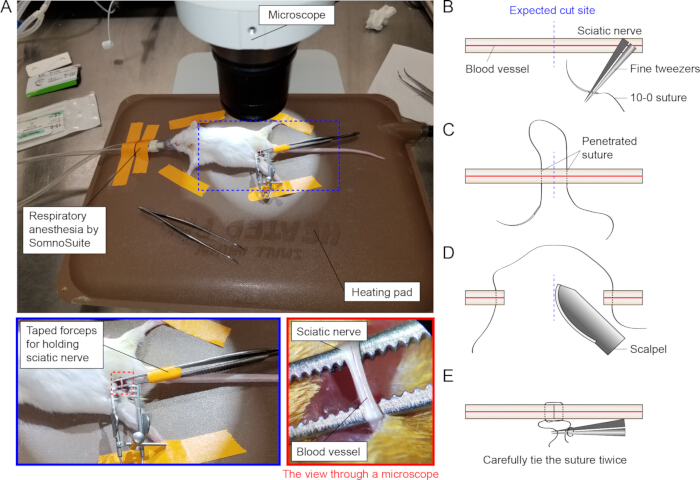
Figure 1: Preparation and procedure for sciatic nerve transection (SNT) and resuture. (A) The anesthetized mouse is positioned on a heating pad under the microscope. Anesthesia is maintained using the SomnoSuite system, and the mouse is secured with tape. The heating pad is placed beneath the microscope to enable visualization of the sciatic nerve. The sciatic nerve is gently lifted with forceps from below to expose it outside the body. (B) A 10-0 nylon suture needle is grasped with tweezers and inserted laterally near the anticipated transection site. (C) The needle is then passed through to the opposite side of the intended transection site. (D) The sciatic nerve is cleanly transected at the marked site using a scalpel in a single motion. (E) The transected ends are carefully rejoined with a double-knot suture using tweezers. Caution is taken to avoid damaging the nerve by excessive tightening or inadvertent pricking. Please click here to view a larger version of this figure.
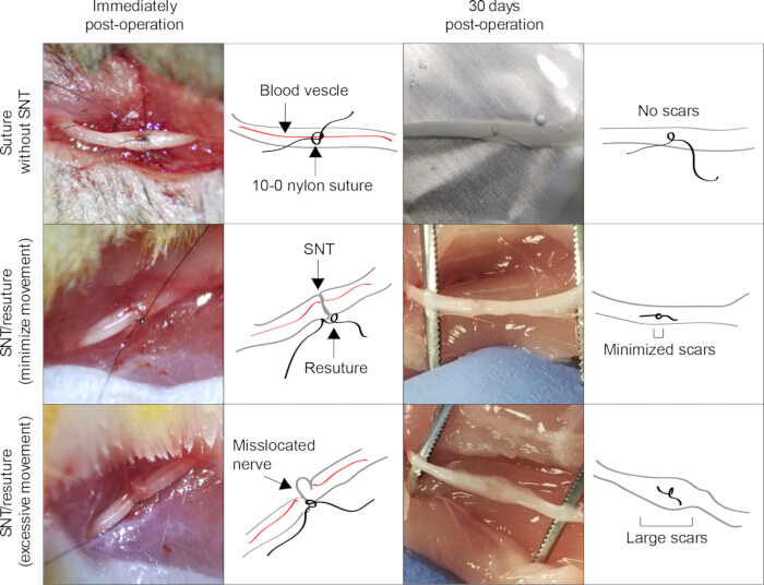
Figure 2: Examples of surgeries with minimal or excessive movement of nerve and follow-up behavioral tests. Representative images indicate a suture without SNT, an SNT/resuture with minimal nerve movement, and an SNT/resuture with excessive nerve movement at the immediate or 30 days post-operation. SNT/resuture with minimal movement resulted in minimal scars in the recovered sciatic nerve lesion, whereas excessive movement resulted in large scars. Please click here to view a larger version of this figure.
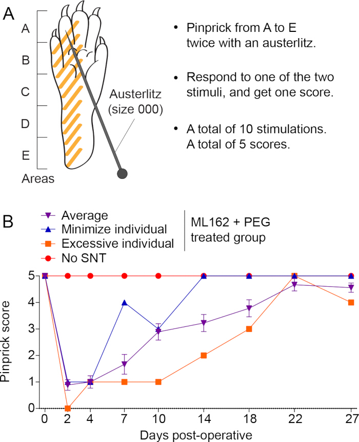
Figure 3: The surgical technique and outcome of returning the severed nerve to its original shape may affect pinprick behavioral test measurements in the early stages. (A) Schematic diagram illustrating the pinprick test method. (B) Quantification of the pinprick test. 8-week-old animals were tested for pinprick response right before the operation (Day 0). Immediately after SNT/resuture, the injured nerves were treated with ML162 + PEG. Mice were then evaluated at different time points for a month. n = 9 for ML162 + PEG group. Error bars represent the standard error of the mean (SEM). The "average" refers to the mean behavioral score of all 9 animals in the group. MMI and EMI are shown to illustrate the range of intra-group variability. Please click here to view a larger version of this figure.
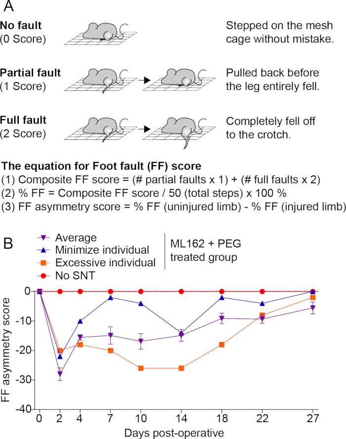
Figure 4: The surgical technique and outcome of returning the severed nerve to its original shape may affect foot fault (FF) asymmetric behavioral test measurements in the early stages. (A) Schematic diagram illustrating the FF asymmetry test method. (B) Quantification of the FF asymmetry test using the same experimental animals from Figure 3B. n = 9 for ML162 + PEG group. Error bars represent the standard error of the mean (SEM). The "average" refers to the mean behavioral score of all 9 animals in the group, with MMI and EMI shown to illustrate surgical variability. Please click here to view a larger version of this figure.
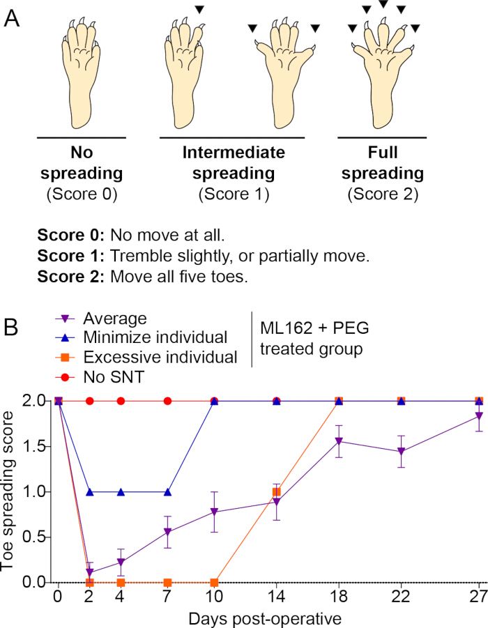
Figure 5: The surgical technique and outcome of returning the severed nerve to its original shape may affect toe spreading behavioral test measurements in the early stages. (A) Schematic diagram illustrating the toe spreading test method. (B) Quantification of toe spreading using the same experimental animals from Figure 3B. n = 9 for ML162 + PEG group. Error bars represent the standard error of the mean (SEM). The "average" reflects the group mean (n = 9), while MMI and EMI illustrate individual differences in surgical outcomes. Please click here to view a larger version of this figure.
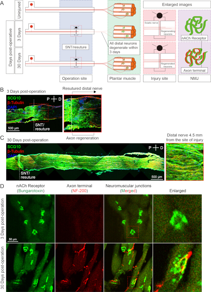
Figure 6: Resuturing a completely severed nerve allows the regrowth of a bundle of neurons and the reinnervation of the neuromuscular junction. (A) Schematic diagram showing the nerve repair process when a severed nerve is resutured. All neurons in the severed distal nerve degenerate within 3 days, and neuron regeneration proceeds from the proximal to the distal part. (B) Neurons regenerating from the proximal to distal sciatic nerves as observed by SCG10 staining. (C) SCG10 staining image showing well-regenerated neurons in the sciatic nerve 30 days post-operation. (D) OCT section of the plantar muscle (12 µm interval) showing completely denervated neuromuscular junctions (NMJs) 3 days post-operative. Reinnervated NMJs were observed in the plantar muscle 30 days post-operation. Bungarotoxin binds to nACh receptors, and NF-200 antibody binds to axon terminals, which are then labeled in green and red, respectively. See Protocol Step 5 for tissue perfusion and processing details. Scale bars: (B,C) 500 µm; (D) 50 µm. Please click here to view a larger version of this figure.
Discussion
Nerve regeneration studies using nerve transection models are widely performed on the mouse sciatic nerve31,39. However, due to its small size and soft texture, the mouse sciatic nerve is difficult to handle gently with surgical tools, and the tissue is prone to tearing during suturing. The microsuture process, often involving excessive manipulation, can lead to nerve deformation and trauma. Even when unnecessary instrument stimulation is minimized, excessive movement of the sutured nerve can still result in misalignment, potentially impairing early-stage functional recovery. These variables compromise the consistency and reliability of experimental outcomes in transection models, highlighting the need for a surgical approach that ensures reproducibility and minimizes nerve damage.
The surgical procedure was designed to control variables while minimizing surgeon intervention. When designing this surgical procedure, the following considerations were considered: (1) Minimize the use of surgical instruments to minimize the stress on the nerve. Avoid pinching the nerve with tweezers as much as possible; (2) Minimize the number of suture knots, since tying knots can also cause sufficient damage to the nerve; (3) Minimize reliance on the surgeon's surgical technique. After extensive evaluation, it was concluded that the most common cause of nerve damage or deformation was the surgical procedure of pulling the severed nerve with an instrument during suturing. Therefore, a method of penetrating and positioning the suture through the nerve before cutting was adopted to eliminate the need for manipulating the nerve with a surgical tool.
To minimize procedural errors such as nerve tearing or suture misalignment, over one month was dedicated to microsurgical training prior to initiating the experimental surgeries. Although severely damaged nerves were not included in the analysis, Figure 2 illustrates examples of both minimal and excessive manipulation outcomes, which can serve as practical visual references for identifying suboptimal results during training or troubleshooting. If the nerve was torn or deformed beyond repair, the animal was excluded from behavioral testing.
The mouse is anesthetized and positioned with its head and dorsal side facing upward, limbs gently spread and secured, so that the exposed sciatic nerve appears horizontally aligned from the surgeon's viewpoint. After identifying an imaginary transection point on the horizontally aligned sciatic nerve, insert a 10-0 nylon suture from bottom to top on the right side of the designated point. Then, insert another 10-0 suture from top to bottom on the left side, forming a horseshoe-shaped loop over the nerve, centered on the imaginary cut site. It is critical that the suture penetrations are made precisely through the center of the sciatic nerve. Once the horseshoe-shaped suture is in place, use a surgical scalpel to transect the nerve at the midpoint of the loop, creating a clean cut surface. To prevent spontaneous sealing of the severed axons and to clear blood from the vessels, rinse the area with calcium-free PBS and blot dry with tissue. Next, gently tie the ends of the suture together to form a knot. This step poses the highest risk of inducing nerve damage or deformation, so it must be performed with utmost care. Fine-tipped tweezers are essential, as 10-0 sutures are extremely thin and difficult to visualize. The surgeon should be well-practiced in manipulating 10-0 sutures using tweezers. This procedure establishes a transection model using only two penetrations and one knot with minimal surgical intervention, thereby reducing the risk of mechanical damage.
This method enhances the reliability of experimental outcomes by ensuring consistent surgical procedures and minimizing nerve damage or deformation caused by the surgeon in a mouse sciatic nerve transection model. However, a limitation of this approach is that, even when the nerve is transected with minimal distortion and restored to its original alignment, the reattached nerve ends might not represent the two fragments of the same axon in most cases.
In future studies, this "loop-before-transection" model may serve as a platform to evaluate candidate neuroregenerative therapies, test genetic factors influencing axonal regrowth, or investigate neuromuscular reinnervation dynamics. Practically, the pre-suture approach minimizes nerve manipulation, reduces variability, and shortens surgical time. Once sufficient training is achieved, procedures can be reliably completed within 15-20 min per animal, making the technique suitable for training and high-throughput use.
The current findings are consistent with prior PEG-fusion studies showing rapid functional recovery in rodent models18,34. By adapting these protocols to address the anatomical challenges specific to mice, a standardized and reproducible microsurgical technique was established that supports early behavioral assessment and expands experimental accessibility25.
Disclosures
Authors have no conflicts of interest to disclose.
Acknowledgements
This work was supported by NIA R01AG070214 to L.C., NIA R01AG071591 to L.C., and NIA 1R01AG085545 to L.C. The content is solely the responsibility of the authors and does not necessarily represent the official views of the NIH.
Materials
| Name | Company | Catalog Number | Comments |
|---|---|---|---|
| #55 Forceps | Dumostar-Biology | 11295-51 | One in each hand. You will need two to grasp a 10-0 nylon suture. |
| 10-0 nylon suture | DemeTECH | NL761000,4F0P | |
| 4-0 nylon suture | Matrix Wizard | 0197 | |
| Anti-beta-tubulin | R&D Systems | MAB1195 | |
| Anti-NF-200 | Millipore Sigma | N4142 | |
| Austerlitz size 000 | For Pinprick test | ||
| CF488 conjugated-alpha-bungarotoxin | Biotium | 00005 | |
| Dissecting scissors | Kent Scientific | ||
| Fine suturing scissors | Kent Scientific | ||
| Iris forceps | Kent Scientific | ||
| K&H Small Animal Heated Pad | K&H pet products | ||
| ML162 | Millipore Sigma | SML2561 | |
| Mouse surgical kit | Kent Scientific | ||
| Needle holder | Kent Scientific | ||
| Polyethylene glycol (PEG) | Millipore Sigma | P5413 | |
| SCG10 | Novus Biologicals | NBP1-49461 | |
| SomnoSuite | Kent Scientific | For isoflurane anesthesia | |
| Splitter forcep | Used to expose the sciatic nerve. |
References
- Akram, R., et al. Axonal Regeneration: Underlying molecular mechanisms and potential therapeutic targets. Biomedicines. 10 (12), 10123186(2022).
- Sutherland, T. C., Geoffroy, C. G. The influence of neuron-extrinsic factors and aging on injury progression and axonal repair in the central nervous system. Front Cell Dev Biol. 8, 190(2020).
- Farah, M. H., et al. Reduced BACE1 activity enhances clearance of myelin debris and regeneration of axons in the injured peripheral nervous system. J Neurosci. 31 (15), 5744-5754 (2011).
- Mahar, M., Cavalli, V. Intrinsic mechanisms of neuronal axon regeneration. Nat Rev Neurosci. 19 (6), 323-337 (2018).
- He, Z., Jin, Y. Intrinsic control of axon regeneration. Neuron. 90 (3), 437-451 (2016).
- Tsao, J. W., George, E. B., Griffin, J. W. Temperature modulation reveals three distinct stages of Wallerian degeneration. J Neurosci. 19 (12), 4718-4726 (1999).
- Vargas, M. E., Barres, B. A. Why is Wallerian degeneration in the CNS so slow. Annu Rev Neurosci. 30, 153-179 (2007).
- Gaudet, A. D., Popovich, P. G., Ramer, M. S. Wallerian degeneration: Gaining perspective on inflammatory events after peripheral nerve injury. J Neuroinflammation. 8, 110(2011).
- Hussain, G., et al. Current status of therapeutic approaches against peripheral nerve injuries: A detailed story from injury to recovery. Int J Biol Sci. 16 (1), 116-134 (2020).
- Raivich, G., Makwana, M. The making of successful axonal regeneration: genes, molecules and signal transduction pathways. Brain Res Rev. 53 (2), 287-311 (2007).
- Neumann, B., Linton, C., Giordano-Santini, R., Hilliard, M. A. Axonal fusion: An alternative and efficient mechanism of nerve repair. Prog Neurobiol. 173, 88-101 (2019).
- Hoy, R. R., Bittner, G. D., Kennedy, D. Regeneration in Crustacean Motoneurons: Evidence for axonal fusion. Science. 156, 251-252 (1967).
- Birse, S. C., Bittner, G. D. Regeneration of giant axons in earthworms. Brain Res. 113, 575-581 (1976).
- Murphy, A. D., Kater, S. B. Specific reinnervation of a target organ by a pair of identified molluscan neurons. Brain Res. 156, 322-328 (1978).
- Deriemer, S. A., Elliott, E. J., Macagno, E. R., Muller, K. J. Morphological evidence that regenerating axons can fuse with severed axon segments. Brain Res. 272, 157-161 (1983).
- Bedi, S. S., Glanzman, D. L. Axonal rejoining inhibits injury-induced long-term changes in aplysia sensory neurons in vitro. J Neurosci. 21 (24), 9667-9677 (2001).
- Neumann, B., Nguyen, K. C., Hall, D. H., Ben-Yakar, A., Hilliard, M. A. Axonal regeneration proceeds through specific axonal fusion in transected C. elegans neurons. Dev Dyn. 240 (6), 1365-1372 (2011).
- Bittner, G. D., et al. effective, and long-lasting behavioral recovery produced by microsutures, methylene blue, and polyethylene glycol after completely cutting rat sciatic nerves. J Neurosci Res. 90 (5), 967-980 (2012).
- Bamba, R., et al. A novel therapy to promote axonal fusion in human digital nerves. J Trauma Acute Care Surg. 81, S177-S183 (2016).
- Borgens, R. B., Shi, R., Bohnert, D. Behavioral recovery from spinal cord injury following delayed application of polyethylene glycol. J Exp Biol. 205, 1-12 (2002).
- Kim, C. Y., et al. Accelerated recovery of sensorimotor function in a dog submitted to quasi-total transection of the cervical spinal cord and treated with PEG. Surg Neurol Int. 7 (Suppl 24), S637-S640 (2016).
- Kim, C. Y., Oh, H., Hwang, I. K., Hong, K. S. GEMINI: Initial behavioral results after full severance of the cervical spinal cord in mice. Surg Neurol Int. 7 (Suppl 24), S629-S631 (2016).
- Liu, Z., et al. Restoration of motor function after operative reconstruction of the acutely transected spinal cord in the canine model. Surgery. 163 (5), 976-983 (2018).
- Shi, R., Borgens, R. B. Acute repair of crushed cuinea pig spinal cord by polyethylene glycol. Am Physiol Soc. 81 (5), 2406-2414 (1999).
- Ko, S. H., et al. GPX modulation promotes regenerative axonal fusion and functional recovery after injury through PSR-1 condensation. Nat Commun. 16 (1), 1079(2025).
- Schmalbruch, H. Fiber composition of the rat sciatic nerve. Anat Rec. 215 (1), 71-81 (1986).
- Swett, J. E., Torigoe, Y., Elie, V. R., Bourassa, C. M., Miller, P. G. Sensory neurons of the rat sciatic nerve. Exp Neurol. 114 (1), 82-103 (1991).
- Rigaud, M., et al. Species and strain differences in rodent sciatic nerve anatomy: Implications for studies of neuropathic pain. Pain. 136 (1-2), 188-201 (2008).
- Savastano, L. E., et al. Sciatic nerve injury: A simple and subtle model for investigating many aspects of nervous system damage and recovery. J Neurosci Methods. 227, 166-180 (2014).
- Bozkurt, A., et al. Aspects of static and dynamic motor function in peripheral nerve regeneration: SSI and CatWalk gait analysis. Behav Brain Res. 219 (1), 55-62 (2011).
- Lee, J. I., et al. A novel nerve transection and repair method in mice: histomorphometric analysis of nerves, blood vessels, and muscles with functional recovery. Sci Rep. 10 (1), 21637(2020).
- Moosmayer, D., et al. Crystal structures of the selenoprotein glutathione peroxidase 4 in its apo form and in complex with the covalently bound inhibitor ML162. Acta Crystallogr D Struct Biol. 77 (Pt 2), 237-248 (2021).
- Ma, C. H., et al. Accelerating axonal growth promotes motor recovery after peripheral nerve injury in mice. J Clin Invest. 121 (11), 4332-4347 (2011).
- Britt, J. M., et al. Polyethylene glycol rapidly restores axonal integrity and improves the rate of motor behavior recovery after sciatic nerve crush injury. J Neurophysiol. 104 (2), 695-703 (2010).
- Bittner, G. D., Schallert, T., Peduzzi, J. D. Degeneration, trophic interactions, and repair of severed axons: A reconsideration of some common assumptions. Neuroscientist. 6 (2), 88-109 (2000).
- Bittner, G. D., Ballinger, M. L., Raymond, M. A. Reconnection of severed nerve axons with polyethylene glycol. Brain Res. 367 (1-2), 351-355 (1986).
- Spaeth, C. S., Robison, T., Fan, J. D., Bittner, G. D. Cellular mechanisms of plasmalemmal sealing and axonal repair by polyethylene glycol and methylene blue. J Neurosci Res. 90 (5), 955-966 (2012).
- Spaeth, C. S., Boydston, E. A., Figard, L. R., Zuzek, A., Bittner, G. D. A model for sealing plasmalemmal damage in neurons and other eukaryotic cells. J Neurosci. 30 (47), 15790-15800 (2010).
- Félix, S. P., Pereira Lopes, F. R., Marques, S. A., Martinez, A. M. Comparison between suture and fibrin glue on repair by direct coaptation or tubulization of injured mouse sciatic nerve. Microsurgery. 33 (6), 468-477 (2013).