The therapeutic application of extracellular vesicles (EVs) has the potential to revolutionize cancer treatment and drug delivery. Chimeric antigen receptor (CAR) cell-derived EVs (CAR-EVs) isolated using ion-exchange chromatography exhibit increased cargo capacity, significantly enhancing their functional efficacy. This study further characterizes CAR-EVs to elucidate their biological activity and therapeutic potential.
Method Article
Enhancing Chimeric Antigen Receptor-Extracellular Vesicles (CAR-EV) Technology: The Future of Cancer Therapy
In This Article
Summary
Abstract
CAR cell therapies have significantly advanced personalized treatment for several hematological malignancies. Currently, seven CAR- cell products are approved by the Food and Drug Administration (FDA) and six by the European Medicines Agency (EMA) for treating lymphoma, multiple myeloma, and chronic lymphocytic leukemia. Several challenges and limitations remain, with cytokine release syndrome (CRS) and immune effector cell-associated neurotoxicity syndrome (ICANS) being the most significant. Cell-free therapies, such as CAR-EVs, offer substantive advantages over their cellular counterparts. These include enhanced tumor infiltration and the potential for repeat administration while minimizing the risks of CRS, ICANS, and other adverse side effects. Additionally, the potency of CAR-EVs can be tuned by engineering the inclusion of cytotoxic agents and function-modifying ribonucleic acids (RNAs). Herein, we report on the development of a scalable CAR-EV platform for producing tunable CAR-EVs. This platform includes the engineering and pre-conditioning of EV producer cells (e.g., CAR-T and CAR-natural killer (CAR-NK) cells), isolation and enrichment of CAR-EVs using a Good Manufacturing Practice (GMP) grade ion-exchange chromatography (IEX) platform, fully automated high-throughput EV subpopulation analysis, and in vitro evaluation of CAR-EV functional cytotoxic activity. The platform has been validated using CAR-NK-EVs and CAR-T-EVs for both hematological and solid tumor cell lines. The CAR-EV platform represents a promising approach for the rapid development of off-the-shelf therapeutic CAR-EVs tailored to specific disease indications, with the potential to reduce adverse side effects associated with CAR-cell-based therapies.
Introduction
CAR-cell-based therapies have been approved for treating hematological malignancies, however, significant challenges and limitations persist1. Notably, the risks of CRS and ICANS remain critical factors when assessing patient suitability for CAR therapy, underscoring the need for continuous advancements in treatment strategies2.
EVs derived from CAR cells (CAR-EVs) have emerged as a more effective and potentially safer allogeneic therapeutic option. Such EVs offer several advantages, including reduced immunogenicity, lower risk of CRS, enhanced tumor infiltration, suitability for repeat dosing, potential for combination therapies, as well as efficient production and storage processes3. Biologically derived EVs can be engineered to express cancer-targeting molecules that bind with high affinity to specific surface receptors on cancer cells. Once modified, these designer EVs present a promising strategy for delivering therapeutic cargo selectively, with the potential to minimize off-target effects. Derived from CAR cells, these weaponized EVs exhibit enhanced functional properties that enable more precise and potent therapeutic delivery. Their inherent features confer multiple advantages that collectively contribute to improved treatment efficacy.
Two principal hurdles to the use of EVs effectively in the clinic are scale and efficacy. The methodologies delineated herein exhibit inherent scalability and are readily adaptable across a broad spectrum of experimental paradigms. Upscaling EV production to clinical-grade manufacturing is an area of active development but requires further optimization to generate sufficient quantities of clinical-grade material4. Importantly, recent studies have demonstrated promising approaches to improving EV efficacy, such as preconditioning parent cells with interleukins (e.g., IL-7, IL-15, IL-21) and generating EVs under hypoxic (1-2% O2) conditions5,6,7.
The aim of this study was to investigate the therapeutic potential of epidermal growth factor receptor (EGFR) and human epidermal growth factor receptor 2 (HER2)-targeting CAR EVs in vitro, using MCF-7 breast cancer cells and K562 myeloid leukemia cells. EVs were isolated and enriched from CAR cell-conditioned media (CM) via IEX, characterized using nanoparticle tracking and Western blot analysis, and subjected to functional assays. CAR EVs containing endogenous cytotoxic cargo effectively induced cell death in vitro. The data obtained establish the feasibility of scalable targeted EV production for therapeutic applications.
Protocol
NOTE: Handle all live cells within a certified biosafety level 2 (BSL-2) cabinet using appropriate PPE (e.g., gloves, lab gowns). Biohazard materials, such as cell pellets containing live cells, must be decontaminated using a final 10% sodium hypochlorite (bleach) solution.
1. Cell culture and CM collection
- Culturing of CAR cells
NOTE: Although the lentiviral-transduced CAR cells used in this study have been outsourced, the protocol remains applicable to CAR-T/NK cells generated in-house via standard cell engineering techniques.- Maintain CAR NK/T cells in xeno-free culture (growth medium supplemented with 100 IU/mL IL-2) at a density of 0.5-1 x 10⁶/mL, in a humidified chamber at 37 °C (5% CO2, 21% O2). Monitor every 2-3 days and limit cell counts to 3 times weekly due to passage capacity constraints.
- Confirm CAR expression via microscopy, ensuring cells are fluorescent (if appropriately tagged). Cells in healthy clumps are indicative of a robust CAR cell culture.
- CM collection
- Once cell cultures reach 80% confluence, assess cell viability and determine live cell density via Trypan blue automated cell counting analysis.
- Centrifuge the cells at 300 × g (room temperature) for 5 min to form a pellet.
- Carefully collect the supernatant and centrifuge at 3,000 × g for 10 min at 4 °C to clarify.
- Store the clarified CM at -80 °C until ready for EV enrichment.
2. EV isolation and enrichment
NOTE: The following methods outline two approaches for extracellular vesicle (EV) enrichment: the IEX method, developed for EV isolation for functional in vitro analysis, and the pan-exosome capture high-throughput method, optimized for downstream characterization studies.
- Enrichment of functional CAR EVs via IEX
NOTE: All volumetric specifications are expressed in terms of Column Volume (CV), reflecting the proportional relationship between reagent volumes and the resin bed volume within the chromatographic column.- Bring the IEX column (see Table of Materials for the details of the column used) to room temperature (RT) by allowing it to stand for 15 min prior to use. Add 10 CV of regeneration buffer, followed by equilibration using 10 CV isotonic equilibration buffer (at pH 7.4) (see Table of Materials for the details of the buffers).
NOTE: Do not allow the column to run dry for extended periods. Always keep the column upright. - Thaw the CM and pass through a 0.22 µm polyethersulfone (PES) filter.
- Add the sample into the equilibrated column (up to 31.2 CV), followed by 10 CV of isotonic wash buffer (at pH 7.4). Add 2.5 CV of hypertonic elution buffer (see Table of Materials) and collect the flow through, as this is the EV eluate sample.
- Perform a buffer exchange on EV eluate using commercially available ultrafiltration centrifugal filters (30 kDa cutoff) and an isotonic solution as the diluent, ensuring a minimum dilution factor of 100x. Target volume after buffer exchange can be determined by the user to suit their experimental parameters.
- Conduct a final filtration step using a 0.22 µm filter to sterilize the enriched EVs for in vitro functional assays.
- Determine CAR-EV size distribution, concentration, and yield using nanoparticle tracking analysis (NTA)8,9.
- Bring the IEX column (see Table of Materials for the details of the column used) to room temperature (RT) by allowing it to stand for 15 min prior to use. Add 10 CV of regeneration buffer, followed by equilibration using 10 CV isotonic equilibration buffer (at pH 7.4) (see Table of Materials for the details of the buffers).
- High-throughput pan-exosome EV isolation (via automated purification system, performed at room temperature (RT)), for characterization of a limited or a large array of samples10,11.
- Prepare three 96 deep-well (DW) plates with 1 mL of 1x PBS per well.
- Create an 'Elution' plate by adding 35 µL of 1% sodium dodecyl sulfate (SDS) buffer to each well.
- Prepare a 'Binding' plate containing 1 mL of the CM sample and 30 µL of EXO-NET pan-exosome capture beads per well.
- Set the following settings on the automated isolation system:
- Step 1: Pick Up Tip: Ensure the isolation protocol starts with the instrument engaging a 96-deep-well tip comb to enable magnetic bead manipulation throughout the isolation process.
- Step 2: Ensure the CM sample and magnetic beads are mixed under continuous slow-speed conditions for 30 min to allow efficient capture of EVs onto the magnetic particles. After the mixing, 5 cycles of 30 s each are included to ensure capture of magnetic beads.
- Step 3: Ensure each of the three wash steps uses a gentle 30 s slow-speed mix to remove non-specifically bound contaminants while preserving the integrity of the bound EVs. As previously, the step finalizes with 5 cycles of 30 s each to capture the magnetic beads.
- Step 4: EV Protein Elution (Lysis): Ensure bound EVs are lysed for protein recovery through a two-phase process. An initial mixing for 30 s (bottom mixing) is performed before an incubation for 7 min and 30 s in a paused state, with the tip position held above the well. Once the time elapses, there is a second 30 s mixing and pausing for another 7 min and 30 s. Finally, 5 cycles of 30 s each are incorporated.
- Step 5: At this point, ensure the protein content is in the buffer, whereas the beads are attached to the tip. Following the elution step, the used tip comb is discarded, completing the protocol.
NOTE: EXO-NET Pan-Exosome Capture beads contain 0.05% (w/w) preservative solution. While not classified as hazardous under Occupational Safety and Health Administration (OSHA) regulations, appropriate personal protective equipment (PPE) should be used when handling, especially in the context of biofluids.
3. Characterization of EVs via western blotting
NOTE: CAR-EVs were characterized by Western blot (Granzyme B and Calnexin), in comparison to cells as a control.
- Prepare cell lysates by adding 10 µL of 1x radioimmunoprecipitation assay (RIPA) buffer (with protease inhibitors) per 1 × 10⁶ cells. Incubate on ice for 30 min, then centrifuge at 14,000 × g (4°C) for 15 min to remove cell debris. EVs can be lysed using the same buffer or 1% SDS.
- Quantify the protein amount using bicinchoninic acid (BCA) assay (or a suitable nano spectrophotometer).
- Assemble a 4-12% SDS-polyacrylamide gel in an electrophoresis apparatus following the manufacturer's protocol. Prepare protein lysates using Laemmli's buffer.
NOTE: Gel systems may vary by brand - Load 5 µg of sample into each well and run gels at 200 V for 20 min.
- Perform a Western transfer using polyvinylidene fluoride (PVDF) membranes, blocking the blots with 5% bovine serum albumin (BSA) in 1x Tris-buffered saline with 0.1% Tween 20 (1x TBST) thereafter.
- Incubate the blots in primary monoclonal antibody (Granzyme B and Calnexin, 1 in 500 dilution in a solution of 5% BSA in 1x TBST), stagnant (non-shaking), and overnight at 4 °C.
- The following day, wash the blots 3x for 5 min each with 10-15 mL of 1x TBST prior to incubating in secondary antibody (species dependent, 1 in 1,000 dilution in a solution of 5% BSA in 1x TBST for 1 h at RT). Detect bands using enhanced chemiluminescence (ECL), with images captured via a charged-coupled device (CCD) digital camera.
4. Functional assay (via MTS)
NOTE: Cytotoxicity was evaluated by in vitro MTS [(3-(4,5-dimethylthiazol-2-yl)-5-(3-carboxymethoxyphenyl)-2-(4-sulfophenyl)-2H-tetrazolium] assay, where cells reduce the tetrazolium compound into a soluble colored formazan product, measurable at 490 nm absorbance. MCF-7 breast cancer, and K-562 blood cancer cells were treated up to 3 days in three independent experiments with increasing concentrations (0-500,000 particles/cell) of CAR-EVs.
- Seed 5,000 recipient cells per well (50 µL/well) in a 96-well plate using a multichannel pipette. Reserve one column for blank medium (for MTS reagent background subtraction). Avoid edge wells, filling them with 200 µL of sterile water.
NOTE: Adherent cells should be plated a day in advance to ensure cellular attachment to the plate surface before treatment. - Add EVs in increasing amounts (e.g., 2.5 x 10⁵ and 4.5 x 10⁵, or 0.5, 0.75, and 1 million EVs per cell). Include controls: 'No EV', 1x PBS 'EV buffer', and a positive cell-kill control (e.g., a target-specific small molecule inhibitor or 0.1% TX-100).
- Incubate cells in a humidified chamber at 37 °C (5% CO2, 21% O2) for 2-3 days.
- At the designated treatment timepoint, examine cells via microscopy. Add the MTS reagent (20 µL for every 100 µL culture per well). Incubate between 1-4 h, measuring the absorbance using a plate reader at 490 nm, hourly.
- Subtract blank medium absorbance reading for data analysis. Calculate statistics using Student's t-test for two-group comparisons, or one-way ANOVA with multiple comparisons for three or more groups.
Results
A typical workflow of CAR T/NK cell culturing, followed by EV isolation, characterization and utilization of EVs in a functional assay is illustrated in Figure 1. CM from culturing of CAR T/NK cells is collected, prior to EV enrichment and downstream characterization (including nanoparticle tracking analysis) and functional activity assessed via a multi-well plate MTS assay (Figure 1).
GFP-tagged CAR-cells can be visualized via microscopy to confirm the expression of the CAR construct in culture. Microscopy should be performed as a routine step prior to and during the collection of the CM to ensure sufficient transduction efficiency. It is important to overlay brightfield and GFP channels, to capture potential autofluorescence when assessing efficiency. Of note, T cells possess a tendency to naturally clump (similarly observed in NK cells; data not shown), and fluorescence can clearly be observed when cells exhibit a clumped phenotype (Figure 2).
Following EV enrichment, EVs can be characterized via NTA and western immunoblotting. Using NTA, analysis of particle yield can confirm an increased concentration in the isolated EVs, compared to CM (Figure 3A). Western blot data further confirms the expression of the cytolytic marker granzyme B in CAR cells and EVs, and the absence of calnexin in the latter, which confirms the absence of cellular material in preparations. HEK293T cells are included as a CAR-negative control (Figure 3B).
Analysis of EV functionality was performed via MTS assay to determine cell viability/proliferation capacity. The effect of EGFR and HER2-targeting CAR-EVs on MCF-7 breast cancer and K562 blood cancer cell viability and growth is determined by measurement of the colorimetric MTS reagent at an absorbance of 490 nm (after removal of background signal). In this example, EGFR targeting CAR-EVs reduced MCF-7 and K562 cell viability by 70% & 40%, respectively. HER-2 targeting CAR-EVs reduced MCF-7 by ~10% (as determined via Student's t-test). Considering the experimental layout, employing a non-parametric test (e.g., Mann-Whitney U) may offer more robust insights in subsequent studies (Figure 4).
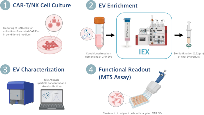
Figure 1: CAR-EV generation, enrichment, EV characterization, and functional readout. Schematic diagram depicting a typical workflow of EV generation, production, and characterization of enriched CAR-EV samples prior to analysis of functionality. Please click here to view a larger version of this figure.
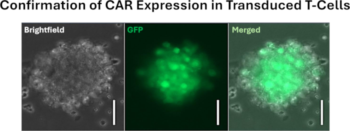
Figure 2: Representative microscopy analysis by confirmation of GFP expression in HER2-targeting CAR-T cells. Images were captured via fluorescence microscopy, using a 20x objective. The white scale bar represents a length of 50 µm. T cells have a natural tendency to grow in clumps prior to detachment into single cells. Please click here to view a larger version of this figure.
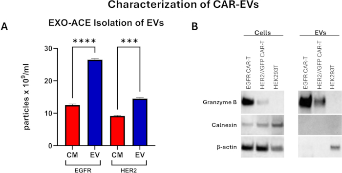
Figure 3: Characterization of CAR cells and EVs. (A) Nano-tracking particle analysis of CAR-T CM and enriched EVs. The particle concentration of EVs was 1.6-2.1-fold greater than that of CM, while protein concentration was reduced by >80%. Statistically significant differences were tested with one-way ANOVA and Tukey's multiple comparison test; *** p≤ 0.001, **** p≤ 0.0001. (B) Western blot analysis of transduced T-cells and enriched CAR-EVs confirmed the presence of cytolytic granzyme B in both CAR cells and EVs, whereas an absence was noted in HEK293T non-CAR cells. Calnexin was not detected in CAR-EV, indicating low contamination by endoplasmic reticulum particles. Protein load per lane = 5 µg. Please click here to view a larger version of this figure.
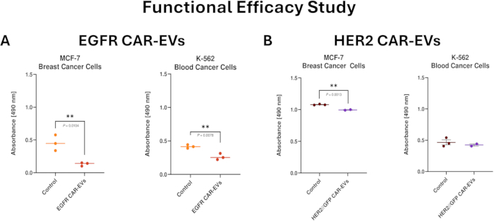
Figure 4: Effect of CAR-EVs on target cell viability (MCF-7 breast cancer cells and K562 chronic myelogenous leukemia cell line). Target cells were treated with a single dose of 4.5 x 10⁵ and 2.5 x 10⁵ EVs/ cell (EGFR or HER2 CARs, respectively) or control (EV buffer) for up to 3 days. (A) EGFR-targeting CAR-EVs cells. (B) HER2-targeting CAR-EVs. A decrease in absorbance reading (indicative of treatment efficacy) was observed in both cell lines. Data represent the mean absorbance ± SD (n = 3), analyzed using a two-tailed unpaired Student's t-test, with p-values indicating statistical significance; ** p≤ 0.01. Please click here to view a larger version of this figure.
Discussion
Combining the proven efficacy of CAR therapy with the promising advantages of extracellular vesicle (EV)-based therapy may improve the effectiveness of cancer treatments, as demonstrated in studies targeting breast and lung cancers12,13. The CAR-EV therapeutic approach addresses current limitations of CAR therapy, such as delivery challenges and associated toxicity, while leveraging the biocompatibility and targeting capabilities of EVs. Integrating these modalities may facilitate the development of new therapeutic strategies for cancer, expanding opportunities for the application of EVs in a clinical setting. In addition, engineering and enrichment of functional EVs provides a safer (via encapsulation of cytolytic cargo) and more precise method (i.e., the addition of targeting domains) for delivering therapeutic cargo, especially to challenging locations such as the tumor core, bone marrow, and areas protected by barriers like the blood-brain or blood-testis barrier14,15,16. Despite the well-established method of EV enrichment using ultracentrifugation, several limitations remain. Notable constraints include: scalability, co-isolation of impurities, and limited post-production yield17. There is a clear need for improved processes for generating therapeutic EVs at scale.
This proof-of-concept study demonstrates that IEX is a scalable and effective method for producing, isolating, and enriching functional CAR-EVs, using clarified CM from CAR-expressing cells as input. IEX enables high-throughput processing and supports both research and cGMP-scale production, allowing for quality control throughout the workflow-from EV generation to final drug product testing18,19. While EV characterization is essential to confirm function, yield can vary significantly due to differences in sample volume, input concentration, and EV-specific properties such as surface charge and binding properties. In-house data using MCF-7 cells shows an average pre-purification yield of ~2.85 × 10⁸ particles/mL, with post-purification concentrations typically ranging from 3 × 10¹¹ to 1 × 10¹² particles/mL. These levels can be adjusted by modifying the resuspension volume during buffer exchange to match downstream needs. Given this variability, users are strongly encouraged to optimize the protocol with CM relevant to their experimental setup, to ensure reproducible and robust yields.
Several steps within the protocol are critical to the successful production of CAR-EVs, with particular emphasis on the sample loading and elution phases. These steps directly influence the efficiency of EV capture by the column and, consequently, the overall yield. The buffer exchange step is crucial, as incomplete removal of excess solutes can compromise EV stability, leading to degradation over time. EVs can then be characterized by using methods such as NTA or proteomic profiling. If CAR-EVs are intended for functional assays, such as cellular uptake studies or therapeutic interventions, sterility is of utmost importance. A final 'terminal' (0.22 µm) sterilization step is essential to prevent microbial contamination, which may confound experimental outcomes or pose biosafety risks. Given these factors, users are strongly advised to exercise particular care during these steps to maintain reproducibility and functional integrity of the final EV preparation.
Several protocol modifications may be necessary to address common issues encountered during CAR-EV production. Low yield is most commonly caused by suboptimal loading of CM, either exceeding or falling short of the column's binding capacity. To determine the optimal input volume, it is advisable to conduct a CM titration study, incrementally increasing the load to identify the point of maximal EV recovery. Filter clogging, typically observed during the final sterile filtration step, often results from excessive sample concentration and viscosity, which can promote EV aggregation. In such cases, dilution of the final EV preparation is recommended. If dilution is not feasible, sterile filtration may be performed prior to buffer exchange; however, this approach necessitates that all subsequent steps be conducted under strict aseptic conditions to maintain sterility.
The development of EVs as cancer therapeutics requires reproducible production processes and robust validation of their efficacy against target cancer cell lines. A common limitation of IEX in EV enrichment is the co-isolation of non-EV particles – a challenge identifiable through downstream analysis of EV-specific markers, and reducible via further CM optimization. Control release characterization of EVs should include: NTA to determine particle number and size, and EV concentration post-enrichment; genetic and transcript analysis of CAR construct expression; determination of cellular and EV protein expression of the CAR, canonical EV biomarkers and cytolytic markers (e.g., granzyme B and perforin); and on- and off- target effects.The incorporation of additional techniques, such as transmission electron microscopy (TEM), will provide morphological validation to complement size-based characterization, supporting a more comprehensive analysis. To maintain quality control, EV products should undergo mycoplasma and endotoxin contamination testing to verify sterility and suitability for clinical production. The employment of suitable formulations for long-term storage of functional CAR-EVs may offer opportunities to enhance their stability, including maintaining quality, quantity, and therapeutic efficacy. Consistent evaluation of functional performance is essential for ensuring product integrity. Notably, the MTS assay utilized in this study offers an indirect evaluation of cytotoxicity via measurement of metabolic activity. Future studies would benefit from integrating standardized cytotoxicity assays, such as lactate dehydrogenase (LDH) release, Annexin V/PI staining, or caspase activation profiling, to delineate cytotoxic effects more accurately. Furthermore, incorporating CAR cell comparators (not incorporated in the current study due to sourcing limitations) either alone or in combination with CAR-EVs, and including parallel treatment of control non-cancer cell lines, could deepen the interpretative value of the findings.
This study highlights the potential use of EGFR/HER2 CAR EVs as a viable therapeutic strategy for specific cancers. Enhancing the efficacy of CAR EVs can be achieved through multiple dosing or pre-activation of parent T-cells to augment cytotoxicity. Moreover, advancements in emerging approaches, such as siRNA cargoes, and improvements in EGFR/HER2 binding capacity present opportunities to refine the design of these therapeutic platforms20,21. The promising application of dual chimeric antigen receptors (dual-CARs) in cell therapy offers an additional opportunity for advancement. Extending the use of dual-CARs by the utilization of their EV counterparts in therapeutics could be a pivotal step in broadening their therapeutic scope and enhancing treatment modalities22,23. Real-time monitoring of CAR EV-driven cancer cell cytotoxicity (e.g., caspase 3/7 activation, impedance reading) provides valuable insights for determining optimal dosage regimens, facilitating streamlined translation into future in vivo applications.
Disclosures
Asari K, Chelvaretnam C, Mom KT, Bhuiyan S, Pham Q, Fitri A, Palma C, Shojaee M, Khanabdali R, Hinch L, and Rice G are employees of INOVIQ Ltd.
Acknowledgements
The authors wish to thank Siena Barton, Hiba Rashid, Sara Nikseresht, Shayalini Wignarajah and Abel Sujeev for their contributions to the laboratory. The authors acknowledge all internal and external collaborators, with this study supported and funded by INOVIQ Ltd.
Materials
| Name | Company | Catalog Number | Comments |
|---|---|---|---|
| Materials | |||
| 1x phosphate buffered saline (PBS), pH 7.4 | Gibco | 10010049 | EV enrichment |
| 1% sodium dodecyl sulfate (SDS) | Sigma | L3771-100G | Dissolve 1g of SDS powder in 10 mL of ultrapure water |
| 1x tris buffered saline with Tween-20 (TBST) | Thermo Scientific Chemicals | J77500.K2 | Dilute with ultrapure water to make a 1x solution (alternatively, prepare a 0.1% polysorbate-20/Tween-20 solution in 1x tris buffered saline) |
| 96-well clear flat bottom polystyrene TC-treated microplate, sterile | Corning | 3595 | In vitro functional assay |
| Bolt Bis-Tris Plus Mini Protein Gels, 4-12%, 1.0 mm, WedgeWell format | Invitrogen | Cell/EV characterization | |
| Bovine serum albumin | Sigma-Aldrich | A9647-100G | Cell/EV characterization |
| Calnexin antibody | Cell Signaling | 2433S | Cell/EV characterization |
| CAR-NK/T cells | ProMab Biotechnologies | Varies | CAR extracellular ligand-binding domain depends on preferred target on recipient cells |
| CellTiter 96 AQueous One Solution Cell Proliferation Assay (MTS) | Promega | G3580 | In vitro functional assay |
| cOmplete, Mini, EDTA-free Protease Inhibitor Cocktail | Roche | 11836170001 | Cell lysis |
| ECL Western blotting detection reagent | Amersham | RPN2232 | Cell/EV characterization |
| Elution buffer | INOVIQ | EV enrichment/isolation | |
| Equilibration buffer | INOVIQ | EV enrichment/isolation | |
| EXO-ACE IEX column | INOVIQ | EV enrichment/isolation for downstream functional assay Patent ID AU2024901931 | |
| EXO-NET pan-exosome capture beads | INOVIQ | EV isolation for downstream characterization Patent ID WO/2014/011673 | |
| Granzyme B antibody | Cell Signaling | 4275 | Cell/EV characterization |
| Human IL-2 IS, premium grade | Miltenyi Biotec | 130-097-744 | Xeno-free culture of T/NK cells |
| iBlot 2 Transfer Stacks, PVDF | Invitrogen | Cell/EV characterization | |
| ImmunoCult-XF (cGMP) | Stemcell Technologies | 100-0956 | Xeno-free culture of T/NK cells |
| K-562 | ATCC | CCL-243 | In vitro functional assay |
| MCF-7 | ATCC | HTB-22 | In vitro functional assay |
| Millex-GP syringe filter unit, 0.22 µm | Merck Millipore | SLGPR33RS | Terminal filtration |
| Regeneration buffer | INOVIQ | EV enrichment/isolation | |
| RIPA buffer (10X) | Cell Signaling Technology | 9806 | Cell lysis |
| Trypan blue solution, 0.4% | Gibco | 15250061 | Assessment of cell viability and density |
| Wash buffer | INOVIQ | EV enrichment/isolation | |
| Equipment | |||
| Automated cell counter | Invitrogen | Countess II FL | Assessment of cell viability and density |
| Benchtop automated extraction instrument | Invitrogen | KingFisher Apex System | High throughput automated purification system See link for protocol details: https://www.inoviq.com/site/technology/resources |
| Fluorescence microscope | Zeiss | Axio Observer 7 | Cell culture |
| Nanoparticle tracking analysis (NTA) | Particle Metrix GmbH | Zetaview PMX-130 | Particle analysis |
| Plate reader | BMG Labtech | SPECTROstar Nano | In vitro functional assay |
| Refrigerated benchtop centrifuge | Beckman Coulter | Avanti J-15R | Cell culture & EV enrichment processing |
| SDS-PAGE mini gel tank | Invitrogen | A25977 | Cell/EV characterization |
| Western blot transfer system | Invitrogen | iBlot 2 | Cell/EV characterization |
References
- Park, J. H., Geyer, M. B., Brentjens, R. J. CD19-targeted CAR T-cell therapeutics for hematologic malignancies: Interpreting clinical outcomes to date. Blood. 127 (26), 3312-3320 (2016).
- Jain, M. D., Smith, M., Shah, N. N. How I treat refractory CRS and ICANS following CAR T-cell therapy. Blood. 141 (20), 2430-2442 (2023).
- Tang, X. J., et al. Therapeutic potential of CAR-T cell-derived exosomes: A cell-free modality for targeted cancer therapy. Oncotarget. 6 (42), 44179-44190 (2015).
- St-Denis-Bissonnette, F., et al. A clinically relevant large-scale biomanufacturing workflow to produce natural killer cells and natural killer cell-derived extracellular vesicles for cancer immunotherapy. JEV. 12 (12), e12387(2023).
- Jiang, Y., et al. Hypoxia enhances the production and antitumor effect of exosomes derived from natural killer cells. Ann Transl Med. 9 (6), 473-473 (2021).
- Decot, V., et al. Natural-killer cell amplification for adoptive leukemia relapse immunotherapy:comparison of three cytokines IL-2, IL-15, or IL-7 and impact on NKG2D, KIR2DL1, and KIR2DL2 expression. Exp Hematol. 38 (5), 351-362 (2010).
- Kim, I. Y., et al. Functional enhancement of exosomes derived from NK cells by IL-15 and IL-21 synergy against hepatocellular carcinoma cells: The cytotoxicity and apoptosis in vitro study. Heliyon. 9 (6), e16962-e16962 (2023).
- Mehdiani, A., et al. An innovative method for exosome quantification and size measurement. J Vis Exp. (95), e50974(2015).
- Bachurski, D., et al. Extracellular vesicle measurements with nanoparticle tracking analysis-an accuracy and repeatability comparison between NanoSight NS300 and ZetaView. JEV. 8 (1), 1596016(2019).
- Khanabdali, R., et al. High throughput surface epitope immunoaffinity isolation of extracellular vesicles and downstream analysis. Biol Methods Protoc. 9 (1), bpae032(2024).
- Salomon, C., et al. Early detection of ovarian cancer: An accurate high-throughput extracellular vesicle test. J Clin Oncol. 43 (16_suppl), 5582-5582 (2025).
- Fu, W., et al. CAR exosomes derived from effector CAR-T cells have potent antitumour effects and low toxicity. Nat Commun. 10 (1), 1-12 (2019).
- Zheng, W., et al. Inhalable CAR-T cell-derived exosomes as paclitaxel carriers for treating lung cancer. J Transl Med. 21 (1), 383(2023).
- Tian, J., et al. Engineered exosome for drug delivery: Recent development and clinical applications. Int J Nanomed. 18, 7923-7940 (2023).
- Chen, Z., et al. Encapsulation and assessment of therapeutic cargo in engineered exosomes:a systematic review. J Nanobiotechnology. 22 (1), 18(2024).
- Basyoni, A. E., Atta, A., Salem, M. M., Mohamed, T. M. Harnessing exosomes for targeted drug delivery systems to combat brain cancer. Cancer Cell Int. 25 (1), 150(2025).
- Coughlan, C., et al. Exosome isolation by ultracentrifugation and precipitation and techniques for downstream analyses. Curr Protoc Cell Biol. 88 (1), e110(2020).
- Zhang, W., et al. Large-scale separation and purification of exosomes using ion-exchange chromatography. Biomed Res Ther. 11 (4), 6348-6356 (2024).
- Fernandes, R. P., et al. Targeted isolation of extracellular vesicles from cell culture supernatant using immuno-affinity chromatography. Sep Purif Technol. 358 (Part A), 130312-130312 (2024).
- Jiang, J., et al. Anti-EGFR ScFv functionalized exosomes delivering LPCAT1 specific siRNAs for inhibition of lung cancer brain metastases. JNB. 22 (1), 159(2024).
- Dobersberger, M., et al. An engineering strategy to target activated EGFR with CAR T cells. Cell Rep Methods. 4 (4), 100728-100728 (2024).
- Shi, M., et al. Bispecific CAR T cell therapy targeting BCMA and CD19 in relapsed/refractory multiple myeloma:a phase I/II trial. Nat Commun. 15 (1), 3371(2024).
- Roddie, G., Amrolia, P. J. Dual targeting CAR-T cells for B-cell acute lymphoblastic leukaemia and B-cell non-Hodgkin lymphoma. Blood Adv. 9 (4), 704-721 (2024).
Reprints and Permissions
