This protocol describes the in vivo reprogramming of mouse cancer cells into type 1 dendritic-like cells within the tumor microenvironment through enforced expression of the transcription factors PU.1, IRF8, and BATF3.
Method Article
Tractable In Vivo Reprogramming of Tumor Cells to Type 1 Conventional Dendritic Cell-like Cells
* These authors contributed equally
In This Article
Summary
Abstract
The efficacy of cancer immunotherapy relies on the recruitment and activation of cytotoxic T cell responses against solid tumors by type 1 conventional dendritic cells (cDC1s). However, the generation of cDC1s for cancer immunotherapy faces significant limitations, including poor cell yield, functional heterogeneity, and susceptibility to immunosuppression in the tumor microenvironment (TME). We recently developed an immunotherapy modality based on in vivo reprogramming of cancer cells into immunogenic cDC1-like cells, which enabled cancer cells to present tumor antigens as cDC1s and elicited polyclonal cytotoxic T cell responses and durable systemic anti-tumor immunity. Here, we describe a tractable protocol to generate cDC1-like cells within the TME by overexpressing the minimal cDC1-specific gene regulatory network-PU.1, IRF8, and BATF3 (collectively referred to as PIB)-in cancer cells, followed by subcutaneous implantation of a mixture of transduced and parental cells. PIB overexpression drives the gradual acquisition of the hematopoietic marker CD45 and the professional antigen presentation complex MHC class II on tumor cells, serving as cell surface readouts for in vivo cDC1 reprogramming. When compared to the reprogramming process in vitro, reprogramming of the YUMM1.7 mouse melanoma model in vivo demonstrated faster kinetics and higher efficiency. cDC1-like cells induced rapid remodeling of the TME by recruiting host immune cells within the first 3 days and leading to the formation of tertiary lymphoid structure by day 9. Reprogrammed cDC1-like cells persisted in tumors for at least 9 days but were undetected at day 15. The in vivo cDC1 reprogramming protocol described here provides a tractable and robust method to effectively transform "immune-cold" tumors into "immune-hot". Overall, it offers a powerful platform to study the mechanisms underlying cDC1-mediated anti-tumor immunity and uncover synergistic combinations with other cancer immunotherapy modalities.
Introduction
Recent advancements in cancer immunotherapy modalities, including immune checkpoint blockade (ICB), adoptive cell therapy (ACT), and vaccines, have shifted the landscape of cancer treatment1. In melanoma, response rates to programmed cell death protein 1 (PD-1) and cytotoxic T lymphocyte-associated protein 4 (CTLA-4) inhibition can reach up to 60%2. Nevertheless, over 40% of melanoma patients fail to respond to ICB, and response rates in less immunogenic cancers, such as breast cancer, microsatellite-stable colorectal cancer, and glioblastoma, remain below 5%, highlighting clinical gaps3,4,5. A major barrier to broad clinical success is the poor activation of cytotoxic T cell responses, which depend on efficient antigen presentation within the tumor microenvironment (TME)6.
Conventional type 1 dendritic cells (cDC1s) are rare, but indispensable for the initiation of tumor-specific immunity, offering three key signals to prime T cell responses: (1) professional processing and presentation of antigens on major histocompatibility complexes (MHC) class I and II, (2) co-stimulation via CD80 and CD86, and (3) pro-inflammatory cytokine signaling including IL-127,8. Among the dendritic cell (DC) subsets, cDC1s excel in cross-presenting tumor cell-associated antigens to prime naïve or re-activate memory CD8+ T cells or drive CD4+ T cell differentiation to type 1 T helper (TH1) cells9. Additionally, cDC1s are the main producers of chemokines, C-X-C motif chemokine ligands 9 and 10 (CXCL9/10), which mediate T cell recruitment to the tumor tissue10,11,12,13. Over the last few years, numerous studies have highlighted that tumor rejection and effective responses to ICB or ACT strongly correlate with the presence of intratumoral cDC1s12,13. Hence, cDC1s have developed as an attractive target for cancer immunotherapy and successfully harnessing their unique features can have significant translational potential.
Current methods to generate cDC1s are based on blood isolation or differentiation from CD34+ progenitors or pluripotent stem cells14,15,16. The predominant clinical strategies rely on isolating a mixture of autologous DC subsets from the blood and stimulating them ex vivo with toll-like receptor agonists or pro-inflammatory cytokines to induce a mature, immunogenic program before reinfusion16. However, these strategies are limited by poor cell maturation, immunosuppressive barriers in the TME, and subset heterogeneity in the infused DC mixture, where only a small proportion of cDC1s is present7. Moreover, isolating only cDC1s for cancer immunotherapy or vaccination is challenging due their extremely low abundance in peripheral blood (<0.3%)16. Additionally, cDC1s can be produced from CD34+ progenitor cells or induced pluripotent stem cells using cytokines and NOTCH signaling. However, these methods require complex and long protocols and yield low numbers and heterogeneous populations of DC subsets17,18,19.
To address these limitations, in vivo reprogramming provides a suitable strategy for cancer immunotherapy to generate cDC1-like cells, which resemble the phenotypic, transcriptional, and functional properties of cDC1s20. Through the overexpression of the cDC1-specific minimal gene regulatory network -- PU.1, IRF8, and BATF3 (collectively referred to as PIB) -- fibroblasts, stromal cells, and cancer cells have been gradually reprogrammed into cDC1-like cells enabling tumor antigen processing and presentation on MHC class I and II, co-stimulatory signaling via CD80 and CD86, and cytokine/chemokine secretion, including CXCL9/10 and IL-1220,21,22,23. cDC1 reprogramming progressed faster and more efficiently across human xenografts in vivo and independently of immunosuppression, generating cDC1-like cells with a mature immunogenic signature20. In syngeneic melanoma tumors, we observed that cDC1-like cells remodeled their TME, induced the formation of tertiary lymphoid structure (TLS), and recruited and expanded polyclonal TH1 and cytotoxic memory T cells, leading to long-term systemic immunity20. Finally, we developed a novel cancer immunotherapy modality based on in situ reprogramming of cancer cells into cDC1-like cells by intratumoral injection of adenoviral vectors20. These findings pave the way for clinical translation of cDC1 reprogramming as a new cancer immunotherapy modality that can harness the functional properties of cDC1s.
Here, we present a tractable protocol to generate cDC1-like cells within the TME based on in vivo cDC1 reprogramming. Lentiviral delivery of PIB to cancer cells and subsequent implantation as a defined mix of transduced cancer cells with untransduced parental cancer cells allows in vivo reprogramming of a controlled fraction of cancer cells within the TME. Additionally, this method is not limited by the in situ delivery efficiency of viral vectors20. We outline the steps for efficient lentiviral vector production, functional titration, and in vivo cDC1 reprogramming using the syngeneic melanoma model, YUMM1.7, as an example. We detail the experimental setup and the gating strategy for analyzing in vivo reprogrammed cDC1-like cells by flow cytometry after tumor dissociation. We also provide the steps to evaluate changes in the TME by performing cryopreservation, sectioning, and immunofluorescence staining of tumor sections, followed by confocal microscopy. Finally, we address how to monitor cDC1-induced immunity by measuring tumor growth and survival. This tractable and scalable approach offers researchers a valuable tool to assess combinatorial treatment strategies with in vivo cDC1 reprogramming, to characterize changes in the TME and TLS neogenesis across cancer models or perform large screens modifying the expression of various immune pathways, for example, antigen presentation or cytokine/chemokine secretion, and unravel their impact on antitumor immunity.
Protocol
Animal experimental procedures were performed in accordance with Swedish regulations after approval from the Swedish Board of Agriculture.
1. Reagent preparation
- To prepare Dulbecco's modified Eagle's medium (DMEM) complete medium, add 10% (v/v) fetal bovine serum (FBS), 1 mM sodium pyruvate, 100 U/mL penicillin (Pen), 2 mM glutamine substitute, and 100 mg/mL streptomycin (Strep).
- To prepare RPMI 1640 complete media, add 10% (v/v) FBS, 2 mM glutamine substitute, 1 mM sodium pyruvate, 50 mM 2-mercaptoethanol, 100 U/mL Pen, and 100 mg/mL Strep.
- To prepare DMEM/F12 complete media, add 10% (v/v) fetal bovine serum (FBS), 1 mM sodium pyruvate, 0.1 mM non-essential amino acids, and 100 U/mL Pen and 100 mg/mL Strep.
- To prepare 1 mg/mL Polyethylenimine (PEI), dissolve 100 mg of PEI powder in 100 mL of sterile deionized water in a glass beaker or conical tube. Place the beaker on a magnetic stirrer and stir at room temperature (RT) to create a vortex until dissolved completely. Adjust the pH to 6.9-7.1, adding hydrochloric acid (HCl)/sodium hydroxide (NaOH) dropwise, and filter the solution through a 0.22 µm PES membrane filter.
- To prepare 1 mM Sodium Butyrate, dissolve 1.10 mg in 10 mL of sterile deionized water.
- To prepare 8 mg/mL Polybrene, dissolve 80 mg of Polybrene powder in 10 mL of sterile deionized water.
- To prepare FACS buffer, add 2% FBS and 1 mM EDTA to phosphate-buffered saline (PBS).
2. Lentivirus production
- Thaw HEK 293T cells by adding 1 mL of cells to 9 mL of prewarmed DMEM complete medium and centrifuge the cells at 350 × g for 5 min. Resuspend the cell pellet in 20 mL of DMEM complete medium and culture cells in a 150 mm plate at 37 °C with 5% CO2.
- Expand and split HEK 293T cells when they reach 80-90% confluency. To split cells, wash once with PBS, add 5 mL of trypsin to dissociate them from the plate, and incubate 5-10 min at 37 °C with 5% CO2.
- Collect cells with 5 mL of DMEM complete medium, centrifuge them at 350 × g for 5 min, and split them 1:6 into new 150-mm plates, 48 h before transfection.
NOTE: The quality of the lentivirus-producing cell line is crucial for producing high titers. Do not allow cells to exceed 95% confluency and avoid seeding them at <30% confluency. - After 48 h, or when cells reach 70-80% confluency, prepare the transfection mix in a 50 mL conical tube. Use the following transfer plasmid: 1) SFFV-PIB-IRES-eGFP-a polycistronic lentiviral vector containing the transcription factors PU.1, IRF8, BATF3, an internal ribosomal entry site (IRES), and an enhanced green fluorescent protein (eGFP) as the fluorescent transduction reporter; or 2) SFFV-eGFP, a control lentiviral vector with a multiple cloning site and eGFP; or 3) SFFV-mCherry, a lentiviral vector expressing mCherry. Add 10 µg of transfer plasmid, 7.5 µg of packaging plasmid psPAX2, and 2.5 µg of envelope plasmid pMD2.G. Top up the tube with Opti-MEM to reach a total volume of 2 mL. Finally, add 60 µL of the transfection reagent PEI (from the 1 mg/mL stock) to the transfection mix.
NOTE: The lentiviral vector SFFV-eGFP is used as a transduction control for reprogramming experiments to account for lentiviral-mediated immunogenicity, while SFFV-mCherry is used to generate fluorescently labeled cancer cell lines. - Vortex the transfection mixes thoroughly for 1 min and incubate for 15 min at RT to allow the formation of DNA-lipid complexes.
NOTE: Confluency significantly influences viral titers. Transfecting cells at less than 70% confluency will result in lower viral titers due to fewer cells receiving the plasmids. - Aspirate the medium of the HEK 293T cells and add 10 mL of DMEM without Pen/Step and with 10% of sterile-filtered FBS.
- Add the transfection mix dropwise to the 150 mm plates and incubate for 16 h at 37 °C with 5% CO2.
- After transfection, replace the medium with 20 mL of DMEM complete medium supplemented with 1 mM Sodium Butyrate, and incubate cells for 24 h at 37 °C with 5% CO2.
NOTE: Sodium butyrate is a class 1 histone deacetylase (HDAC) inhibitor and can enhance gene expression in HEK 293T cells by inducing histone hyperacetylation, which opens up the chromatin structure and facilitates transcription of viral packaging and transfer plasmids. Transfection efficiency can be assessed by checking eGFP expression using a fluorescence microscope. - Collect the lentivirus-containing supernatant 48 h and 60 h post transfection into 50 mL conical tubes. After the first collection, add 12 mL of prewarmed DMEM complete medium per 150 mm plate for the second collection. Store the collections at 4 °C, filter them through a 0.45 µm low-protein-binding PES filter and combine them.
- To concentrate the virus, pipette 37 g of lentivirus-containing medium into 38.5 mL Open-Top Thinwall Polypropylene Tubes. Measure the weight of the tubes using a bench scale.
NOTE: Measure the weight of the lentivirus-containing medium rather than its volume, as weight provides greater precision for balancing. The weight of the tubes must be well balanced to avoid damaging the ultracentrifuge. - Place the tubes into the ultracentrifugation buckets, close the lids, and place the buckets into a Swinging-bucket rotor. Centrifuge at 25,000 × g for 90 min at 4 °C.
- After centrifugation, remove the tubes from the buckets and aspirate the supernatant.
NOTE: The same tubes can be filled up repeatedly and used for a total number of three centrifugation rounds to produce a bigger lentivirus pellet per tube. - Place the Open-Top Thinwall Polypropylene Tubes upside down for 5 min on a laboratory wipe or similar absorbent material to allow excess liquid to drain and dry the pellet.
- Add 200 µL of ice-cold DMEM supplemented with HEPES or PBS to each pellet. Place the microcentrifuge tubes into 50 mL conical tubes and close the lid to maintain sterility.
- Incubate the pellet overnight at 4 °C to allow resuspension.
- Combine the resuspended pellets, aliquot, and store them at -80 °C for up to a year.
NOTE: To avoid loss of titer by repeated rounds of freeze-thawing, use smaller aliquot volumes (e.g., 50-200 µL).
3. Functional virus titration
NOTE: This step should be repeated for each batch of lentivirus to determine the optimal viral dose required to achieve a 1:1 ratio of transduced (eGFP+) to untransduced (eGFP-) cells.
- Thaw and expand cancer cells in appropriate culture media (e.g., the YUMM1.7 murine melanoma cell line in DMEM/F12 complete medium).
NOTE: Cells should undergo at least one round of division before a reprogramming experiment. Passages of cells should be similar between experiments. - One day before tumor establishment, raise cancer cells by aspirating their culture media, washing once with PBS, and adding 2 mL or 5 mL of trypsin per 100 mm plate or 150 mm plate. Incubate for 5-10 min at 37 °C with 5% CO2 and collect the cells by adding 8 mL or 5 mL complete media per 100 mm or 150 mm plate, respectively, into 50 mL conical tubes.
- Centrifuge cells at 350 × g for 5 min to remove trypsin and resuspend in complete medium.
- Take a 10 µL aliquot of cells, add 10 µL of Trypan Blue, and count viable cells using a hemocytometer.
- Transfer 6 × 10⁵ cells to a new 50 mL conical tube containing 12 mL of complete medium and add 12 µL of 8 mg/mL polybrene to the cell suspension to reach a working concentration of 8 µg/mL.
- Pipette 2 mL of the cell suspension into each well on a 6-well tissue culture plate.
NOTE: If necessary, increase cell numbers to 1.2-1.8 x 10⁶ per 6-well plate. - Add increasing volumes of lentivirus to each well to determine the optimal volume needed to achieve a 1:1 ratio of transduced (eGFP+) to untransduced (eGFP-) cells.
NOTE: Add the recommended lentivirus volumes of lentivirus of 1, 2, 4, 8, 16, and 32 µL in triplicates. Perform the functional titration for the empty control lentiviral vector SFFV-eGFP and SFFV-mCherry. - Incubate the cells for 24 h at 37 °C with 5% CO2.
- On day 0, remove the lentivirus-containing media, replace with fresh media, and incubate for 72 h at 37 °C with 5% CO2.
- On day 3, check the cells for eGFP expression under a fluorescence microscope.
NOTE: This step provides a rapid verification of cell transduction but does not replace the analysis performed by flow cytometry in step 3.14 to determine the exact ratio of eGFP+ to eGFP- cells.
In some cancer cells (e.g., B16 mouse melanoma cells) the eGFP signal may be low and difficult to detect using a fluorescence microscope. It is recommended to proceed directly to flow cytometry. - Aspirate the medium, wash the cells once with PBS, and add 500 µL trypsin per well. Incubate for 5-10 min at 37 °C with 5% CO2.
- Raise the cells in each well with 1.5 mL of FACS buffer and pipette the cell suspension into 5 mL polystyrene tubes. Take an aliquot and count viable cells using a hemocytometer and Trypan blue staining.
- Centrifuge the cell suspension at 350 × g for 5 min, resuspend the pellet in 100 µL of FACS buffer, and proceed with flow cytometry analysis.
- Quantify the precise ratio of transduced (eGFP+) to untransduced (eGFP-) cells by flow cytometry. For in vivo testing, use the lentivirus volume that yields closest to 1:1 ratio.
NOTE: To accurately determine the ratio of eGFP+ to eGFP- cells, include non-transduced cancer cells as negative controls for gating.
4. Subcutaneous tumor establishment for in vivo reprogramming
- Thaw and expand cancer cells in their appropriate culture medium for generating mCherry-expressing cells.
NOTE: mCherry expression facilitates the distinction of cancer cells during flow cytometry analysis from other populations in the tumor, for example, endogenous stromal and immune cells. - Perform steps 3.2-3.4 and resuspend cells in complete media at a concentration of 105 cells per mL.
- Add 8 µg/mL of polybrene and the determined volume of SFFV-mCherry lentivirus to the cell suspension to reach >90% transduction efficiency.
NOTE: Alternatively, if achieving >90% transduction compromises cell viability, sort mCherry+ cells by fluorescence-activated cell sorting (FACS) 72 h after transduction and expand sorted cells in culture. - Plate 5 mL of the cell suspension (containing 5 × 10⁵ cells) per 100 mm plate and incubate cells at 37 °C with 5% CO2 for 24 h.
- After 24 h, replace the medium with 10 mL of complete medium and expand the cells by culturing them at 37 °C with 5% CO2 for at least 7 days before starting reprogramming experiments.
- After expansion, follow steps 3.2-3.4 to prepare the cell suspensions for subsequent transduction with the previously determined volume of SFFV-PIB-IRES-eGFP, resulting in the closest 1:1 ratio of transduced (eGFP+) to untransduced (eGFP-) cells on day 3.
- Add 8 µg/mL of polybrene to the cell suspension, along with the previously determined volume of lentivirus to the cell suspension in 50 mL conical tubes. Mix well without creating bubbles.
- Pipette 2 mL of the cell suspension (containing 105 cells) per well in a 6-well tissue culture plate and incubate for 24 h at 37 °C with 5% CO2.
NOTE: Alternatively, plate the cells in 5 mL of transduction media in 100 mm tissue-culture plates. However, note that transduction efficiency may be reduced in larger volumes and should be empirically determined as described in steps 3.1-3.13.
It is recommended to transduce at least 10% more cells to compensate for potential cell loss during the transduction process and to ensure sufficient volume for pipetting or injection during tumor establishment. - After 24 h, remove the lentivirus-containing medium, wash twice with PBS, add 500 µL of trypsin to each well, and incubate for 5-10 min at 37 °C with 5% CO2.
- Check under a light microscope whether the cells dissociated completely from the plates and then collect them with complete media into 50 mL conical tubes.
- Pellet the cells by centrifuging them for 5 min at 350 × g, wash the pellet with PBS, and centrifuge again for 5 min at 350 × g. Take an aliquot and count viable cells using a hemocytometer and Trypan blue staining.
- Resuspend the pellet in ice-cold PBS to reach a concentration of 2-4 × 10⁷ cells per mL, which corresponds to 1-2 × 10⁶ cells in 50 µL of injection volume per tumor.
NOTE: Importantly, these numbers have been validated for YUMM1.7 melanoma tumors to isolate in vivo reprogrammed cDC1-like cells for flow cytometry analysis. For other cell lines and for other experimental readouts, such as tumor growth and survival experiments, the numbers need to be empirically tested. For tumor growth and survival experiments, lower cell numbers-ranging from 100,000 to 250,000 cells per tumor-are recommended. - Relocate to the work area where animal experiments are performed and keep the cells on ice.
NOTE: After cell preparation, perform cell injections as quickly as possible as cell viability is reduced over time. - Prepare a 60 µL anesthesia solution per mouse by diluting Xylazine (3 mg/kg per mouse) and Ketamine (135 mg/kg per mouse) in PBS.
- Anesthetize CD45.1 mice by injecting 60 µL of anesthetic intraperitoneally. Randomize the mice and keep them on a warming pad to avoid a drop in body temperature. Apply eye crème on their eyes to prevent ocular dehydration during anesthesia.
NOTE: It is critical to use CD45.1 mice to enable distinction between in vivo reprogrammed cDC1-like cells expressing the wild-type CD45.2 allele and endogenous immune cells. If the cell line is CD45.1 C57BL/6 mice (CD45.2) can be used as recipients. Mice should be sex- and age-matched between experimental groups and randomized before tumor establishment. In this study, 8-week-old female mice were used. - Wait 5-10 min until all mice are fully anesthetized.
- Shave the right flank of the mice using a razor; optionally, mark them using an ear clipper. Disinfect the area and swipe away the remaining hair with 70% ethanol.
- Resuspend cancer cells by pipetting up and down using a P1000 pipette. Load the cell suspension into a 0.5 mL 30 G syringe.
- Gently lift the skin of the shaved flank of the mouse using two fingers and insert the tip of the needle where the skin forms a crease.
NOTE: Be cautious to avoid puncturing your fingers with the needle. - Release the skin, hold the syringe in place with one hand, and use two fingers of the other hand to stretch the skin of the mouse.
- Slowly inject 50 µL of the cancer cell suspension to create an oval bulge (subcutaneous swelling) at the injection site.
NOTE: If a bulge does not form, the suspension may have leaked into the surrounding subcutaneous tissue, which can lead to inconsistent or failed tumor formation. - Post procedure, monitor all mice wake up to ensure survival after anesthesia.
- Maintain the mice in a controlled temperature environment with 23 ± 2 °C and a fixed 12 h light/dark cycle and free access to food and water.
NOTE: If you wish to monitor tumor growth, proceed with steps 4.24-4.26. Otherwise, skip these steps and proceed directly to the next sections. - To monitor tumor growth, gently restrain the mouse with one hand, allowing clear visualization of the tumor.
- Using an electronic caliper, measure the tumor's length (l), width (w), and height (h). Calculate the tumor volume (V) using the formula: V = (l x w x h) / 2.
- Measure tumor dimensions every two to three days. When the tumor reaches a volume that meets the humane endpoint criterion (e.g., 1,500 mm³), euthanize the animal in accordance with ethical and institutional guidelines.
5. Tumor isolation for flow cytometry analysis
- Prepare scissors, forceps, FACS buffer, and 12-well non-tissue culture plates with ice-cold RPMI complete medium for collection of tumor samples.
- Prepare the tumor digestion solution by mixing 30 µL of 100 mg/mL collagenase D and 3 µL of 10 mg/mL DNAse I in 3 mL of RPMI complete medium per tumor (to reach a working concentration of 1 mg/mL collagenase D and of 10 µg/mL DNAse I).
NOTE: If digestion efficiency is suboptimal, add 2-5 mM CaCl2 to support collagenase activity. - Euthanize the mice by cervical dislocation or CO2 asphyxiation on the day of isolation.
NOTE: If CO2 euthanasia is chosen, introduce CO2 at 30–70% chamber volume/min and confirm death by an approved secondary method (e.g., cervical dislocation). - Excise subcutaneous tumors with scissors and forceps and place each tumor into a well of the 12-well non-tissue culture plate with ice-cold RPMI. Try to minimize excision of non-tumor tissue.
- Cut tumor samples into 1-2 mm3 pieces using scissors, collect the pieces into 50 mL conical tubes containing 3 mL of digestion solution, and incubate at 37 °C while shaking at 200-250 rpm for 30-45 min.
NOTE: Vortex or pipette every 15 min to facilitate the digestion process. - Following digestion, vigorously pipette the mixture for 1 min using a 5 mL serological pipette. Filter the cell suspension through a 70 µm cell strainer into a new 50 mL conical tube.
- Use the plunger of a 10 mL syringe to smash non-digested pieces against the 70 µm cell strainer and rinse the strainer with 10 mL of RPMI complete medium.
- Centrifuge the cell suspension at 350 × g for 5 min at 4 °C, resuspend in 1 mL of FACS buffer, and count viable cells.
- Transfer the cell suspension to 5 mL polystyrene tubes, pellet cells at 350 × g for 5 min, and resuspend 106 viable cells per 100 µL of FACS buffer.
- Separate cells for single color (SC) and FMO controls into separate polystyrene tubes.
- Prepare an antibody staining master mix containing rat serum (1:100) to block unspecific binding; a fixable viability dye (1:100) to exclude dead cells in the analysis; and fluorescently labelled antibodies anti-CD45.1, anti-CD45.2, and anti-MHC-II at their optimal concentration. Add 100 µL of the master mix to each sample and incubate for 20 min at 4 °C in the dark.
- Wash the samples 2x with 2 mL of FACS buffer and proceed with flow cytometry analysis.
- Quantify the percentages of cDC1-like cells expressing CD45.2 and/or MHC-II, by gating in transduced (eGFP+) cancer cells (CD45.1-mCherry+).
NOTE: This protocol can be used to perform immunophenotyping of tumors after in vivo cDC1 reprogramming by complementing the staining panel with antibodies against endogenous lymphoid and/or myeloid immune cell populations.
6. Tumor freezing and sectioning for confocal microscopy analysis
- Prepare for tumor isolation by following steps 5.1 and 5.3. Excise subcutaneous tumors with scissors and forceps.
- Fix tumors in freshly prepared 4% paraformaldehyde (PFA) in PBS at 4 °C with gentle shaking. Adjust fixation time according to tumor size, using 1 h per mm³ of tissue as a guideline.
- Transfer tumors into 15% sucrose in PBS and incubate at 4 °C while gently shaking the tissues until they sink to the bottom of the tube. Then, move them to 20% sucrose in PBS under the same conditions.
- Once tumors have settled at the bottom of the tube, incubate them in a 1:1 mixture of 20% sucrose in PBS and OCT compound for 30 min at 4 °C with shaking.
- Remove the mixture, embed the tissues in a cryomold with 100% OCT and snap-freeze using liquid nitrogen.
NOTE: Always wear appropriate personal protective equipment (cryogloves, face shield, and lab coat) to prevent cold burns or injury from rapid vapor expansion. Work in a well-ventilated area to avoid asphyxiation risk.- Store blocks at -80 °C in closed containers to prevent dehydration.
7. Immunofluorescence staining of tumor sections
- Using a cryostat, cut the tumors into approximately 8 µm-thick sections and mount them on microscope glass slides. If needed, store the slides at -80 °C.
NOTE: To preserve tissue integrity, keep the blocks and slides with mounted tumor cuts on dry ice during the transport to and from the cryostat. Ensure the cryostat is maintained at -20 °C during sectioning. - Thaw the slides in PBS for 5 min and fix them in a 4% PFA solution for 10 min at RT.
- Following fixation, wash the slides in PBS for 5 min.
NOTE: Ensure the tissues remain hydrated; always keep them in liquid throughout all subsequent staining steps. If the experiment needs to be paused, slides can be stored in PBS at RT for up to a 3 hours. Avoid storing the slides in PBS overnight. - For antigen retrieval, fill up a steamer with water and turn it on. Place a jar containing deionized water and a jar containing 10 mM Sodium Citrate (pH 6) into the steamer and cover the opening with aluminum foil. Without removing the lid, insert a thermometer through the vents of the steamer lid, pierce the aluminum foil, and monitor the temperature, ensuring that the solutions reach ~99 °C.
- When the buffers reach ~99 °C, dip the slides in deionized water for 10 s and then quickly transfer them to the antigen retrieval solution.
- Incubate the slides at ~99 °C for 15 min.
- Wash the slides in 1x PBS for 3 x 5 min.
- Use a Hydrophobic Barrier Pap Pen to delineate each tissue section, forming a hydrophobic perimeter that prevents any liquid added to the tissues from leaking.
NOTE: Subsequent PBS washes may disrupt the hydrophobic perimeter surrounding the tissues and require renewal. Draw it again as needed. - Block and permeabilize tissues with 150 µL of blocking buffer (5% serum and 0.5% Triton X-100 in 1x PBS) for 60 min at RT. For all staining steps use serum from the same species as the host of the secondary antibody.
- Wash slides in 1x PBS for 3 x 5 min.
- Incubate tissues with 20 µg/mL of the primary anti-CD45 antibody in blocking buffer (5% serum and 0.5% Triton X-100 in 1X PBS) overnight at 4 °C.
NOTE: To use other antibodies, empirically validate antibody concentration. - Wash slides in 1x PBS for 3 x 5 min.
- Incubate the tissue sections with the goat anti-rat secondary antibody conjugated with AlexaFluor-647 at a 1:250 dilution in blocking buffer (5% serum) for 2 h in the dark at RT.
NOTE After adding the secondary antibody, perform all subsequent steps in the dark to prevent photobleaching of the fluorophores. - Wash slides in 1x PBS for 3 x 5 min.
- Incubate tissues with 1 µg/mL of DAPI and 20 µg/mL of AlexaFluor-594 conjugated antibodies in blocking buffer (5% serum) for 1 h in the dark at RT.
- Wash slides in 1x PBS for 3 x 5 min.
- Add a drop of mounting solution onto each tumor section, then apply a few drops of mounting medium to the coverslip. Quickly invert the coverslip onto the slide to mount it and ensure the absence of bubbles near the tumors.
- Allow the slides to dry overnight and image them by confocal microscopy.
Results
A schematic overview of the tractable in vivo reprogramming protocol is presented in Figure 1. First, we produced the polycistronic lentiviral vector SFFV-PIB-IRES-eGFP (PIB-eGFP), encoding for the cDC1-instructing reprogramming factors PU.1, IRF8, BATF3, followed by an internal ribosomal entry site (IRES) and enhanced green fluorescent protein (eGFP) in the HEK 293T packaging cell line. To control for lentivirus-mediated immunogenicity, we produced an empty SFFV-MCS-eGFP (eGFP) vector, containing a multiple cloning site (MCS) and IRES-eGFP, but excluding the reprogramming factors. Following viral production, we harvested the lentivirus-containing supernatant and concentrated it by ultracentrifugation. We functionally titrated the virus to determine the amount required to achieve a 1:1 ratio of transduced (eGFP+) to untransduced (eGFP-) cells, which we evaluated on day 3 post transduction. Then, we used the resulting virus amount to transduce the selected cancer cell line on day -1. After 24 h, we collected the cells and implanted them subcutaneously to establish tumors. This strategy ensured that transduced cells reprogrammed into cDC1-like cells in vivo, within the TME. At indicated time points post engraftment, we excised and processed tumors for downstream analyses, including flow cytometry of dissociated tumor cells, to quantify in vivo reprogramming efficiency, or immunofluorescence staining of cryopreserved tumor sections to characterize changes in the TME.
First, to ensure the production of high lentiviral titers, we monitored HEK 293T cells before transfection, targeting to reach 70-80% confluency and 24 h later, we assessed the transfection efficiency through eGFP expression by fluorescence microscopy (Figure 2A). Next, we collected virus-containing medium 48 h and 60 h post transfection, ultracentrifuged it, and stored in aliquots at -80 °C (Figure 2B). Then, we determined the optimal viral amount required to achieve a 1:1 ratio of eGFP+ to eGFP- cells in YUMM1.7 melanoma cells. We seeded 105 YUMM1.7 cells per well in a 6-well plate, transduced the cells with increasing volumes of lentivirus, and replaced the virus-containing supernatant with fresh DMEM/F12 complete media after 24 h (Figure 2C). On day 3, we assessed eGFP expression by fluorescence microscopy, before proceeding to quantify the precise percentages of eGFP+ cells by flow cytometry (Figure 2C). To quantify the ratio of eGFP+ to eGFP- cells, we gated in live, single cells to exclude dead cells and doublets (Figure 2D). The optimal volume of the produced lentivirus batch was determined both for PIB-eGFP and the control eGFP virus as 2 µL per 105 YUMM1.7 cells, resulting in 50.0 ± 0.3% and 63.0 ± 0.9% eGFP+ cells, respectively (Figure 2E).
Next, to evaluate in vivo reprogramming efficiency, we generated a fluorescently labelled (mCherry+) cancer cell line (YUMM1.7-mCherry). This ensured that we could discriminate between cancer cells and stromal or immune cells by gating in live mCherry+ cells in the flow cytometry analysis (Figure 3A,B). We then transduced YUMM1.7-mCherry cells with 2 µL of PIB-eGFP or control eGFP virus per 105 cells to achieve a 1:1 eGFP+ to eGFP- ratio. We subcutaneously implanted 1.6 × 10⁶ cells per tumor in CD45.1 mice and kept an aliquot of cells to culture in vitro for comparison. We evaluated reprogramming efficiency by flow cytometry, gating live mCherry+ eGFP+ and CD45.1- cells to exclude host immune cells that have engulfed eGFP or mCherry (Figure 3B). From days 1 to 9, we observed the gradual acquisition of CD45.2 and/or MHC-II expression, marking partially reprogrammed cDC1-like cells (CD45.2+ MHC-II- or CD45.2- MHC-II+) and fully reprogrammed cDC1-like cells (CD45+ MHC-II+) (Figure 3C). We validated that reprogramming of mouse cancer cells in vivo resulted in higher percentages of cDC1-like cells at day 9 compared to in vitro conditions (9.54 ± 1.54% CD45+ MHC-II+ in vivo vs. 4.72 ± 1.1 % CD45+ MHC-II+in vitro, P = 0.018). The kinetics of reprogramming were also accelerated as we observed a higher percentage of cDC1-like cells as early as day 4 (8.7 ± 3.3% CD45+ MHC-II+in vivo vs. 0.3 ± 0.1 % CD45+ MHC-II+in vitro, P = 0.05), indicating that the presence of environmental cues promote faster and more efficient reprogramming towards cDC1-like cells19. While in vitro reprogrammed cDC1-like cells were present until day 15, in vivo cDC1-like cells persisted for 9 days, but were undetected by day 15, suggesting depletion from the TME by the host immune system (Figure 3C).
Lastly, we aimed to characterize early changes of the TME as a result of in vivo cDC1 reprogramming in the first 9 days. We performed immunofluorescence staining of cryopreserved and sectioned tumor tissues from both eGFP and PIB-eGFP tumors after 3 and 9 days of in vivo reprogramming, focusing on the infiltration of immune cells marked by the pan-hematopoietic marker CD45. Notably, cDC1-like cells recruited significantly more CD45 cells to the TME readily on day 3, compared to eGFP tumors (49.6 ± 17.0 vs. 8.0 ± 5.8 eGFP mean fluorescence intensity of CD45 staining per tumor area; Figure 4A,B). Interestingly, we detected TLS within the TME of tumors after 9 days of in vivo reprogramming. This suggests that cDC1-like cells can rapidly remodel the TME from an "immune-cold" to an "immune-hot" environment within 3 days, which then subsequently leads to TLS formation on day 9.
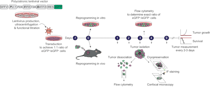
Figure 1: Protocol overview to generate cDC1-like cells by in vivo reprogramming of cancer cells. Lentiviral vectors SFFV-PIB-IRES-eGFP-encoding PU.1, IRF8, and BATF3 in a polycistronic cassette under the control of the spleen focus-forming virus promoter (SFFV) and eGFP after an internal ribosome entry site (IRES) to track transduced cells by flow cytometry and fluorescence microscopy-are produced in the HEK 293T packaging cell line. Then, lentiviral vectors are ultracentrifuged and functionally titrated to determine the amount of virus required for transducing cancer cells in a ratio of 1:1 transduced eGFP+ to untransduced eGFP- cells. For in vivo reprogramming experiments, cancer cells are plated with the optimal amount of lentivirus on day -1. The next day, this 1:1 mix of transduced GFP+ and untransduced GFP- cells is washed and collected for subcutaneous tumor establishment in mice. Parallel in vitro cultures are maintained to validate transduction on day 3 by flow cytometry. Animals are monitored every 2-3 days for tumor growth and survival. Tumors are collected at defined time points, dissociated and stained for surface marker expression for flow cytometry analysis. Alternatively, whole tissues are fixed, cryopreserved, and sectioned for immunofluorescence staining and confocal microscopy analysis. In vivo reprogramming efficiency is evaluated by flow cytometry analysis, while changes in the tumor microenvironment are characterized by confocal imaging. Please click here to view a larger version of this figure.
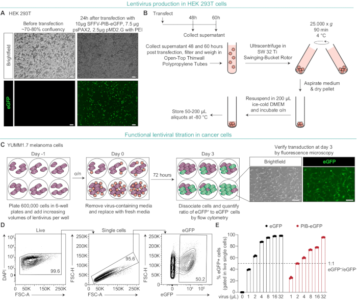
Figure 2: Production and functional titration of lentivirus for in vivo cDC1 reprogramming. (A) When HEK 293T cells reach ~70-80% confluency, they are transfected with a mixture containing 10 µg of transfer plasmid either SFFV-PIB-eGFP (PIB-eGFP) or empty lentiviral vector control SFFV-eGFP (eGFP), along with 7.5 µg of psPAX2, and 2.5 µg of mPD2.G and 60 µL of 1 mg/mL PEI. Brightfield and fluorescence microscopy images of HEK 293T cells showing eGFP expression 24 h post transfection, indicating successful plasmid delivery. Scale bars = 200 µm. (B) Schematic representation of the ultracentrifugation workflow to concentrate lentivirus. The lentivirus-containing supernatant of HEK 293T cells is collected 48 h and 60 h post transfection, filtered through 0.45 µm low-protein binding PES filter, and 37 g are weighed in Open-Top Thinwall Polypropylene Tubes using a bench scale. Tubes are ultracentrifuged in a Swinging-Bucket Rotor, the supernatant is aspirated, and the pellet dried for 5 min. Pellets are then resuspended in ice-cold DMEM containing HEPES and incubated overnight before aliquoting and storing at -80 °C. (C) Experimental strategy of the functional titration to determine the optimal viral dose for achieving a 1:1 ratio of GFP+ to GFP- cells. Brightfield and fluorescence microscopy images showing eGFP expression in YUMM1.7 melanoma cells, 72 h after transduction, indicating successful delivery of the reprogramming factors and eGFP. Scale bars = 100 µm. (D) Representative flow cytometry plots showing gating strategy to quantify the ratio of eGFP+ cells. Live cells were gated for viability dye negative population (DAPI) followed by doublet exclusion. (E) Flow cytometry quantification of transduction efficiency with increasing viral doses to determine the optimal volume for achieving 1:1 GFP+/GFP- cells. For both PIB-eGFP and eGFP lentivirus, 2 µL volume per 105 cells achieved the desired ratio. Abbreviations: cDC1s = type 1 conventional dendritic cells; DAPI = 4',6-diamidino-2-phenylindole; PIB = PU.1, IRF8, and BATF3; PEI = polyethylenimine. Please click here to view a larger version of this figure.
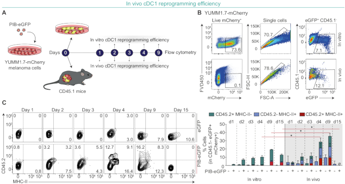
Figure 3: Assessing in vivo cDC1 reprogramming efficiency. (A) Schematic representation of the experimental workflow for evaluating in vivo cDC1 reprogramming efficiency. Cancer cells are initially transduced with a lentivirus expressing mCherry to generate a fluorescently labeled cell line (YUMM1.7-mCherry), followed by transduction with the optimized dose of PIB-eGFP lentivirus on day -1. Cells are then injected subcutaneously with a 1:1 ratio of eGFP+/eGFP- cells on day 0 to establish tumors in CD45.1 mice and allow PIB-transduced cells to reprogram into cDC1-like cells within the TME. Tumors are then isolated at indicated time points, dissociated, and stained for CD45.1, CD45.2, and MHC-II using flow cytometry. This allows for the exclusion of CD45.1 host immune cells to quantify in vivo cDC1 reprogramming efficiency by expression of CD45.2 and MHC-II. Parallel in vitro cultures are kept throughout the time points as a comparison to in vivo reprogramming. (B) Representative flow cytometry plots illustrating the gating strategy on day 9. Cells were gated on mCherry+ and GFP+ to identify successfully transduced cancer cells. Host immune cells were excluded by gating out CD45.1+ cells. (C) Quantification of in vivo reprogramming efficiency (left) of YUMM1.7-mCherry cells by flow cytometry as the percentage of CD45+ MHC-II+ cells (completely reprogrammed) and CD45.2+ HLA-DR- or CD45.2- HLA-DR+ cDC1-like cells (partially reprogrammed) gated within eGFP+ mCherry+ cells. Representative flow cytometry plots (right) showing the gradual acquisition of CD45.2 and MHC-II expression in PIB-eGFP-transduced YUMM1.7-mCherry cells throughout the indicated time points. eGFP-transduced YUMM1.7-mCherry cells were used as a control and in vitro reprogrammed cDC1-like cells were used for comparison. Data in (C) indicate mean ± SD of 3-5 biological replicate experiments. Statistical analysis in (C) was performed for CD45+ MHC-II+ cells using the Mann-Whitney test. *P < 0.05. Abbreviations: cDC1s = type 1 conventional dendritic cells; PIB = PU.1, IRF8, and BATF3; TME = tumor microenvironment. Please click here to view a larger version of this figure.
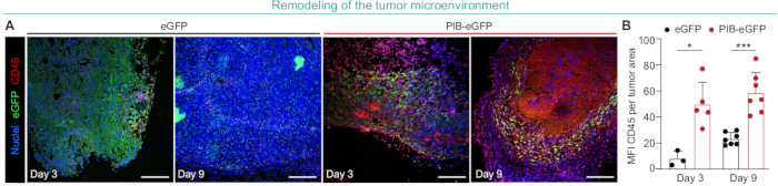
Figure 4: Analyzing immune infiltration in the tumor microenvironment following in vivo cDC1 reprogramming. (A) Representative confocal images showing CD45+ cells in YUMM1.7 tumors after subcutaneous implantation of PIB-eGFP- or control eGFP-transduced cells (1:1 ratio of transduced to parental cells) on days 3 and 9. Scale bars = 500 µm. (B) Quantification of the MFI for CD45 staining in PIB-treated and control eGFP-transduced YUMM1.7 tumors on days 3 and 9. Data in (B) indicate mean ± SD of 3-6 biological replicate experiments. Statistical analysis in (B) was performed using the Mann-Whitney test. *P < 0.05 and ***P < 0.001. Abbreviations: cDC1s = type 1 conventional dendritic cells; PIB = PU.1, IRF8, and BATF3; MFI = mean fluorescence intensity. Please click here to view a larger version of this figure.
Discussion
In this protocol, we describe a tractable method to generate cDC1-like cells based on in vivo reprogramming of cancer cells from solid tumors. Our results demonstrate that the overexpression of PU.1, IRF8, and BATF3 drives the gradual acquisition of a cDC1 phenotype in the TME until day 9, whereafter cDC1-like cells are depleted from tumors. Ultimately, this leads to an increase in the infiltration of CD45 cells into the TME by day 3, causing the remodeling of an "immune-cold" TME to become "immune-hot".
Current protocols to generate cDC1 include enrichment of autologous DCs from peripheral blood, differentiation from CD34+ hematopoietic progenitor cells, or differentiation from pluripotent stem cells14,15,16,17,18,19. However, these methods present limiting factors: insufficient maturation, reduced antigen presentation and cytokine/chemokine production capacity in vivo, susceptibility to immunosuppression, high DC heterogeneity, and low numbers of generated cells7. Here, we provide a protocol that allows the scalable generation of cDC1-like cells in vivo for functional and mechanistic studies.
With this method, we tested whether the expression of the transcription factor combination PU.1, IRF8, and BATF3 was sufficient to drive in vivo reprogramming. This protocol enables precise control over delivery efficiency, as varying the dose of genetically modified cells implanted directly influences treatment efficacy. We have previously shown that 2% transduced cells, which correspond to 0.15% CD45+ and MHC-II+ cDC1-like cells, are sufficient to elicit anti-tumor immunity20. Furthermore, the rapid immune infiltration observed on day 3 suggests that partially reprogrammed cDC1-like cells are functionally active in vivo, as there is no substantial increase in fully reprogrammed cDC1-like cells at this time point. In the future, it will be interesting to unravel the functional role of partially versus fully reprogrammed cDC1-like cells in anti-tumor efficacy using modified vectors containing a suicide gene to induce cell death in selected cell populations at defined time points24. This protocol can further be used to explore the combination with epigenetic adjuvants, which can increase reprogramming efficiency25. Finally, this method could also be adapted to generate other DC subtypes in vivo, launching different types of immune responses, for example, anti-viral or tolerogenic responses14. In addition, we can harness in vivo reprogramming as a platform to perform barcoded screens for transcription factor combinations to generate diverse immune cell types. This possibility could increase the flexibility of treatments by leveraging diverse functional properties of specific immune subsets.
In our previous study, we demonstrated that the immunosuppressive in vivo environment does not hinder the reprogramming of human cancer cells into immunogenic cDC1s20. In this article, we show that in mice, reprogramming is more rapid and efficient in vivo than in vitro, suggesting that the enhanced reprogramming observed in vivo is a conserved phenomenon across both species. However, one of the limitations of this method is the lack of the in vivo environment during initial vector delivery; thus, it cannot discern whether environmental cues impact in vivo delivery efficiency. This should be determined in situ by comparing different moieties. Adenoviral vectors, which we have identified to be more efficient than lentiviral vectors for in situ delivery20 could be compared to non-viral moieties such as linear, circular, and self-amplifying RNAs or small molecule cocktails26,27.
We have previously shown that cDC1 reprogramming endows cancer cells with pro-inflammatory cytokine and lymphocyte-recruiting chemokine secretion and professional antigen processing and presentation on MHC-I and MHC-II in melanoma models23,28. This results in remodeling of the TME from an "immune-cold" to an "immune-hot" state by inducing TLS formation, which increases the infiltration of cytotoxic effector T cells, NK cells, and B cells, and reduces immunosuppressive populations such as regulatory and exhausted T cells20. In patients, positive responses to immunotherapy have been linked to the presence of TLS within tumors, where sustained T and B cell priming against tumor antigens generates tumor-specific antibody responses29,30. In the future, we envision employing this tractable in vivo cDC1 reprogramming protocol to unravel the mechanisms of induced TLS neogenesis, TME remodeling and anti-tumor immunity across different cancer types and characterize whether the production of tumor-specific antibodies is stimulated.
We have previously observed synergism between in vivo cDC1 reprogramming and ICB with anti-PD-1 or anti-CTLA-420. This suggests that the "immune-hot" TME created by cDC1 reprogramming could provide a favorable environment for possible combinations with other immunotherapies such as ACT including T cell receptor (TCR)-T cells, chimeric antigen receptor (CAR)-T cells, or tumor-infiltrating lymphocytes (TILs). Another advantage of this method is the possibility to perform large unbiased combinatorial or CRISPR screens, in which immunological receptors, cytokines, chemokines, or lymphotoxins could be overexpressed or depleted in cDC1-like cells in resistant tumor types, thus unraveling the role of distinct pathways in enhancing cDC1-mediated immunity. In conclusion, the current protocol provides a valuable resource for a broad scientific community, since in vivo cDC1 reprogramming is a flexible and powerful platform for mechanistic studies and therapeutic innovation in cancer immunotherapy.
Disclosures
C.-F.P. has equity interest and serve in management positions at Asgard Therapeutics AB, which develops cancer immunotherapies based on in vivo DC reprogramming technologies. C.-F.P is an inventor on granted patents US 11,345,891, JP 7303743, and CN ZL201880005047.3 and patent applications WO 2018/185709 and WO 2022/243448 (together with E.A.) held by Asgard Therapeutics, which cover the cell-reprogramming approach described here.
Acknowledgements
We thank Mariana Lopes and Camille Chatelain, members of the Cell Reprogramming in Hematopoiesis and Immunity Laboratory, for editing and proofreading the manuscript.
This study was accomplished within the context of the National ATMP Research School funded by the Swedish Research Council. The projects discussed here were co-funded by the European Research Council under the European Union's Horizon 2020 research and innovation program (866448-TrojanDC and 101189370), Novo Nordisk Fonden (NNF22C0079466), European Innovation Council (101130218-RESYNC), Cancerfonden (23 2932 Pj), the Swedish Research Council (2020-00615), Swedish Innovation Agency (2024-02178), ALF (44410), FCT (2022.02338.PTDC), and Plano de Recuperação e Resiliência de Portugal pelo fundo NextGenerationEU (C644865576-00000005). The Knut and Alice Wallenberg Foundation, the Medical Faculty at Lund University, and Region Skåne are acknowledged for generous financial support.
Materials Availablity:
Constructs and vectors used for reprogramming are available from Asgard Therapeutics under a material transfer agreement with the company. All other data needed to evaluate the conclusions in the paper are present in the main text or the supplementary materials.
Materials
| Name | Company | Catalog Number | Comments |
|---|---|---|---|
| 0.45 μm low-protein binding filter, 150 mL Bottle Top Vacuum Filter | Fisher scientific | 15983297 | |
| 2-Mercaptoethanol 50 mM | ThermoFisher Scientific | 31350010 | |
| Anti-mouse CD45 Monoclonal Antibody (clone: 30-F11), Rat IgG2b, κ | ThermoFisher Scientific | 14-0451-82 | |
| B6.SJL-PtprcaPepcb/BoyCrl (CD45.1) | The Jackson Laboratory | 002014 | |
| BD Micro-Fine U-100 Insulin Syringes (0.5 mL) | Fisher Scientific | 11395751 | |
| C57BL/6J mice | The Jackson Laboratory | 000664 | |
| Collagenase D | Sigma Aldrich | 11088882001 | |
| Corning Falcon Cell Strainer For Use With 50ml Conical Tubes | Sigma Aldrich | CLS352340 | |
| Corning cover glasses 24 x 50 mm | Sigma Aldrich | CLS2975245 | |
| DAPI (4',6-diamidino-2-phenylindole, dihydrochloride) | ThermoFisher Scientific | D1306 | |
| DNase I | Sigma Aldrich | 10104159001 | |
| eBioscience Fixable Viability Dye eFluor 450 | Invitrogen | 65-0863-14 | |
| eBioscience IHC Antigen Retrieval Solution - Low pH (10x) | ThermoFisher Scientific | 00-4955-58 | |
| Fetal Bovine Serum (FBS) | ThermoFisher Scientific | A5256701 | |
| Fluoromount-G Mounting Medium | ThermoFisher Scientific | 00-4958-02 | |
| GFP Antibody [Alexa Fluor 594] | Novus Biologicals | NB600-308AF594 | |
| Gibco DMEM/Nutrient Mixture F-12 (DMEM/F-12) | ThermoFisher Scientific | 31331028 | |
| Gibco MEM Non-Essential Amino Acids Solution (100x) | ThermoFisher Scientific | 11140035 | |
| Gibco Opti-MEM I | ThermoFisher Scientific | 11058021 | |
| Gibco Roswell Park Memorial Institute (RPMI) 1640 Medium | ThermoFisher Scientific | 52400025 | |
| Gibco Sodium Pyruvate (100 mM) | ThermoFisher Scientific | 11360070 | |
| Gibco Trypan Blue Solution, 0.4% | ThermoFisher Scientific | 15250061 | |
| GlutaMAX Supplement | ThermoFisher Scientific | 35050061 | |
| Goat anti-rat IgG (H+L) Cross-Adsorbed Secondary Antibody, Polyclonal, Alexa Fluor 488 | ThermoFisher Scientific | A-11006 | |
| HEK 293T cells | ATTC | CRL-11268 | |
| Hexadimethrine bromide (Polybrene) | Sigma Aldrich | H9268 | |
| HyClone Dulbecco's Modified Eagle Medium (DMEM) with high glucose | Cytiva | SH30243.01 | |
| HyClone Penicillin Streptomycin 100x Solution (Pen/Strep) | Cytiva | SV30010 | |
| HyClone Phosphate Buffered Saline solution (PBS) | Cytiva | SH30028.02 | |
| Ketaminol vet. Solution for injection 100 mg/mL | MSD Animal Health Sweden | QN01AX03 | |
| Microtome Blade - C35 | pfm medical | 207500005 | |
| Mouse anti-CD45.2 (104), Surface, APC, Monoclonal | Biolegend | 109814 | |
| Mouse anti-MHCII (M5/114.15.2), Surface, PE-Cy7, Monoclonal | Biolegend | 107630 | |
| Normal Goat Serum | Abcam | ab7481 | |
| Normal Rat Serum (sterile) | Abcam | ab7488 | |
| Open-Top Thinwall Polypropylene Tubes | Beckman Coulter Life Sciences | 326823 | |
| Paraformaldehyde, 16% w/v aq. soln., methanol free | ThermoFisher Scientific | 043368.9M | |
| pFUW-tetO-Batf3, a monocistronic cassette encoding mouse Batf3 | Addgene | 139837 | |
| pFUW-tetO-Irf8, a monocistronic cassette encoding mouse Irf8 | Addgene | 139838 | |
| pFUW-tetO-Pu.1, a monocistronic cassette encoding mouse Pu.1 | Addgene | 139839 | |
| Plasmid pMD2.G | Addgene | 12259 | |
| Plasmid psPAX2 | Addgene | 12260 | |
| Plasmid SFFV-eGFP (eGFP) | In house cloning | - | |
| Plasmid SFFV-mCherry (mCherry) | In house cloning | - | |
| Plasmid SFFV-PIB-eGFP, a polycistronic cassette encoding mouse PU.1, IRF8, and BATF3 (PIB) | In house cloning | - | Constructs and vectors used for reprogramming are available from Asgard Therapeutics under a material transfer agreement with the company |
| Polyethylenimine (PEI), Linear, MW 25000, Transfection Grade | Polysciences | 23966 | |
| ReadyProbes Hydrophobic Barrier Pap Pen | ThermoFisher Scientific | R3777 | |
| Sodium Butyrate | Sigma Aldrich | 303410 | |
| Sucrose | Sigma-Aldrich | S7903 | |
| SuperFrost Plus Adhesion Microscope Slides | Epredia | J1800AMNZ | |
| Tissue-Tek O.C.T. Compound | Sakura Finetek | 4583 | |
| TrypLE Express Enzyme (1x) no phenol red | ThermoFisher Scientific | 12604021 | |
| TT Cryomold Intermediate, square (15 x 15 x 5 mm) | Sakura Finetek | 4566 | |
| UltraPure 0.5 M EDTA, pH 8.0 | Invitrogen | 15575020 | |
| Xysol vet. Solution for injection 20 mg/mL | VM Pharma | QN05CM92 | |
| YUMM1.7 cells | Antineo |
References
- Oliveira, G., Wu, C. J. Dynamics and specificities of T cells in cancer immunotherapy. Nat Rev Cancer. 23 (5), 295-316 (2023).
- Larkin, J., et al. Five-year survival with combined nivolumab and ipilimumab in advanced melanoma. N Engl J Med. 381 (16), 1535-1546 (2019).
- Adams, S., et al. Pembrolizumab monotherapy for previously untreated, PD-L1-positive, metastatic triple-negative breast cancer: cohort B of the phase II KEYNOTE-086 study. Ann Oncol. 30 (3), 405-411 (2019).
- Patel, M. R., Falchook, G. S., Hamada, K., Makris, L., Bendell, J. C. A phase 2 trial of trifluridine/tipiracil plus nivolumab in patients with heavily pretreated microsatellite-stable metastatic colorectal cancer. Cancer Med. 10 (4), 1183-1190 (2021).
- Reardon, D. A., et al. Randomized phase 3 study evaluating the efficacy and safety of nivolumab vs bevacizumab in patients with recurrent glioblastoma: CheckMate 143. Neuro Oncol. 19 (suppl_3), iii21-iii21 (2017).
- Sharma, P., Hu-Lieskovan, S., Wargo, J. A., Ribas, A. Primary, adaptive, and acquired resistance to cancer immunotherapy. Cell. 168 (4), 707-723 (2017).
- Heras-Murillo, I., Adán-Barrientos, I., Galán, M., Wculek, S. K., Sancho, D. Dendritic cells as orchestrators of anticancer immunity and immunotherapy. Nat Rev Clin Oncol. 21 (4), 257-277 (2024).
- Heras-Murillo, I., et al. Immunotherapy with conventional type-1 dendritic cells induces immune memory and limits tumor relapse. Nat Commun. 16 (1), 3369(2025).
- Theisen, D. J., et al. WDFY4 is required for cross-presentation in response to viral and tumor antigens. Science. 362 (6415), 694-699 (2018).
- Meiser, P., et al. A distinct stimulatory cDC1 subpopulation amplifies CD8+ T cell responses in tumors for protective anti-cancer immunity. Cancer Cell. 41 (8), 1498-1515.e10 (2023).
- Spranger, S., Dai, D., Horton, B., Gajewski, T. F. Tumor-residing Batf3 dendritic cells are required for effector T cell trafficking and adoptive T cell therapy. Cancer Cell. 31 (5), 711-723.e4 (2017).
- Hubert, M., et al. IFN-III is selectively produced by cDC1 and predicts good clinical outcome in breast cancer. Sci Immunol. 5 (46), eaav3942(2020).
- Mahadevan, K. K., et al. Type I conventional dendritic cells facilitate immunotherapy in pancreatic cancer. Science. 384 (6703), eadh4567(2024).
- Ascic, E., Pereira, C. -F. Transcription factor-mediated reprogramming to antigen-presenting cells. Curr Opin Genet Dev. 90, 102300(2025).
- Silk, K. M., et al. Cross-presentation of tumour antigens by human induced pluripotent stem cell-derived CD141+ XCR1+ dendritic cells. Gene Ther. 19 (10), 1035-1040 (2012).
- Nair, S., Archer, G. E., Tedder, T. F. Isolation and generation of human dendritic cells. Curr Protoc Immunol. Chapter 7, 7.32.1-7.32.23 (2012).
- Makino, K., et al. Generation of cDC-like cells from human induced pluripotent stem cells via Notch signaling. J Immunother Cancer. 10 (1), e003827(2022).
- Balan, S., et al. Large-scale human dendritic cell differentiation revealing Notch-dependent lineage bifurcation and heterogeneity. Cell Rep. 24 (7), 1902-1915.e6 (2018).
- Kirkling, M. E., et al. Notch signaling facilitates in vitro generation of cross-presenting classical dendritic cells. Cell Rep. 23 (12), 3658-3672.e6 (2018).
- Ascic, E., et al. In vivo dendritic cell reprogramming for cancer immunotherapy. Science. 386 (6719), eadn9083(2024).
- Rosa, F. F., et al. Direct reprogramming of fibroblasts into antigen-presenting dendritic cells. Sci Immunol. 3 (30), 1-16 (2018).
- Rosa, F. F., et al. Single-cell transcriptional profiling informs efficient reprogramming of human somatic cells to cross-presenting dendritic cells. Sci Immunol. 7 (69), eabg5539(2022).
- Zimmermannova, O., et al. Restoring tumor immunogenicity with dendritic cell reprogramming. Sci Immunol. 8 (85), eadd4817(2023).
- Bouquet, L., et al. RapaCaspase-9-based suicide gene applied to the safety of IL-1RAP CAR-T cells. Gene Ther. 30 (9), 706-713 (2023).
- Huangfu, D., et al. Induction of pluripotent stem cells from primary human fibroblasts with only Oct4 and Sox2. Nat Biotechnol. 26 (11), 1269-1275 (2008).
- Rojas, L. A., et al. Personalized RNA neoantigen vaccines stimulate T cells in pancreatic cancer. Nature. 618 (7963), 144-150 (2023).
- Guan, J., et al. Chemical reprogramming of human somatic cells to pluripotent stem cells. Nature. 605 (7909), 325-331 (2022).
- Ferreira, A. G., Zimmermannova, O., Kurochkin, I., Ascic, E., Åkerström, F., Pereira, C. F. Reprogramming cancer cells to antigen-presenting cells. Bio Protoc. 13 (22), 1-25 (2023).
- Cabrita, R., et al. Tertiary lymphoid structures improve immunotherapy and survival in melanoma. Nature. 577 (7791), 561-565 (2020).
- Ng, K. W., et al. Antibodies against endogenous retroviruses promote lung cancer immunotherapy. Nature. 616 (7957), 563-573 (2023).
Reprints and Permissions
