Our patented, personalized 3D-printed headgear provides rapid and reliable delivery of in-office and remotely delivered multi-electrode transcranial electrical stimulation (ME-tES).
Method Article
Personalized 3D-printed Headgear for Multi-electrode Transcranial Electrical Stimulation
In This Article
Summary
Abstract
We present an overview of a novel, patented, approach for creating personalized 3D-printed headgear that enables multi-electrode transcranial electrical stimulation, even in the home. A structural magnetic resonance imaging (MRI) scan is used to optimize a montage that targets key brain areas/networks and is then uploaded to a secure server to design customized headgear that is ultimately 3D-printed. The headgear allows rapid multi-electrode placement (<1 min per electrode) with comparable accuracy as manual 10-10 measurements. We performed over 1,400 sessions across 63 older adults with neurological injury or disease. We describe an efficient training program for study partners (e.g., spouses) that enabled a 100% success rate and over 900 remotely administered sessions. Study partners and participants reported high levels of confidence and satisfaction with the experience. Because the electrode locations are based on personalized computational modeling, the headgear is ideal for techniques requiring precision (e.g., temporal interference stimulation, transcranial focused ultrasound, transcranial magnetic stimulation).
Introduction
Transcranial electrical stimulation (tES) holds significant potential for treating cognitive, emotional, and motor deficits arising from neurologic injury and disease, especially considering the favorable tolerability and safety profiles1,2,3,4. Specifically, a seminal safety review found only the well-established side effect profile (e.g., tingling, itching, burning sensation) but no evidence of unexpected adverse events across 33,000 sessions, regardless of population2. Likewise, we recently reported an absence of safety events and virtually identical side effect profiles for active and sham transcranial direct current stimulation (tDCS) in 3,046 sessions with 292 older adults across the dementia spectrum3. Such strong safety and tolerability have led to remote tES delivery using conventional dual-electrode montages being widely available5,6, thanks in part to materials (e.g., premeasured head straps, prewetted sponges) that facilitate rapid and reliable electrode placement. The combination of these factors facilitated remotely-supervised tES clinics that treat a range of neurological and psychiatric conditions7 but that primarily rely on the traditional large (e.g., 25 cm2-35 cm2) pad-based approach.
Far fewer studies have investigated the use of remote high-definition/multi-electrode (ME) tES8,9 despite evidence that it can provide more focal stimulation delivery10,11,12, be personalizeed13, and have greater, longer-lasting effects14 than traditional pad-based stimulation. The relative paucity of work in this area likely reflects the greater needs of ME-tES given the distinct materials (e.g., multiple small electrodes, use of gel instead of saline-soaked sponges), potential for greater montage complexity, and the associated need for more precise electrode placement. Herein, we present a patented, personalized, 3D printed headgear that overcomes many of these challenges and enables rapid, reliable ME-tES setup and delivery. This approach overcomes potential limitations of existing options such as neoprene caps that can be easily mispositioned, may be ill-fitting, and have little flexibility when moving hair to ensure optimal electrode contact. We also provide evidence supporting the headgear's use for remote ME-transcranial direct current stimulation (ME-tDCS) in older adults across the dementia spectrum. Taken together, our results strongly support the benefits of the personalized headgear and hold promise for other forms of neuromodulation as well as methods that evaluate neurophysiology.
Protocol
The sections below refer to a) study team members, who are trained research staff, typically with baccalaureate degrees, that are overseen by a licensed Clinical Psychologist who is Board Certified in Clinical Neuropsychology, and b) study partners, who are typically spouses or adult children of participants, none of whom had any prior experience with tES. There are currently no clearly defined criteria for those overseeing tES, though having an advanced degree (e.g., Ph.D., M.D.) and being appropriately trained in methodology and well-informed about safety and tolerability are reasonable requirements and consistent with those driving research in this area.
Across all sessions (in office and remote), study team members administered a standard tolerability questionaire15 at the end of each session. Study team members also monitored all sessions for any safety issues and started each session asking about any changes in health or medications since the last session.
1. Headgear creation (Figure 1)
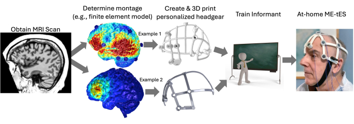
Figure 1: Process for creating personalized headgear and delivering multi-electrode transcranial electrical stimulation at home. Abbreviations: ME-tES = multi-electrode transcranial electrical stimulation; MRI = magnetic resonance imaging. Please click here to view a larger version of this figure.
- Obtaining head measurements:
NOTE: The personalized headgear currently requires a structural magnetic resonance imaging (MRI) scan since this provides highly accurate, 3D measurements of each person's head.- Acquire structural (i.e., T1) scan using parameters that align with several national-level research studies (e.g., SCAN, ADNI). Collect whole-head, high resolution, T1-weighted anatomical image using spoiled-gradient-recalled acquisition (SPGR) in steady-state imaging (TR/TE = 6.95/2.92 ms, flip angle = 8°, field of view = 256 × 256 mm2, matrix size = 256 x 256; slice thickness = 1 mm; 208 sagittal slices; voxel size = 1 x 1 x 1 mm3).
- If no access to such high-resolution MRI scans and MRI scans with other (sometimes lower) resolutions are widely available, use clinical-grade T1 MRI scans with a range of voxel sizes (e.g., 0.98 mm3; 0.94 × 0.94 × 3.00 mm3). Use a clinical FLAIR sequence when the T1 is of insufficient quality.
- In cases of poor tissue segmentation and .STL file creation errors, use the convolutional neural network software (e.g., SynthSR) or comparable software to transform scans into a synthetic, bias-corrected 1 mm isotropic MP-RAGE T116,17. Attempt this step when slice thickness of the clinical T1 exceeds the parameters needed to create accurate headgear.
NOTE: Alternative approaches for acquiring the necessary 3D head measurements (e.g., 3D head scanning) are being developed and will be implemented once available.
- Acquire structural (i.e., T1) scan using parameters that align with several national-level research studies (e.g., SCAN, ADNI). Collect whole-head, high resolution, T1-weighted anatomical image using spoiled-gradient-recalled acquisition (SPGR) in steady-state imaging (TR/TE = 6.95/2.92 ms, flip angle = 8°, field of view = 256 × 256 mm2, matrix size = 256 x 256; slice thickness = 1 mm; 208 sagittal slices; voxel size = 1 x 1 x 1 mm3).
- Perform MRI-based computational modeling (optional):
- Use computational modeling software (e.g., ROAST18) to develop personalized computational model of electrical current flow using the T1 MRI scan. Ensure electrode locations adhere to the standard 10-10 or 10-5 system.
- Headgear development
- Upload the participant's T1 MRI scan to a secure server for scalp segmentation and ultimate headgear creation.
- Specify the number and location of electrode holders using the standard 10-10 (or 10-5) system to ensure the headgear matches the "ground truth" modeling. Wait for the software to automatically a) place crossbars connecting the electrode holders to form a rigid shell that allows the user to reposition hair around the support bars and b) identify anatomical landmarks (e.g., nasion, inion, auricular) to place "orienting tabs" that overlay these regions and allow the user to rapidly place the headgear.
NOTE: The majority of scans did not require manual correction. Soft tissue deformation can be observed around the ears, face, and neck, which are below the headgear shell, and small discrepancies (several millimeters) can be accounted for with the spacers and the subtle flexibility of the nylon frame itself. In some exceptional cases (e.g., misaligned subject positions or low-resolution noisy anisotropic MRI's) correction can be applied in the form of a manually estimated rotation matrix and/or the super resolution upscaler.
- 3D printing:
- Upload the 3D model file (e.g., ".STL") to an appropriate 3D printer (e.g., a selective laser sintering (SLS) printer with medical-grade nylon-12 (PA12) or stereolithography (SLA) printers with resin).
- Once printed, ensure rough edges are smoothed using sandpaper when necessary.
- Placing the headgear:
- Place orienting tabs at appropriate locations on the head.
- Secure headgear using hook and loop straps under chin for bilateral headgear or around the head for unilateral headgear.
- Apply soft spacers as necessary to compensate for minor gaps between the electrode holders and scalp. Check to ensure all other electrode holders remain in contact with the scalp after adding spacers.
- Fill electrode holders with conductive gel.
- Place electrode in holder, ensuring contact with the gel. Apply cap to secure electrode.
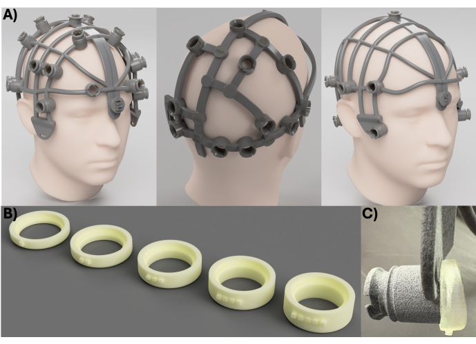
Figure 2: Personalization of ME-tES headgear. (A) Additional examples of personalized ME-tES headgear that show the flexibility of the approach. (B) Five sizes of flexible spacers are used as needed to ensure flush contact with the scalp. (C) Example of spacer placed on end of electrode holder. Abbreviation: ME-tES = multi-electrode transcranial electrical stimulation. Please click here to view a larger version of this figure.
2. Headgear validation
NOTE: Varying data from the first 63 participants who completed ME-tDCS using the headgear are reported. Not all data were available for all participants due to protocol changes or pragmatic factors (e.g., omitting or lack of time to acquire optional questionnaires). All participants completed the scheduled number of sessions. These participants were all diagnosed with neurological injuries or diseases that included dementia of the Alzheimer's type (n = 22), amnestic mild cognitive impairment (n = 13), primary progressive aphasia (n = 12), posterior cortical atrophy (n = 11), behavioral variant frontotemporal dementia (n = 2), anoxic brain injury (n = 2), and Lewy body dementia (n = 1). The mean age was 68.9 years and 50.8% were female.
- Validating headgear measurement accuracy:
- Hand measure and mark the location of each electrode on the participant's head.
- Place the headgear as in step 1.5. Mark the location of each electrode holder. Remove the headgear.
- Measure the difference between the hand-measured location and the headgear location (in mm).
3. Training for remote ME-tES
NOTE: The following rigorous training program for study partners and associated efficacy data are shown below. Training initially required 5 days but was modified to embrace a skill mastery approach given the high success rate in the first few participants. Complete standard tolerability questionnaires after each session15.
- Day 1: Have an experienced study team member explain the components of the headgear (e.g., orienting tabs; electrode holders) and demonstrate the proper placement to the study partner.
- Have the team member ensure each electrode holder is flush with the scalp so that there are no gaps that would allow the gel to leak. If gaps exist, have the team member apply the flexible spacers to the bottom of the electrode holder (Figure 2C) and recheck all other electrode holders, placing additional spacers as needed.
- Have the team member demonstrate best practices such as moving hair from under electrode holders, properly filling the holder with gel (i.e., avoiding/removing air bubbles), placing the electrodes, and applying the corresponding holder cap.
- Have the team member demonstrate how to operate the ME-tES device, check the battery charge, and evaluate electrode impedance. Provide examples of potential issues and problem-solving.
- Perform the session as appropriate for the study.
- Have the team member demonstrate proper rotations, removal, washing, and drying methods (for electrode care, see19).
NOTE: Confidence data are presented from a subset of study partners (n=33) and participants (n = 34) about their ability to set-up and perform stimulation using an anchored Likert scale after they observed the study team member perform this first session (Table 1).
- Day 2: Have the study partner perform all procedures while supervised by a study team member.
- Have the study partner mark the nasion and both auricular points to ensure the headgear orienting tabs are properly placed on the participant's head.
- Have the study team member mark the location of each electrode holder and remove the headgear.
- Have the study team member replace the headgear and mark the location of each electrode holder to evaluate the accuracy of the study partner relative to the team member (Figure 3B). Note differences and provide corrective feedback as necessary.
- Have the study partner perform the entire setup (i.e., ensuring flush fit with scalp, filling the holder with gel, placing the electrode), activate the device, evaluate impedances, and ultimately clean the equipment; all under supervision of the team member.
- Days 3-5: Use a standardized checklist to have the study team member monitor the study partner during each step of the setup process. Provide corrective feedback and tips as necessary until the study partner demonstrates mastery of the entire setup and cleaning process.
NOTE: On the final day, accuracy data were again acquired to determine the accuracy of the headgear placement for the study partner versus the study team member (Figure 3B).- Provide a carrying case with all necessary supplies (e.g., headgear, gel, syringes, electrodes), proper care and maintenance manual, and a second case with the ME-tES device to the study partner at the end of the session.
NOTE: Study partners (n = 31) and participants (n = 30) provided confidence ratings at the end of the final training session (Table 1).
- Provide a carrying case with all necessary supplies (e.g., headgear, gel, syringes, electrodes), proper care and maintenance manual, and a second case with the ME-tES device to the study partner at the end of the session.
- Remote (at home) ME-tES:
- For each scheduled session, have the study partner log into a secure video call with a study team member and then perform all setup activities.
- Have the study partner report impedances. Once acceptable, have the study team member provide a code for the study partner to enter into the ME-tDCS unit.
NOTE: Many ME-tDCS units will require a single-use code to start stimulation, which prevents unscheduled stimulation sessions and optimizes participant safety. - Have the study team member perform the required session activities via video call, including safety and tolerability (e.g., side effect) reporting.
- Once the session is complete, have the study partner follow the care and maintenance checklist and charge the stimulator batteries.
NOTE: Study partners (n = 24) and participants (n = 24) provided confidence ratings after the prescribed number of remote sessions.
Results
Stimulation parameters and total sessions:
We successfully created 100% of attempted headgear (63/63), demonstrating a reliable and efficient pipeline for personalized headgear. Each headgear included between 5 and 22 electrodes, depending on the participant and study (we note that fewer or more electrode locations are also possible). We delivered per channel amplitudes of 4 mA or less and total scalp-based current across all electrodes of less than 16 mA. In total, we performed 1,449 sessions using the headgear.
Validation results:
There was an average of 8.6 mm difference between headgear electrode locations with hand measurement of the same locations across the first 54 headgear (Figure 3A). This difference (i.e., <1 cm) is well within the expected margin of error arising from hand measurement since 1) hand measurement is inherently variable, especially across days and when performed by different study team members, 2) measurement skills/experience can vary across study team members, 3) Individual differences in head size/shape and hair create measurement error, 4) complex montages interact with #1-3 to create variability and require extensive setup times. In fact, this process led us to identify and correct drift in the study team's measurement process (i.e., the headgear provided an objective ground truth). The rigid-body nature of the headgear (though material was optimized to balance comfort, flexibility, and rigidness) and orienting tabs (e.g., nasion) essentially eliminate error in electrode location and, as noted above, the locations are based on the same process as computational modeling software. Headgear placement requires <1 min; total setup time with 20 electrodes is ~10 min (including filling the holders, placing the electrode, measuring impedances); a substantial reduction compared to hand measurement (typically ~45 min depending on the electrode locations).
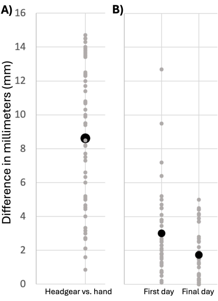
Figure 3: Training for ME-tES headgear. (A) Average difference between hand measurement and headgear (in mm). NOTE: We identified several inaccuracies associated with hand measurement during the comparison process that led to procedural modifications (e.g., measuring tape placement, auricular points). (B) Average difference between study partner-placed headgear and trained study team member on the first and final training days. Black circles represent overall group mean; gray circles represent individual participants. Abbreviation: ME-tES = multi-electrode transcranial electrical stimulation. Please click here to view a larger version of this figure.
Training results:
Study partners accurately placed the headgear on the first attempt, as evidenced by an average of only 3 mm difference relative to where our study team placed it. This difference was 1.74 mm by the final training day (Figure 3B). Importantly, 100% of informants successfully completed training within the 5 available days.
The trained study partners performed 929 sessions in participant's home (range: 5-61 sessions/participant). There were no safety issues. Tolerability was evaluated using the standard questionnaires but is not reported here due to our prior findings of comparable sensory experiences between active and sham ME-tDCS3,15. Table 1 shows that participants and study partners reported high levels of confidence after the first session, which further increased to near ceiling levels by the final training session. Moreover, participants and study partners reported near ceiling levels of satisfaction with the remote ME-tDCS experience following their final session.
Table 1: Study partner and participant experiences using the headgear and performing and receiving remote ME-tDCS. Measures acquired after first session, final training session (~5th), and final ME-tDCS session. Abbreviation: ME-tDCS = multi-electrode-transcranial direct current stimulation. Please click here to download this Table.
Discussion
Our personalized headgear provides a user-friendly method for remote ME-tES delivery, even in older adults with neurologic injury and disease. Our data demonstrate the automated electrode placements for the headgear were comparable to hand measurements. Importantly, the shared MRI-based electrode placement means that tES delivered using the headgear mirrors that shown via computational models. The personalized orienting tabs ensure proper placement, and the crossbar design allows the user(s) to access electrode locations and mitigate potential confounds, especially with respect to moving hair to ensure clear access to the scalp. These benefits are notable advantages relative to existing approaches (e.g., neoprene caps; hand-measured head netting) that are potentially slower, less accurate, and prone to poor contact due to hair, especially in remote settings (e.g., home).
Our remote training program was extremely effective in training study partners to administer ME-tDCS as all those who expressed an interest were able to meet performance criteria. Specifically, study partners accurately placed the headgear after a single observation (3 mm difference; 1.74 mm difference after multiple sessions) and quickly learned to perform all set-up and cleaning activities. Study partners and participants reported very high confidence even after a single day of training and reported near ceiling levels of confidence by the final training day. Both study partners and participants were highly satisfied with the overall experience and expressed interest in additional tES studies (Table 1). Thus, our headgear fills a critical translational gap by making ME-tES available outside of the office setting. Note that these conclusions are based on the above-noted steps, which are critical for users to follow. However, study teams may need to tailor the training approach for other populations.
We are actively working to overcome several potential limitations. For example, the current MRI requirements (e.g., 1 mm3 voxels) may not be available for all participants. Fortunately, our methods appear robust given the initial success using clinical-quality MRI scans (both T1 and FLAIR) to develop the headgear. This challenge may be further mitigated as we refine 3D head scanning methods that may ultimately render MRI scans unnecessary. We have also created "generic" headgear (e.g., 56 cm circumference) that could provide another option relative to hand measuring or neoprene caps. As is the case for such existing approaches, hairstyles may impede accurate headgear placement and, in such cases, teams will need to either work with participants to alter hairstyles or alter their montage. As with all devices and interventions, cost may be an important consideration, and we encourage potential users to consider their needs relative to the strengths and limitations of all available approaches.
The personalized nature and flexibility of the headgear means that electrode holders can easily be modified in any number of ways. For example, we already have templates to support traditional tES pads. Likewise, several emerging neuromodulation techniques integrate personalized computational modeling to target specific brain regions. Our shared electrode positioning with such modeling approaches means our headgear is ideally suited to support precision-based techniques, such as temporal interference stimulation and transcranial focused ultrasound as well as personalized transcranial magnetic stimulation. Finally, we have also developed modifications that enable electroencephalogram and functional near-infrared spectroscopy in isolation as well as with concurrent neuromodulation. In sum, our headgear provides a highly flexible platform that increases access and sustainability while minimizing participant and study team burden.
Disclosures
The headgear is currently patented under Neuropsychological and Neurological Rehabilitation Headgear Device. U.S. Application No.: 17/188,437 Inventor, Patent No. 11865328, and includes modifications for the other neuromodulation and neurophysiology approaches noted above. The product is licensed to Soterix Medical Inc. and the lead author (BMH), University of Michigan, and Department of Veteran's Affairs may receive royalties from sales. ARG, KM, JM, and AD are employees of Soterix Medical Inc. No AI was used in any portion of this work. Potential conflicts were mitigated by ensuring all data were collected by study team members who were unrelated to the patent, license, or Soterix Medical Inc. No member of Soterix Medical, Inc. had any role in study planning or data acquisition, nor did they have access to raw data.
Acknowledgements
The contents of this manuscript do not represent the views of the Department of Veterans Affairs or the United States Government. Grant support from the National Institute on Aging via R35AG072262 and R01AG058724 [both to BMH] is also acknowledged.
Materials
| Name | Company | Catalog Number | Comments |
|---|---|---|---|
| Electrode gel | Signagel | n/a | |
| Headgear | Soterix Medical Inc. | https://soterixmedical.com/research/remote | |
| MxN 10 REMOTE | Soterix Medical Inc. | https://soterixmedical.com/research/remote |
References
- Antal, A., et al. Low intensity transcranial electric stimulation: Safety, ethical, legal regulatory and application guidelines. Clin Neurophysiol. 128 (9), 1774-1809 (2017).
- Bikson, M., et al. Safety of transcranial direct current stimulation: Evidence based update 2016. Brain Stimul. 9 (5), 641-661 (2016).
- El Jamal, C., et al. Tolerability and blinding of high-definition transcranial direct current stimulation among older adults at intensities of up to 4 mA per electrode. Brain Stimulation. 16 (5), 1328-1335 (2023).
- Lefaucheur, J. P., et al. Evidence-based guidelines on the therapeutic use of transcranial direct current stimulation (tDCS). Clin Neurophysiol. 128 (1), 56-92 (2017).
- Charvet, L. E., et al. Remotely-supervised transcranial direct current stimulation (tDCS) for clinical trials: Guidelines for technology and protocols. Front Syst Neurosci. 9, 26(2015).
- Charvet, L. E., Shaw, M. T., Bikson, M., Woods, A. J., Knotkova, H. Supervised transcranial direct current stimulation (tDCS) at home: A guide for clinical research and practice. Brain Stimul. 13 (3), 686-693 (2020).
- Pilloni, G., et al. Tolerability and feasibility of at-home remotely supervised transcranial direct current stimulation (RS-tDCS): Single-center evidence from 6,779 sessions. Brain Stimul. 15 (3), 707-716 (2022).
- Brechet, L., et al. Patient-tailored, home-based non-invasive brain stimulation for memory deficits in dementia due to Alzheimer's disease. Front Neurol. 12, 598135(2021).
- Van De Winckel, A., et al. Home-based transcranial direct current stimulation plus tracking training therapy in people with stroke: An open-label feasibility study. J Neuroeng Rehabil. 15 (1), 83(2018).
- Sharma, G., Chowdhury, S. R. 2019 IEEE Conference on computational intelligence in bioinformatics and computational biology (CIBCB). , 1-5 (2019).
- Borckardt, J. J., et al. A pilot study of the tolerability and effects of high-definition transcranial direct current stimulation (HD-tDCS) on pain perception. J Pain. 13 (2), 112-120 (2012).
- Datta, A., et al. Gyri-precise head model of transcranial direct current stimulation: Improved spatial focality using a ring electrode versus conventional rectangular pad. Brain Stimul. 2 (4), 201-207 (2009).
- Rogers, G., Joodaki, M., Hopf, A., Santarnecchi, E., Guzman, R., Muller, B., Osmani, B. Increasing target engagement via customized electrode positioning for personalized transcranial electrical stimulation: A biophysical modeling study. NeuroImage. 311, 121206(2025).
- Kuo, H. -I., et al. Comparing cortical plasticity induced by conventional and high-definition 4× 1 ring tDCS: A neurophysiological study. Brain stimul. 6 (4), 644-648 (2013).
- Reckow, J., et al. Tolerability and blinding of 4x1 high-definition transcranial direct current stimulation (HD-tDCS) at two and three milliamps. Brain Stimul. 11 (5), 991-997 (2018).
- Iglesias, J. E., et al. Synthsr: A public ai tool to turn heterogeneous clinical brain scans into high-resolution T1-weighted images for 3D morphometry. Sci Adv. 9 (5), eadd3607(2023).
- Iglesias, J. E., et al. Joint super-resolution and synthesis of 1 mm isotropic MP-RAGE volumes from clinical MRI exams with scans of different orientation, resolution and contrast. Neuroimage. 237, 118206(2021).
- Huang, Y., Datta, A., Bikson, M., Parra, L. C. Realistic volumetric-approach to simulate transcranial electric stimulation-roast-a fully automated open-source pipeline. J Neural Eng. 16 (5), 056006(2019).
- Hampstead, B. M., Ehmann, M., Rahman-Filipiak, A. Reliable use of silver chloride HD-tDCS electrodes. Brain Stimul. 13 (4), 1005-1007 (2020).
Reprints and Permissions
