This protocol presents a guide for the precise and patient-centered application of repetitive transcranial magnetic stimulation for treatment-resistant major depression. Details are provided on procedures for accurate coil localization, adjustable stimulus dosing, rigorous safety measures, and strategies that maximize comfort, reproducibility, and long-term clinical effectiveness across acute and maintenance sessions.
Method Article
Conventional Repetitive Transcranial Magnetic Stimulation for Depression: A Step-by-Step Protocol
In This Article
Summary
Abstract
Repetitive Transcranial Magnetic Stimulation (rTMS) is a non-invasive approach for brain stimulation, with proven effectiveness and efficacy to treat Major Depressive Disorder (MDD), particularly for episodes that do not respond to antidepressant medication. A structured patient-centered Transcranial Magnetic Stimulation (TMS) protocol, focused on precision, comfort, and safety, should be provided in TMS clinics treating MDD. The procedure begins with the patient seated comfortably in a reclining chair, equipped with head and foot support. A pillow is placed around the patient's neck to ensure head stability and comfort throughout the session. A lycra cap is placed on the patient's head, and the treatment area is marked on the cap to ensure the correct placement of the electromagnetic coil. Two main strategies, relying on the head landmarks for each individual patient, have been widely used to identify the treatment area: the 5 cm rule and the Beam F3 method. To initiate the treatment, the coil is positioned on the treatment target to deliver a series of electromagnetic pulses. Patients hear a clicking sound and feel a light tapping sensation on the scalp. For acoustic protection, earplugs are provided before the session begins. The stimulation itself typically causes no discomfort, and patients can resume their normal daily activities after the session. However, driving is discouraged immediately after treatment sessions. The rTMS protocol to treat MDD consists of 20 to 30 daily sessions over four to six weeks, monitored by regular consultations with a psychiatrist, to track progress and adjust if needed. Maintenance sessions may follow to sustain clinical benefits. This step-by-step protocol ensures comfort, consistency, and effective treatment delivery for patients undergoing rTMS therapy.
Introduction
Transcranial Magnetic Stimulation (TMS) is a non-invasive neuromodulation technique based on the principle of electromagnetic induction1. If an electric current passes through a TMS coil, it generates an electromagnetic field perpendicular to the current's direction1. When this field interacts with conductive material, such as neural tissue, it induces electrical activity1. This enables targeted modulation of neuronal activity in discrete regions of the cerebral cortex, opening new avenues for both neuroscientific research and clinical applications2. The evolution of TMS technology led to the development of repetitive TMS (rTMS), involving the delivery of repeated electromagnetic pulses to a target cortical region at a predetermined frequency3. High-frequency rTMS (≥ 5 Hz) typically facilitates cortical excitability, whereas low-frequency rTMS (≤ 1 Hz) suppresses it3. More complex protocols for rTMS, such as intermittent theta-burst stimulation (iTBS) and continuous theta-burst stimulation (cTBS), also allow for modulation of brain activity and have been integrated into clinical practice3.
The therapeutic potential of rTMS for psychiatric disorders was first explored in the 1990s, with studies demonstrating its effects in patients with medication-resistant depression4,5. Since then, numerous clinical trials and meta-analyses have established rTMS as a safe and effective treatment for neuropsychiatric disorders6. In 2008, the U.S. Food and Drug Administration (FDA) cleared high-frequency rTMS for episodes of treatment-resistant depression (TRD)7,8,9,10, marking the first official neuropsychiatric application of the technique11. Subsequent regulatory decisions followed, including clearance of iTBS for TRD in 2018, following evidence of equivalent efficacy to the conventional TMS protocol, and of rTMS for obsessive-compulsive disorder (OCD) in 2019, as well as designation of rTMS as a breakthrough therapy for bipolar depression11. The European regulatory framework, through the Conformité Européenne (CE) Mark, has similarly certified multiple TMS devices and protocols for clinical use11.
Evidence-based recommendations from expert consensus and clinical guidelines have reinforced the therapeutic role of rTMS in depression12. Robust clinical data support its efficacy in TRD, with response rates ranging from 39.5% to 70% and remission rates between 16.6% and 76.9%13. Importantly, the antidepressant effects of TMS have been shown to persist, with 53% of responders maintaining improvements at six months and 46% at one-year post-treatment14. The left dorsolateral prefrontal cortex (L-DLPFC) is the cleared target for depression treatment, though alternative protocols involving right DLPFC stimulation or bilateral approaches have also been explored15. Conventional treatments offer daily stimulation sessions across several weeks, while more recent accelerated protocols have been developed with multiple sessions per day for a shorter period16. In this work, adding to existing guidelines17, we aim to provide a step-by-step guide for application of conventional rTMS to treat a major depressive episode, incorporating elements such as structured ramping strategies, predefined psychometric assessment timelines, and detailed comfort and safety measures, within a clinical setting that ensures consistency, effective treatment delivery, and adherence to regulatory clearances or recommendations11. Hence, we aim to provide guidance for clinical application of TMS to treat depression, towards mitigating variability in application of treatments, while improving tolerability, consistency, and real-world implementation.
Protocol
This protocol provides a standardized approach to treating depression using a conventional protocol for Transcranial Magnetic Stimulation (TMS). TMS sessions are categorized into three types: First Session, Regular Sessions, and Assessment Sessions. Procedures of this protocol are performed at the Champalimaud Foundation using a MagVenture MagPro X100 magnetic stimulator, with a Cool-B65 figure-of-eight coil. SINGER safety Ear Plugs B38 are provided to all participants and TMS technicians.
1. Prescription of TMS (performed by prescribing physician)
- Assess eligibility for TMS treatment during clinical interview, detailing current and past medical history:
- Confirm diagnosis of current major depressive episode for approved use.
- Screen for risk of side-effects (e.g., seizures) and treatment contraindications17,18. Absolute contraindications to rTMS are the presence of electronic or metallic implants in the brain. While not absolute reasons to withhold treatment, careful risk-benefit assessment is required in cases of uncontrolled active medical illness requiring immediate attention, electronic or metallic implants in the body that are not compatible with electromagnetic fields, epilepsy, pregnancy, breastfeeding, women planning pregnancy, and psychotic or mood disorders requiring immediate hospitalization.
- Obtain informed consent clearly explaining procedures, risks, including seizures, and any off-label use17,18.
- Prescribe TMS in a shared decision-making process with the patient, considering approved, off-label, and emerging indications11.
2. TMS clinic and staff
- Use equipment with regulatory clearance, including the TMS stimulator, the TMS coil, and the TMS cooling system.
- Have life-support and emergency plans for syncope and seizures19,20.
- Ensure all staff are trained in TMS mechanisms, risks, and emergency management, with at least basic life support skills17,18,21.
- Include a licensed physician who supervises treatment, who must be accessible, at least by phone, during sessions17,18,21.
3. First TMS session
- Arrival and administrative check-in (administrative staff)
- Greet the patient upon arrival at the TMS Clinic.
- Confirm necessary documentation for admission, including TMS prescription, completed informed consent documentation, and safety check.
- Introduce the patient to the TMS technician.
- Have the TMS technician explain the general procedures.
- Provide the patient with a clipboard containing the following assessment materials to evaluate sociodemographic variables and collect self-reported psychometric data (by TMS technician). i) Sociodemographic questionnaire: Self-report questionnaire to collect information such as age, gender, education, occupation, handedness, mode of transport to and from treatment, and habits, among others, found relevant for each specific context. ii) Beck Depression Inventory-II (BDI-II): A 20-item scale that assesses the severity of depressive symptoms, completed by the patient22. Other self-report scales to assess depression severity are available. It is recommended that at least one self-report scale should include one or more items to assess suicidal ideation and/or intent. iii) Visual Analog Scale (VAS): A 10 cm line used to assess the patient's mood, where 0 represents the worst possible mood and 10 represents the best possible mood.
- Pre-session device check (by TMS technician)
- Verify and confirm the integrity of the stimulator and coil.
- Confirm integrity of active cooling (if applicable).
NOTE: Routine technical inspection and calibration of the TMS device are recommended. The service schedule should follow the manufacturer's guidelines and may vary depending on the age and condition of the equipment. Consult the device provider for specific recommendations.
- Introduction to TMS (by TMS technician)
- Call the patient to the treatment room. Normally, the session is conducted with only the technician and the patient. Upon explicit consent from the patient, chaperones (e.g., family members) or trainees may be present.
- Introduce the TMS technique, including an explanation of known mechanisms of action in lay terms.
- Discuss potential side effects23.
- Demonstration (by TMS technician):
- Invite the patient to experience the stimulation on the forearm, ensuring the coil is positioned correctly and that the patient is informed about the brief tapping or tingling sensation.
- Demonstrate the sensation of TMS on the patient's forearm, usually the inner side, midway between the wrist and elbow, using a single pulse at low intensity (e.g., 30-40% of maximum stimulator output).
NOTE: This familiarization step helps reduce anxiety, build rapport, and increase comfort during the procedure. Avoid this demonstration in patients with electronic implants (e.g., pacemakers, neurostimulators) or in pregnant women. While peripheral exposure to electromagnetic fields is considered safe, it may pose theoretical risks18. In such cases, use verbal explanation and reassurance instead of direct stimulation, or stimulate a different region, farther from the electric device or the abdomen (e.g., ankle).
- Clinical interviews (by TMS technician): Conduct semi-structured clinical interviews to assess the severity of symptoms. Psychometric instruments serve to establish baseline parameters to monitor treatment progress. The following are two examples of clinician-administered scales:
- Montgomery-Asberg Depression Rating Scale (MADRS): Use it to evaluate the severity of depressive symptoms24. MADRS (and BDI-II) can be used to assist in assessing treatment outcome. While the definitive assessment of outcome is performed collaboratively by the patient and the prescribing physician, quantitative definitions of response and remission are relatively consensual in the available literature25.
NOTE: A partial responder is defined as a patient who experiences at least 25% reduction in depressive symptoms, while a full responder has 50% reduction or more. These definitions are used in both clinical and research settings24. Remission is defined as the state in which depressive symptoms have been reduced to a level considered within the normal range, indicating near or complete resolution of the depressive episode, usually operationalized as a total score below an instrument-specific threshold (e.g., MADRS ≤ 10)16,26. - Young Mania Rating Scale (YMRS): Use it to evaluate the severity of manic or hypomanic symptoms. Mania is a concern among patients with a known diagnosis of bipolar disorder, but it can also occur in patients without a previous diagnosis of bipolar disorder. Scales such as the YMRS are used to monitor the occurrence of mania as a potential side-effect of treatment.
NOTE: Once baseline assessments are completed, the TMS technician initiates all procedures that require the use of the TMS device. Clinical assessments are repeated at variable intervals depending on available resources and patient needs. Self-report scales and/or clinical interviews are repeated at least after TMS sessions have been completed, to assist in the assessment of response to treatment.
- Montgomery-Asberg Depression Rating Scale (MADRS): Use it to evaluate the severity of depressive symptoms24. MADRS (and BDI-II) can be used to assist in assessing treatment outcome. While the definitive assessment of outcome is performed collaboratively by the patient and the prescribing physician, quantitative definitions of response and remission are relatively consensual in the available literature25.
4. Full TMS procedures (performed by TMS technician)
NOTE: Full TMS procedures, including determination of the motor threshold, are performed in the first TMS session and can be repeated at variable intervals across TMS centers depending on available resources and patient needs. Some centers conduct a single full session at the beginning of the acute TMS treatment phase, while others perform all assessments daily17,27. An evidence-based approach is to repeat all assessments every five sessions (i.e., weekly), typically at sessions 6, 11, 16, 21, 26, to allow for the definition of TMS intensity for the following week27.
- Initial procedures: Systems using helmet-like coils11 have a particular protocol, described in detail by Feifel and Pappas28. The following steps are used for TMS systems with figure-of-eight coils, which are predominant in most countries.
- Place a lycra cap on the patient to mark areas of interest on the scalp. Confirm the cap is aligned according to the patient's eyebrows and the upper part of the ears, ensuring consistent reference points for cap placement in subsequent sessions.
- Provide earplugs to the patient to minimize discomfort from the stimulation sounds. Use your own earplugs during TMS procedures.
- Register these procedures in the patient's clinical chart.
- Determining the motor hotspot: This step involves identifying the stimulation hotspot for the primary motor cortex (MH) of the hand region in the left hemisphere.
- Use a measuring tape to determine and mark the mid-sagittal line, extending from the nasion (the top of the nose bridge) to the inion (the prominent bump at the back of the skull).
- Mark the intertragus line by placing one end of the tape at one tragus (point just in front of the ear) and stretching it across to the other side.
- Consider the intersection of these two lines as the cranial vertex, from which additional measurements are taken to locate the motor cortex hotspot.
- Mark a triangle by extending a line 5 cm to the left along the intertragus line and 5 cm anteriorly along the mid-sagittal line, both starting at the cranial vertex. The third side is drawn diagonally to link the free extremities of the first 2 lines. The reference point to initiate the search for the hand motor hotspot search is located 2.5 cm along the diagonal line, measured from the left-most vertex of the triangle.
- Mark 4 additional points positioned 0.5 cm anterior, posterior, lateral, and medial from this reference point to facilitate exploration.
- Use the above 5 points as locations where the anterior edge of the TMS coil is placed during the motor hotspot detection procedure.
- To start the motor hotspot search, instruct the patient to rest the arms and hands on the armrests of the chair or on his/her lap, ensuring they are visible and relaxed.
- Place the handle of the figure-of-eight TMS coil on the head of the patient at a 45° angle relative to the sagittal midline (handle directed posteriorly and to the left).
- Apply pulses maintaining the coil in the position mentioned above. To familiarize the patient with the procedure, deliver initial pulses at lower intensities (e.g., 20-30%), while assessing patient's sensory experience. Confirm visible muscle contraction of the contralateral hand muscles.
- If no motor response is observed at these initial intensities in the marked reference points from the previous step, increase the stimulation intensity in 5% increments until a motor response is elicited, defined as a visible contraction of the right hand (i.e., the hand contralateral to the stimulated hemisphere).
NOTE: Depending on the patients' anatomy, the TMS technician aims to determine specifically the area responsible for muscle contraction of the abductor pollicis brevis and/or first digit interosseus (contraction of the first two digits)29,30. - Once a motor response is detected, explore the several marked reference points to identify the site where the motor response is maximal.
- Mark and use additional reference points as needed, at different cardinal positions and/or spaced more than 0.5 cm (e.g., 1cm). The hotspot is the location where the contraction exhibits the highest magnitude.
- Once the optimal stimulation site is identified, trace a concave line following the anterior edge of the coil and mark the center of this line.
- If no motor response is observed at these initial intensities in the marked reference points from the previous step, increase the stimulation intensity in 5% increments until a motor response is elicited, defined as a visible contraction of the right hand (i.e., the hand contralateral to the stimulated hemisphere).
- Confirm MH at regular intervals. If MH refinement is necessary during a session or during a treatment cycle, mark the new MH in the patient's cap and register the reason for refinement in the clinical chart, as per protocol (e.g., weekly MH assessments) or due to another reason.
- Resting motor threshold (rMT) determination: After finding the MH, assess the rMT. This is the minimum dose required to elicit a contraction of the hand in at least 5 out of 10 pulses delivered to the MH.
- Begin this process after an accommodation period.
- Deliver pulses at 50% of the machine's maximum intensity. Confirm that a visible muscle contraction of the contralateral hand muscles is occurring at least 5 out of 10 pulses.
- If a hand contraction is not elicited in at least 5 out of 10 pulses, increase the intensity by 2% points until this is achieved.
- If this occurs, decrease the intensity by 1% until the minimum intensity that elicits a motor response is found, considering the criterion of a response in at least 5 out of 10 pulses.
- Register rMT in the patient's clinical chart for further use in the following sessions.
- If rMT re-assessment is necessary during the session or during the treatment cycle, register the new rMT and the reason for re-assessment, namely per protocol (e.g., weekly rMT assessments) or due to an unexpected reason (e.g., change in medication that potentially impacts rMT).
NOTE: This procedure is tailored for TMS devices where stimulation intensity is expressed as a percentage of the device's maximum output. For devices that represent intensity using different units, appropriate adaptations are made.
- Treatment site (TS) determination: After identifying the MH and determining the rMT, proceed to locate the TS. There are several methods to determine the TS including neuronavigated methods, as described by Dunlop et al.31. Here, the focus is on non-neuronavigated approaches, which rely on scalp-based measurements and have been widely utilized and supported in clinical practice17. Two methods have been advocated: the 5 cm-rule and Beam F3.
- 5 cm rule: From the midpoint of the concave line drawn after determining the motor hotspot, extend a para-sagittal line anteriorly and mark the stimulation spot for the left DLPFC at 5-6 cm (most frequently 5.5 cm) on this line.
- Beam F3: Use this strategy to locate the F3 position of the 10-20 electroencephalogram system on the scalp, an alternative landmark for the L-DLPFC. Input specific patient's head measurements into a freely available computer software that provides instructions to mark F3 in accordance with the patient's head size (see Table of Materials)32.
- Obtain 3 measurements using a tape measure: i) Measure the nasion-inion distance from the nasion (the top of the nose bridge) to the inion (the prominence at the back of the skull). ii) Measure the intertragus distance from the left to the right tragus. iii) Measure the head circumference by passing the tape measure over the inion at the level of the eyebrows.
- Input these measures in the software interface to generate two outputs. First, the distance to the left of the midline along the head circumference (Point X). Second, the distance from the vertex along a line intersecting Point X (Point Y). Point Y is the L-DLPFC TS as determined by the Beam F3 method. Use these two distances to mark the TS.
- Determine TS at regular intervals. If TS refinement is necessary during the session or during the treatment cycle, mark the new TS on the patient's cap and register the reason for refinement in the patient's clinical chart, per protocol (e.g., weekly TS assessments) or due to another reason.
- Treatment protocol: Currently, two conventional protocols for stimulation of the L-DLPFC are approved and/or cleared by regulatory agencies for treatment of depression, and available in most TMS centers as acute/induction TMS treatment schedules, with sessions delivered daily over 20-30 daily sessions on weekdays11
- High frequency rTMS: Deliver pulses with intensity at 120% of the rMT, and frequency of 10 Hz (3000 pulses per session, in sequential cycles of 4 s on and 26 s off, with a total duration of 37.5 min).
- iTBS: Deliver pulses with intensity at 120% rMT, in 50 Hz triplet bursts, repeated at 5 Hz (600 pulses per session, in sequential cycles of 2 s on and 8 s off, with a total duration of 3 min and 9 s).
- During session: Ensure that the coil remains correctly located on the head over the TS throughout the session. If the patient experiences any discomfort, assist in normalizing the sensation and adjust the intensity if necessary. Session may be paused or discontinued at the patient's request.
- Post-session procedures
- Instruct the patient to stand up slowly, while monitoring signs of dizziness or imbalance.
- Evaluate the presence of discomfort23. For mitigation measures, suggest use over-the-counter analgesics or contact with the attending physician, in case of severe or persisting symptoms.
5. TMS treatment sessions (performed by TMS technician)
- Pre-session check
- Ask the patient if they experienced any side effects from the previous session.
- Ask the patients for any changes in medication that might affect the rMT. Upon significant changes to medication, re-assess the rMT.
- Hotspot confirmation
- Put the marked lycra cap back on the patient's head and check the MH to ensure the previously marked regions remain accurate.
- If adjustments are necessary, follow the procedures described for the first session to redefine the MH.
- Treatment
- Place the coil at the TS and administer the stimulation according to the selected treatment protocol and the latest rMT assessment.
- Verify the position of the coil on the head of the patient during the session to ensure its accuracy.
- Ramping: During initial treatment sessions, some patients experience scalp discomfort or muscle twitching that prevents immediate delivery of the full target stimulation intensity specified in the TMS protocol. In such cases, implement ramping to enhance patient comfort and tolerability. Ramping in TMS refers to the gradual increase of stimulation intensity over the first sessions, usually the first week of treatment (5 sessions).
- Instead of starting at the full prescribed intensity, begin stimulation at a lower percentage of the motor threshold (e.g., 100% rMT).
- Increases the stimulation intensity within session and/or across sessions, until the target intensity is reached, i.e., 120% of rMT.
NOTE: This approach helps reduce initial discomfort, improve patient adherence, and minimize side effects such as scalp pain or headaches. Ramping is particularly beneficial for individuals new to TMS and those with heightened sensitivity to stimulation.
- Post-session procedures: Follow the procedures outlined above (step 4.7).
6. Physician assessment (performed by prescribing physician)
NOTE: Depending on the physical and human resources available at each TMS center, medical appointments with the TMS physician are scheduled throughout the acute TMS treatment phase.
- Assess patient's progress, address any side effects, and discuss potential mitigation strategies17,23.
- After the patient has completed 4 weeks of treatment, adjust the session frequency based on clinical response.
- After the acute treatment phase, evaluate the patient's overall progress and discuss the next steps in treatment.
- If clinically indicated, recommend transition to a maintenance phase according to center protocols and resources, and patient preference14.
Results
The efficacy of transcranial magnetic stimulation for the treatment of major depression episodes has been consistently confirmed in multiple meta-analyses, with response rates ranging from 40% to 70% and remission rates from 17% to 77%13. Consequently, its clinical and research applications have been expanding33. However, significant variability in treatment outcomes persists13, possibly due to differences in protocols across TMS centers12. Here, we provide a structured, step-by-step guide that promotes consistency across centers, towards enhancing clinical efficacy and patients' quality of life. Additionally, standardizing protocols facilitates more robust clinical data collection, contributing to research in real-world settings24,27,34.
As an example, our TMS center routinely applies this standardized protocol and has contributed to a real-world evidence study evaluating the efficacy of TMS for depression24. This study, which analyzed data from over 500 patients across several centers in multiple countries, compared treatment outcomes between older and younger adults24. At the Champalimaud Foundation, the study was approved by the Champalimaud Ethics Committee, and informed consent was obtained from all participants. Between May 2019 and April 2021, 35 patients treated at our site met eligibility criteria for inclusion. For this study, in addition to eligibility for treatment with rTMS for TRD, participants were excluded if age or motor threshold data were missing, the treatment device was changed mid-course, fewer than 10 sessions were completed, or the average interval between sessions was ≥ 2.5 days. The mean age was 48.6 ± 16 years, and 51.4% were female. The most common indication for referral was unipolar depression (80%), with 20% of patients diagnosed with bipolar depression. The majority of patients (88.6%) received iTBS over the L-DLPFC. In cases where the left-sided protocol was not tolerated, a right-sided (R-DLPFC) low-frequency protocol used (11.4%). The rMT was assessed weekly, consistent with procedures from our previous research27. The mean baseline rMT was 59.4 ± 14.0%, and the end-of-treatment rMT was 58.1 ± 16.8%. Most patients (80%) completed the full six-week acute treatment, with 8.6%, 2.9%, 5.7%, and 2.9% completing only five, four, three, and two weeks, respectively. No dropouts were attributed to protocol-related issues (e.g., coil discomfort, inability to reach target rMT), and non-completion resulted from dropout due to unspecified reasons. The clinical outcomes are illustrated in Figure 1. Mean MADRS reduction was 9.13 (± 9.82 SD), which was significantly different from zero (one-sample t-test, p < 0.001). Response and remission rates were not substantially different from previously reported evidence supporting the efficacy of TMS in the treatment of depressive disorders13. Regarding technician variability, we used a two-way mixed-effects model to calculate the intraclass correlation coefficient (ICC) for absolute agreement in rMT assessment between the first two weekly rMT measurements. Analyses were performed separately for measurements taken by the same technician (intra-rater) and by different technicians (inter-rater). Both ICC values indicated excellent reliability (ICCIntra = 0.92, 95% CI 0.75-0.98, F[11,11] = 23.79; p < 0.001; ICCInter = 0.88, 95% CI 0.75-0.95; F[22,22] = 6.4; p < 0.001).
Regarding the results of the overall sample, while both age groups demonstrated similar overall antidepressant responses, older adults exhibited a delayed response compared to their younger counterparts24. These findings suggest that extending the duration of TMS treatment may be necessary to achieve comparable therapeutic outcomes in older patients and aligns with previous research confirming the effectiveness of TMS in older populations, while highlighting the potential for age-specific adaptations in treatment protocols35.
Integrating these findings into clinical practice requires careful consideration, as they directly support key aspects of the TMS protocol outlined in this work. Specifically, they emphasize the need for regular and extended monitoring of treatment progress, as well as the critical role of effectively managing patient concerns and expectations regarding the varying effects of TMS36. Moreover, they highlight the importance of developing individualized treatment plans that incorporate comprehensive and routine assessments of multiple determinants, including socio-demographic factors (e.g., age), clinical variables (e.g., concomitant medication regimens), and treatment-related parameters (e.g., motor hotspot, motor threshold, and treatment site determination)17. These factors play a crucial role in optimizing both clinical efficacy and treatment tolerance6.
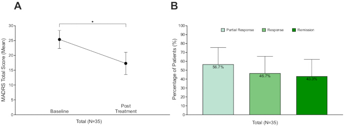
Figure 1: Antidepressant effect after 6 weeks of treatment. (A) Severity of depressive symptoms was assessed using the Montgomery-Asberg Depression Rating Scale (MADRS). Mean MADRS reduction was 9.13 (± 9.82 SD), which was statistically significantly different from zero (one-sample t-test, p < 0.001). Error bars correspond to a 95% Confidence Interval. (B) A partial responder was defined as a patient who experiences at least a 25% reduction in the severity of depression, while a responder was considered if 50% or greater reduction of symptom severity was observed24. Finally, remission was defined as a total score of MADRS ≤ 1016,26. Clinical outcomes were similar to what has been previously reported13. Error bars correspond to a 95% Confidence Interval.Please click here to view a larger version of this figure.
Discussion
The implementation of a standardized TMS protocol in clinical settings ensures consistency in treatment delivery, optimizes patient outcomes, and enhances the reliability of therapeutic interventions for depression17. The protocol outlined in this study provides a structured framework for clinicians and technicians, addressing key procedural elements such as patient preparation, motor hotspot determination, and treatment site localization. By maintaining adherence to these steps, clinicians can reduce variability in treatment application14,37,38, thereby potentially increasing the likelihood of achieving optimal clinical outcomes. Furthermore, integrating psychometric assessments at predefined intervals allows for systematic tracking of patient response, ensuring that treatment adjustments can be made based also on empirical data. In contrast to existing guidelines23, this protocol specifies structured ramping strategies to optimize patient comfort during initial sessions and incorporates predefined psychometric assessment timelines to guide systematic, data-driven treatment adjustments.
Decision-making about the use of TMS to treat an episode of depression should be a collaborative decision between the patient and the prescribing clinician. In most circumstances, the clinician is a psychiatrist, already responsible for patient care, or to whom the patient was referred for assessment of the potential use of TMS. Indications with regulatory approval include TRD, with and without co-morbid anxiety, but also OCD and substance use disorders, with recent clearance of a protocol for smoking cessation11. Protocols cleared by regulatory agencies for depression include high-frequency L-DLPFC stimulation11,39. Off-label uses may be considered based on clinical evidence and expert guidelines. Additionally, promising data also support potential future use for other psychiatric syndromes, namely negative symptoms of schizophrenia and post-traumatic stress disorder, still lacking regulatory approval11,12.
While the focus of this article is on a protocol for conventional rTMS, other protocols are also available. The selection of a specific TMS protocol should be guided by the attending physician, considering both patient and clinician preferences, the specific TMS device available, and the level of experience of the clinical staff. An accelerated TMS protocol (Stanford Neuromodulation Treatment) has been cleared by the FDA for treatment-resistant depression11,16, but is not widely accessible in most TMS centers, despite being a very promising treatment strategy, namely regarding speed of onset36,37. Other protocols, such as low-frequency rTMS targeting the R-DLPFC or bilateral stimulation involving both the L- and R-DLPFC, have demonstrated clinical efficacy6,12. However, these approaches currently hold lower recommendation levels12 and have not yet received regulatory clearance or approval11. Low-frequency rTMS of the R-DLPFC, which may be required when high-frequency stimulation is not tolerated and/or the L-DLPFC is not accessible, is conducted with procedures for determining MH, rMT, and TS as described above, but adapted for the right hemisphere and left hand.
Ethical and regulatory issues should govern TMS use in clinical practice17,18. A fundamental requirement is obtaining informed consent, which must be free and prior to treatment, based on providing all relevant information about procedures, potential risks, and discomforts in an understandable way17,18. Informed consent should clearly discuss the possibility of the occurrence of side effects, particularly a seizure, and clinicians must ensure patients understand the information provided17,18. Full disclosure of the treatment plan, including whether off-label protocols are used, is critical in the informed consent process17,18. Safety is another critical consideration17,18. While generally safe and well-tolerated, the most serious acute adverse effect is the occurrence of a seizure, which is extremely rare when treatment is delivered within recommended parameters11,17,18. Certain conditions and medications can increase the risk of seizures, such as a personal history of epilepsy, brain lesions, administration of drugs lowering seizure threshold, sleep deprivation, and alcoholism18. Other adverse effects, mostly mild and self-limited, can include scalp pain, headache, muscle twitching, dizziness, nausea, anxiety, insomnia, hypomania, and tinnitus11,17,18,39. Absolute contraindications include the presence of metallic hardware in close contact with the coil, such as cochlear implants or implanted pulse generators, due to the risk of inducing device malfunction18. Other conditions, like pregnancy or severe heart disease, involve increased or uncertain risk18.
Patient comfort and adherence40,41,42 play crucial roles in treatment success43. The protocol includes specific measures to improve tolerability, such as initial familiarization with TMS sensations, the use of earplugs to enhance auditory safety, and the implementation of ramping strategies to gradually increase stimulation intensity. These adaptations are particularly valuable for individuals who experience discomfort during early sessions, potentially improving patient retention and overall treatment effectiveness43. The inclusion of structured post-session evaluations also enables the early detection and management of side effects, reinforcing patient safety and engagement throughout the treatment course43.
In addition to TMS equipment with local regulatory approval or clearance (e.g., CE mark in the EU, FDA in the US), sites where rTMS is delivered should be equipped with appropriate life-support equipment, and access to emergency medical facilities is also advised in medical environments where outpatient TMS treatments are delivered18. TMS clinics must have an explicit plan to address syncope and seizures, and every member of the TMS team must be familiar with it18. For suspected syncope, lying the patient down in a supine position with legs elevated is appropriate18. In case of seizures, efforts should focus on preventing complications like aspiration, turning the patient on one side (left lateral decubitus position is desirable) once movement ceases18. TMS-induced seizures are typically brief and self-limited, delayed recovery of consciousness beyond 30 s warrants further medical evaluation18. While life-support equipment should be available in TMS clinical centers18 for outpatient clinical settings applying standard protocols, the consensus of the Clinical TMS Society is that intravenous access, cardiac defibrillators, suction, and oxygen are not necessary17.
Appropriate training for personnel is a crucial step in setting up a TMS clinic19. The required credentials and training depend on the site and the local regulatory requirements, as well as the type and purpose of the TMS application17,18,19. At our center, this includes completion of a certified TMS training program and supervised performance of TMS. Trainees should demonstrate proficiency in MH localization and rMT determination, accurate coil positioning, and adherence to safety procedures, before independent operation. Periodic re-assessment and refresher training are also recommended to maintain standardization. Particularly when rTMS is used as treatment for a medical condition, a licensed physician, serving as the medically responsible clinician, supervises application of treatments, which can be carried out by a trained medical assistant17,18,19. TMS technicians, especially those without medical training, should have basic knowledge of brain physiology, TMS mechanisms, potential risks, and physiological changes induced by treatment17,18,19. It is also recommended that TMS technicians have cardiopulmonary or basic life support training17,18. Additionally, personnel skilled in the management of syncope and seizure should be present in TMS clinics17,18. Finally, for routine clinical treatment sessions, a physician should be accessible in case of an emergency17,18.
Another critical component of effective TMS administration is the precise determination of the stimulation site17. Here, we highlight the importance of using structured non-neuronavigated approaches such as the 5 cm rule and the Beam F3 method to identify the L-DLPFC as the primary target for depression treatment44,45. While non-neuronavigated methods are practical, they nevertheless have limitations such as inherent inter-individual anatomical variation, with implications for precision, accuracy, and reproducibility45,46,47,48. On the other hand, while neuronavigation offers the highest accuracy49, limited availability in clinical settings may limit access if other methods are not used17. Moreover, neuronavigation did not offer a consistent clinical advantage when compared to non-neuronavigated methods50. The step-by-step protocol presented here provides detailed guidance on the latter approaches, ensuring that even without neuronavigation, treatment sites can be determined consistently and reliably27. Future research should explore how progress in imaging technology and real-time neuronavigation could further enhance the precision and efficacy of TMS interventions.
The structured assessment sessions incorporated into the protocol provide an evidence-based approach to monitor treatment efficacy and patient response. Regular psychometric assessment using standardized instruments such as the BDI-II and MADRS allows clinicians to track improvements and determine whether modifications to the treatment plan are necessary24,27. Moreover, periodic reassessments of the rMT ensure that stimulation parameters remain optimal, accounting for any physiological changes that may occur over the course of treatment27,51. While this is expected to contribute significantly to the safety of rTMS, recent evidence indicates that neither motor threshold and its stability, nor treatment intensity, reliably predict clinical outcomes27,52. Finally, baseline assessments, such as the YMRS, may be used to identify patients at risk of mania, hypomania, or mixed states50. In such cases, treatment adjustments may include closer clinical monitoring and coordination with the prescribing physician, medication adjustments (e.g., optimizing mood stabilizers) to mitigate the risk of mood switching, and consideration of the need to postpone rTMS treatment50. This systematic approach underscores the importance of objective, data-driven decision-making in TMS therapy.
Depending on the resources and protocols of each TMS center, as well as patient preference, treatment may transition to a maintenance phase TMS protocol if this is considered of clinical interest14. This decision is typically based on clinical response to TMS and risk of relapse, and follows a pre-defined schedule (e.g., weekly, or biweekly sessions) that can be adjusted according to ongoing psychiatric evaluation. While no standardized protocol exists for transitioning to maintenance, a gradual reduction in session frequency is commonly employed (e.g., three sessions per week, then two sessions per week, and finally weekly) until the target maintenance schedule (e.g., biweekly) is reached14,30. Symptom prompted maintenance protocols have also been described, where short cycles of rescue TMS session are applied when symptoms relapse14,30. Currently, there is no evidence to support a given type of maintenance strategy over another14,30.
Even with a structured protocol, challenges may occur during rTMS administration. Difficulty eliciting or locating the motor hotspot can often be addressed by verifying coil orientation and contact, exploring adjacent scalp positions, increasing intensity in small steps, and ensuring that the patient is relaxed17,18,36. If a patient does not tolerate stimulation, strategies include ramping intensity over initial sessions, slightly adjusting coil placement, or using over-the-counter analgesics17,18,36. In case of session interruptions due to equipment issues, patient discomfort, or unforeseen events, the session should be paused, the problem resolved, and stimulation resumed only once correct coil positioning and patient readiness are re-confirmed. Coil instability can be minimized with proper head and neck support, consistent cap alignment, and coil-holding devices17,18. Variability in motor threshold measurements should prompt reassessment and review of technique and equipment if changes are abrupt17,18,24,36. Auditory discomfort is best prevented with well-fitted, regularly replaced earplugs17,18. Applying these measures improves comfort, accuracy, and treatment consistency.
While our goal is to offer practical guidance for the clinical use of TMS in depression, several limitations merit note. First, we rely on non-neuronavigated scalp targeting (5 cm rule, Beam F3). These methods are accessible and standardized but depend on individual anatomy and can reduce placement precision. Neuronavigation may improve coil-placement accuracy, but it is not universally available and has not shown a consistent clinical advantage in treatment outcomes. Second, while rMT-based dosing is essential for safety and standardization, it does not reliably predict antidepressant efficacy. Nevertheless, to date, no alternative dosing approach has shown sufficiently consistent results to replace rMT as the dosing anchor. Finally, we present maintenance treatment as optional because schedules vary across centers and no single strategy has proven superior. Future work should define more specific, evidence-based maintenance schedules.
In conclusion, the protocol outlined in this study provides a comprehensive, step-by-step framework for application of rTMS in the treatment of depression, balancing standardization with individualized care. Variability in clinical outcomes is addressed through the use of consistent targeting methods, clear stimulation parameter definitions, and structured procedural steps, while still allowing flexibility for patient-specific adjustments. In resource-rich settings, integration of neuronavigation and targeting based on functional imaging may further enhance precision and reproducibility. Future studies should focus on optimizing stimulation parameters, refining targeting strategies, and evaluating the long-term efficacy of structured TMS protocols, including the role of maintenance sessions and personalized adjustments in sustaining therapeutic benefits beyond the acute phase.
Disclosures
AJO-M is recipient of a grant from Schuhfried GmBH for norming and validation of cognitive tests, and was national coordinator for Portugal of trials of psilocybin therapy for treatment-resistant depression, sponsored by Compass Pathways, Ltd (EudraCT number 2017-003288-36 and 2020-001348-25), and of esketamine for treatment-resistant depression, sponsored by Janssen-Cilag, Ltd (EudraCT NUMBER: 2019-002992-33); has received payment, honoraria, or support for attending meetings and participating in advisory boards from MSD, Neurolite AG, Janssen Pharmaceuticals, Angelini Pharma, and the European Monitoring Centre for Drugs and Drug Addiction; has received consultancy fees from Bioprojet Pharma and NaturalX Health Ventures (all outside the submitted work); is Vice President of the Portuguese Society for Psychiatry and Mental Health; is head of the Psychiatry Working Group for the National Board of Medical Examination at the Portuguese Medical Association and Portuguese Ministry of Health; is President of the Ethics Committee for the Public Institute for Addictive Behaviors and Dependence; and is President of the Scientific Council of the Portuguese Obsessive Compulsive Disorder Foundation. The remaining authors declare that they have no potential conflicts of interest involving this work, including relevant financial activities outside the submitted work and any other relationships or activities that readers could perceive to have influenced, or that give the appearance of potentially influencing what is written. None of these agencies had a role in the design and conduct of the study, in the collection, management, analysis, and interpretation of the data, in the preparation, review, or approval of the manuscript, nor in the decision to submit the manuscript.
Acknowledgements
GC is supported by a 2023 NARSAD Young Investigator Grant from the Brain & Behavior Research Foundation. AJO-M is supported by a Starting Grant from the European Research Council (grant agreement no. 950357), and by the PsyPal project (grant agreement no. 875358), both funded by the European Union's Horizon 2020 Research and Innovation Programme. GC and AJO-M are supported by a Proof-of-Concept Grant from the European Research Council (grant agreement no. 101158262). The content of this study is solely the responsibility of the authors and does not necessarily represent the official views of any of the funding agencies.
Materials
| Name | Company | Catalog Number | Comments |
|---|---|---|---|
| BA9 BA8 BA43 Location System | Will Beam & Jeff Borckardt | Web Interface Developed 6/7/2010 | https://clinicalresearcher.org/F3/ |
| Computer Desktop/Laptop | Irrelavant | Not applicable | Required to access BeamF3 website |
| Confortable Chair | Irrelavant | Not applicable | For the patient to seat while all TMS procedures are performed |
| Cool-B65 Coil | Magventure | 9016E0491 | TMS coil |
| Microsoft Excel | Microsoft | Office 365 Personal | Useful to store TMS patients clinical data |
| Paper | Irrelevant | Not applicable | Required to print the psychometric instruments and the score sheets |
| Pen | Irrelevant | Not applicable | Required to complete the psychometric instruments |
| Printer | Irrelevant | Not applicable | Required to print the psychometric instruments and the score sheets |
| Transcranial Magnetic Stimulation (TMS) MagPro X100 | Magventure | 9016D0041 | TMS operaring system |
References
- Lefaucheur, J. P. Transcranial magnetic stimulation. Handb Clin Neurol. 160, 559-580 (2019).
- Citri, A., Malenka, R. C. Synaptic plasticity: multiple forms, functions, and mechanisms. Neuropsychopharmacology. 33, 18-41 (2008).
- Klomjai, W., Katz, R., Lackmy-Vallée, A. Basic principles of transcranial magnetic stimulation (TMS) and repetitive TMS (rTMS). Ann Phys Rehabil Med. 58, 208-213 (2015).
- Pascual-Leone, A., Rubio, B., Pallardó, F., Catalá, M. D. Rapid-rate transcranial magnetic stimulation of left dorsolateral prefrontal cortex in drug-resistant depression. Lancet. 348, 233-237 (1996).
- George, M. S., et al. Daily repetitive transcranial magnetic stimulation (rTMS) improves mood in depression. Neuroreport. 6, 1853-1856 (1995).
- Mutz, J., et al. Comparative efficacy and acceptability of non-surgical brain stimulation for the acute treatment of major depressive episodes in adults: systematic review and network meta-analysis. BMJ. 364, l1079(2019).
- Bessa, J., et al. Real-world evidence from a European cohort study of patients with treatment resistant depression: country-specific data for Portugal. J Affect Disord Rep. 17, 100791(2024).
- Heerlein, K., et al. Real-world evidence from a European cohort study of patients with treatment resistant depression: healthcare resource utilization. J Affect Disord. 298, 442-450 (2022).
- Heerlein, K., et al. Real-world evidence from a European cohort study of patients with treatment resistant depression: treatment patterns and clinical outcomes. J Affect Disord. 290, 334-344 (2021).
- Heerlein, K., et al. Real-world evidence from a European cohort study of patients with treatment resistant depression: baseline patient characteristics. J Affect Disord. 283, 115-122 (2021).
- Cotovio, G., Ventura, F., Rodrigues da Silva, D., Pereira, P., Oliveira-Maia, A. J. Regulatory clearance and approval of therapeutic protocols of transcranial magnetic stimulation for psychiatric disorders. Brain Sci. 13, 1029(2023).
- Lefaucheur, J. P., et al. Evidence-based guidelines on the therapeutic use of repetitive transcranial magnetic stimulation (rTMS): an update (2014-2018). Clin Neurophysiol. 131 (2), 474-528 (2020).
- Thompson, L. Treating major depression and comorbid disorders with transcranial magnetic stimulation. J Affect Disord. 276, 453-460 (2020).
- Senova, S., Cotovio, G., Pascual-Leone, A., Oliveira-Maia, A. J. Durability of antidepressant response to repetitive transcranial magnetic stimulation: systematic review and meta-analysis. Brain Stimul. 12, 119-128 (2019).
- Lefaucheur, J. P., et al. Evidence-based guidelines on the therapeutic use of repetitive transcranial magnetic stimulation (rTMS): an update (2014-2018). Clin Neurophysiol. 131, 474-528 (2020).
- Cole, E. J., et al. Stanford neuromodulation therapy (SNT): a double-blind randomized controlled trial. Am J Psychiatry. 179 (2), 132-141 (2022).
- Perera, T., et al. The Clinical TMS Society consensus review and treatment recommendations for TMS therapy for major depressive disorder. Brain Stimul. 9, 336-346 (2016).
- Rossi, S., Hallett, M., Rossini, P. M., Pascual-Leone, A. Safety of TMS Consensus Group. Safety, ethical considerations, and application guidelines for the use of transcranial magnetic stimulation in clinical practice and research. Clin Neurophysiol. 120, 2008-2039 (2009).
- Morales, I. G., et al. Emergency management of epileptic seizures: a consensus statement. Emergencias. 32, 353-362 (2020).
- Furlan, L., et al. Syncope in the emergency department: a practical approach. J Clin Med. 13, 3231(2024).
- Ventura, F., Cotovio, G., Frias, P., Oliveira-Maia, A. J. Training in transcranial magnetic stimulation: experience from a clinical and research centre. Rev Port Psiquiatr Saúde Ment. 9, 77-84 (2023).
- Almeida, S., et al. Criterion and construct validity of the Beck Depression Inventory (BDI-II) to measure depression in patients with cancer: the contribution of somatic items. Int J Clin Health Psychol. 23, 100350(2023).
- Rossi, S., et al. Safety and recommendations for TMS use in healthy subjects and patient populations, with updates on training, ethical and regulatory issues: expert guidelines. Clin Neurophysiol. 132 (1), 269-306 (2021).
- Cotovio, G., Boes, A. D., Press, D. Z., Oliveira-Maia, A. J., Pascual-Leone, A. In older adults the antidepressant effect of repetitive transcranial magnetic stimulation is similar but occurs later than in younger adults. Front Aging Neurosci. 14, 919734(2022).
- Nierenberg, A. A., DeCecco, L. M. Definitions of antidepressant treatment response, remission, nonresponse, partial response, and other relevant outcomes: a focus on treatment-resistant depression. J Clin Psychiatry. 62, 5-9 (2001).
- Cole, E., et al. accelerated intelligent neuromodulation therapy for treatment-resistant depression (SAINT-TRD). Brain Stimul. 12, 402(2019).
- Cotovio, G., et al. Day-to-day variability in motor threshold during rTMS treatment for depression: clinical implications. Brain Stimul. 14 (5), 1118-1125 (2021).
- Feifel, D., Pappas, K. Treating clinical depression with repetitive deep transcranial magnetic stimulation using the Brainsway H1-coil. J Vis Exp. (116), e53858(2016).
- Seybert, C., et al. Replicability of motor cortex-excitability modulation by intermittent theta burst stimulation. Clin Neurophysiol. 152, 22-33 (2023).
- Faro Viana, F., et al. Reducing motor evoked potential amplitude variability through normalization. Front Psychiatry. 15, 1279072(2024).
- Dunlop, K., et al. MRI-guided dmPFC-rTMS as a treatment for treatment-resistant major depressive disorder. J Vis Exp. (102), e53129(2015).
- Beam, W., Borckardt, J. J., Reeves, S. T., George, M. S. An efficient and accurate new method for locating the F3 position for prefrontal TMS applications. Brain Stimul. 2, 50-54 (2009).
- Yang, J., et al. Status and trends of TMS research in depressive disorder: a bibliometric and visual analysis. Front Psychiatry. 15, 1432792(2024).
- Bouaziz, N., et al. Real-world transcranial magnetic stimulation for major depression: a multisite, naturalistic, retrospective study. J Affect Disord. 326, 26-35 (2023).
- Valiengo, L., et al. Repetitive transcranial magnetic stimulation for major depressive disorder in older adults: systematic review and meta-analysis. J Gerontol A Biol Sci Med Sci. 77 (4), 851-860 (2022).
- Xu, Y., Zhang, Y., Zhao, D., Tian, Y., Yuan, T. F. Growing placebo response in TMS treatment for depression: a meta-analysis of 27-year randomized sham-controlled trials. Nat Ment Health. 1, 792-809 (2023).
- Kar, S. K. Predictors of response to repetitive transcranial magnetic stimulation in depression: a review of recent updates. Clin Psychopharmacol Neurosci. 17, 25(2019).
- Valiengo, L., et al. Repetitive transcranial magnetic stimulation for major depressive disorder in older adults: systematic review and meta-analysis. J Gerontol A Biol Sci Med Sci. 77, 851-860 (2022).
- Chen, L., et al. Treating depression with repetitive transcranial magnetic stimulation: a clinician's guide. Am J Psychiatry. 182 (6), 525-541 (2025).
- Rossi, S., et al. Safety and recommendations for TMS use in healthy subjects and patient populations, with updates on training, ethical and regulatory issues: expert guidelines. Clin Neurophysiol. 132, 269-306 (2021).
- Stultz, D. J., Osburn, S., Burns, T., Pawlowska-Wajswol, S., Walton, R. Transcranial magnetic stimulation (TMS) safety with respect to seizures: a literature review. Neuropsychiatr Dis Treat. 16, 2989-3000 (2020).
- Wieczorek, T., et al. Transcranial magnetic stimulation (TMS) in treatment of psychiatric disorders: review of current studies. Psychiatr Pol. 55, 565-583 (2021).
- Mallon, S., Walker, K., Bayley, Z., Griffiths, C. Practitioner perspectives on best practice in non-treatment factors that support the delivery of repetitive transcranial magnetic stimulation (rTMS) for depression. J Psychiatr Ment Health Nurs. 29, 463(2022).
- Trapp, N. T., Pace, B. D., Neisewander, B., Ten Eyck, P., Boes, A. D. A randomized trial comparing Beam F3 and 5.5 cm targeting in rTMS treatment of depression demonstrates similar effectiveness. Brain Stimul. 16, 1392-1400 (2023).
- Trapp, N. T., et al. Reliability of targeting methods in TMS for depression: Beam F3 vs. 5.5 cm. Brain Stimul. 13, 578-581 (2020).
- Herwig, U., Padberg, F., Unger, J., Spitzer, M., Schönfeldt-Lecuona, C. Transcranial magnetic stimulation in therapy studies: examination of the reliability of "standard" coil positioning by neuronavigation. Biol Psychiatry. 50, 58-61 (2001).
- Sack, A. T., et al. Optimizing functional accuracy of TMS in cognitive studies: a comparison of methods. J Cogn Neurosci. 21, 207-221 (2009).
- Mir-Moghtadaei, A., et al. Concordance between Beam F3 and MRI-neuronavigated target sites for repetitive transcranial magnetic stimulation of the left dorsolateral prefrontal cortex. Brain Stimul. 8, 965-973 (2015).
- Caulfield, K. A., et al. Neuronavigation maximizes accuracy and precision in TMS positioning: evidence from 11,230 distance, angle, and electric field modeling measurements. Brain Stimul. 15, 1192-1205 (2022).
- Hebel, T., et al. A direct comparison of neuronavigated and non-neuronavigated intermittent theta burst stimulation in the treatment of depression. Brain Stimul. 14, 335-343 (2021).
- Cotovio, G., et al. Reply: variability in motor threshold. Brain Stimul. 14, 1523-1524 (2021).
- Lee, N. A., et al. Motor threshold parameters do not predict repetitive transcranial magnetic stimulation and intermittent theta burst stimulation outcomes in major depressive disorder. J Affect Disord. 372, 144-148 (2025).
Reprints and Permissions
