This protocol demonstrates how the Src homology 2 domain-containing 5'-inositol phosphatase (SHIP)-deficient mouse model of Crohn disease (CD)-like ileal inflammation and fibrosis can be used to test novel therapeutics for CD.
Method Article
Evaluating Therapeutic Interventions in the SHIP-deficient Mouse Model of Crohn Disease-like Ileitis and Fibrosis
* These authors contributed equally
In This Article
Summary
Abstract
Crohn disease (CD) is a chronic, relapsing inflammatory condition characterized by segmental, transmural inflammation that can affect any part of the gastrointestinal tract. A hallmark of CD is impaired intestinal barrier function, and intestinal fibrosis is a common complication. SHIP-deficient (SHIP-/-) mice spontaneously develop gut barrier dysfunction, ileal inflammation, and fibrotic pathology, recapitulating key features of CD-like ileitis. The SHIP-/- mice serve as a valuable model for testing anti-inflammatory and anti-fibrotic therapies in CD.
Here, we describe methods to evaluate therapeutic interventions in this model, using dexamethasone as an example of an effective therapy. We provide a step-by-step protocol for characterizing disease and treatment response, including in vivo assessment of gut permeability measured by FITC-dextran, as well as gross pathological examination and detailed histological analyses. Histological assessments incorporate hematoxylin and eosin (H&E) staining for inflammation, Masson's trichrome staining for fibrosis, and Alcian blue/Periodic Acid-Schiff (PAS) staining for goblet cell hyperplasia and hypertrophy. Additionally, cytokines and inflammatory markers in ileal tissue are quantified to assess the inflammatory state. Together, these methods provide a comprehensive framework for evaluating treatment efficacy in the SHIP-/- mouse model of CD-like ileitis. This protocol is broadly applicable to investigators studying gut inflammation, mucosal healing, and intestinal fibrosis.
Introduction
The two main types of inflammatory bowel disease (IBD) are Crohn disease (CD) and ulcerative colitis (UC), both of which are characterized by chronic gastrointestinal (GI) inflammation that can follow a relapsing-remitting or progressive course1. Although IBD has historically been considered a disease of Westernized nations, its incidence is increasing rapidly in developing countries across South America, Asia, and Africa2. Concurrently, prevalence continues to rise in North America, Europe, and Australia, with an almost 50% increase in both incidence and prevalence reported between 1990 and 2019, underscoring the growing global health burden of IBD2,3. Despite significant research efforts, the underlying cause of IBD remains unknown. Current evidence suggests a complex interplay between genetic susceptibility, immune system dysfunction, and environmental triggers, including alterations in the microbiome4,5.
There are key differences between CD and UC. In UC, inflammation is confined to the colon and is characterized by continuous involvement that is limited to the mucosal layer6,7. In contrast, CD is characterized by discontinuous and transmural inflammation that can affect any part of the GI tract, most commonly in the terminal ileum6,7. A hallmark complication of CD is intestinal fibrosis, which contributes to the formation of strictures and bowel obstruction8. Fibrosis is defined by excessive deposition of extracellular matrix components, such as collagen, and thickening of the intestinal muscle layers9,10. Approximately 30% of people with CD develop stricturing disease within 10 years of diagnosis11. Currently, no therapies directly target fibrosis, an unmet clinical need that may be addressed using suitable preclinical models to study this complication9,12.
To better understand the pathogenesis of IBD and facilitate the development of new therapies, numerous mouse models have been established12. While a great number of colitis models exist, only a few recapitulate ileal inflammation and CD-associated fibrosis12. Among these are the Src homology 2 domain-containing 5'-inositol phosphatase-deficient (SHIP-/-) described herein, as well as the SAMP1/YitFc mice and the TnfΔARE mice. These models collectively reflect the complexity of human CD as each is driven by distinct cellular and molecular mechanisms. Disease in SAMP1/YitFc mice results from a polygenic predisposition affecting epithelial barrier function and immune regulation13,14, whereas TnfΔARE mice carry a deletion of AU-rich elements in the TNF mRNA, leading to increased mRNA stability and TNF overproduction15,16. Though valuable, these models have limitations. The SAMP1/YitFc model suffers from low breeding efficiency, which poses challenges in generating sufficient cohort sizes for studies17. In addition, the TnfΔARE model requires up to 24 weeks to develop notable fibrotic pathology, which makes it expensive and challenging to do intervention studies16.
SHIP is a negative regulator of the phosphatidylinositol-3-kinase (PI3K) signaling pathway and is predominantly expressed in hematopoietic cells. It functions by removing the 5' phosphate group from phosphatidylinositol 3,4,5-trisphosphate, thereby attenuating downstream PI3K signaling18. The SHIP-/- mice offer several advantages for preclinical studies. These mice are relatively easy to breed and display signs of fibrosis as early as 8 weeks of age19. The accelerated fibrotic remodeling in this model is likely driven by heightened inflammatory responses due to a dysregulated PI3K pathway, which leads to immune cell hyperactivation and subsequent rapid tissue remodeling. Most importantly, we and Kerr's group have independently shown that SHIP-/- mice develop ileitis with features that closely resemble human CD19,20. Supporting the relevance of this model, a subset of individuals with CD have reduced SHIP activity in both ileal biopsies and PBMCs21. Moreover, those with low SHIP activity tend to experience a more severe disease course, often requiring multiple surgeries22.
SHIP-/- mice begin to develop spontaneous, discontinuous intestinal inflammation localized to the distal ileum as early as 4 weeks of age, with inflammation present in all mice by 6 weeks of age20. By 8 weeks of age, mice have established fibrotic pathology, characterized by thickening of the muscularis externa and extensive collagen accumulation in the submucosa and between the muscle layers19. Inflammation in this model is driven by macrophage-derived interleukin-1β (IL-1β)21. Both depletion of macrophages using clodronate-containing liposomes and pharmacologic blockade of IL-1 signaling with an IL-1 receptor antagonist (anakinra) ameliorated intestinal inflammation in SHIP-/- mice21. In addition to ileitis, SHIP-/- mice exhibit lung inflammation, which we have used to evaluate off-target effects of therapies designed for localized intestinal delivery23. Others have reported that caspase-8 inhibition with Z-IETD-FMK significantly reduces both ileal and lung inflammation in SHIP-/- mice24, further supporting lung pathology as an additional feature for assessing systemic inflammatory control.
This methodological paper revisits our recently published work on successful treatment with dexamethasone23 and provides a detailed description of key technical procedures, with additional considerations for using mouse models in pharmacological testing. Specifically, we highlight standard parameters for evaluating drug efficacy in models of IBD, including assessment of gut permeability, gross pathology, histopathology, and pro-inflammatory mediators and effectors, and discuss how to integrate these data to make conclusions about overall therapeutic efficacy.
Protocol
Studies involving animals should be performed in compliance with ethical protocols approved by the Institutional Animal Care and Use Committee and in accordance with National Research Councils' Guidelines on Animal Care. All animal procedures in this protocol followed the ethical guidelines of the Canadian Council on Animal Care and were approved by the University of British Columbia Animal Care Committee (protocol A21-0212).
NOTE: To assess the therapeutic impact of the drug on intestinal pathology of SHIP-/- mice, include SHIP+/+ as healthy controls for comparison. For therapeutic intervention, initiate the treatment at 6 weeks of age, when the disease is fully established in the model. Here, both SHIP+/+ and SHIP-/- mice received daily oral gavage of dexamethasone at 3 mg/kg in 0.5% β-cyclodextrin (vehicle) or the vehicle alone for 2 weeks, in a volume of 10 µL/g.
1. FITC-dextran assay to measure epithelial barrier permeability
- Fast mice for 4 h prior to the assay.
- Dissolve 4 kDa FITC-dextran in sterile 1x PBS to a final concentration of 80 mg/mL.
- Prepare enough for 150 µL per mouse, plus 50 µL extra for the standard curve.
NOTE: Minimize light exposure of FITC-dextran solution and plasma samples throughout the procedure. - Gently restrain (scruff) each mouse and administer 150 µL of the FITC-dextran solution via oral gavage.
NOTE: Start a timer immediately after administering the first gavage. Wait 10-15 min between each subsequent mouse to allow sufficient time for euthanasia and downstream procedures to be performed in a staggered manner, minimizing variability due to timing differences. - Maintain mice without food for 4 h.
- Euthanize mice using 5% isoflurane (inhalant anesthetic delivered with 1.5 L oxygen/min) followed by carbon dioxide in accordance with standard institutional procedure. Collect blood via cardiac puncture with 25 G needles and 1 mL syringes.
- Immediately add 10 µL of acid-citrate-dextrose solution to every 100 µL of collected blood as an anticoagulant. Mix thoroughly by inversion.
- Centrifuge at 1500 x g for 10 min at 4 °C and carefully transfer plasma (supernatant) to labeled 1.7 mL microcentrifuge tubes. Keep samples on ice and protected from light until analysis.
- Dilute each plasma sample 1:2 and 1:10 with 1x PBS.
- Add 100 µL of each dilution to an opaque 96-well plate in duplicates.
- Perform 1:2 serial dilutions of the original 80 mg/mL FITC-dextran stock using 1x PBS to achieve the following concentrations: 3000, 1500, 750, 375, 187.5, 93.8, 46.9, 23.4, and 0 ng/mL. Add 100 µL of each concentration to the 96-well plate in duplicate.
- Measure fluorescence at 485 nm excitation and 535 nm emission using a microplate reader.
2. Gross pathology assessment
- Following blood collection for the FITC-dextran assay, carefully excise the entire small intestine.
- Gently remove any attached fat and connective tissue.
NOTE: Be careful when handling the tissue, as diseased regions can be fragile and prone to tearing. - Lay the intestine flat on a sheet of blank white paper for optimal contrast. Visually inspect the tissue for macroscopic signs of disease. Inflammation is typically localized to the distal 10 cm of the small intestine in the SHIP-/- mouse.
NOTE: Key macroscopic features to assess include color changes, with regions showing visible reddening or hyperemia relative to adjacent normal-appearing tissue. Additionally, examine for tissue thickening, as affected areas may appear swollen, edematous, or rigid upon gentle palpation. Look for the presence of blood, which may appear as visible hemorrhagic spots, dark red discoloration, or adherent blood. - Align the intestine next to a ruler for scale and take a photo to document the gross pathological findings.
3. Histological assessment
- Remove intestinal contents by gently flushing the lumen with 1x PBS using a syringe. Do not apply too much force to the tissue, as this may distort villus and crypt architecture.
- Identify and harvest approximately 1 cm of the distal ileum that is most representative of the gross pathology. Include visible affected regions, if present. For comparisons (SHIP+/+ vs. SHIP-/-), ensure that tissues are collected from matched anatomical locations.
NOTE: For initial evaluation of treatment efficacy, preparing the ileum as a Swiss roll is recommended. This method provides a global visualization of disease and reduces sampling bias. - Lay the tissue flat in a histology cassette, placing it between sponges to prevent folding.
- For hematoxylin & eosin (H&E) and Masson's trichrome staining, fix the tissue in 10% neutral buffered formalin overnight at 4 °C, using at least 10-20 times the tissue volume (about 5 mL per cassette) and ensuring that all cassettes are fully submerged. Transfer cassettes to 70% ethanol for storage, keeping them fully submerged until processing.
NOTE: 10% formalin, a fixative containing 3.7% formaldehyde, is a potent respiratory irritant, a dermal sensitizer, and is classified as a human carcinogen. Inhalation, ingestion, or skin contact can result in acute toxicity, while chronic exposure may increase cancer risk. All procedures involving formalin should be performed in a certified chemical fume hood while wearing appropriate personal protective equipment (PPE), including a lab coat, chemical-resistant gloves, and eye protection. Collect waste in containers clearly labeled "Formalin Waste" for proper disposal according to institutional hazardous waste protocols. All materials contaminated with formalin (e.g., pipette tips, tissues, gloves) should be treated as chemically hazardous waste and discarded following local Environmental Health and Safety (EHS) procedures. - For Alcian blue/PAS staining, fix the tissue in Carnoy's solution (60% ethanol, 30% chloroform, and 10% glacial acetic acid) at 4 °C overnight, using approximately 5 mL per cassette or 10-20 times the tissue volume. Transfer cassettes to 100% ethanol for storage until processing.
NOTE: Carnoy's solution is composed of 60% ethanol, 30% chloroform, and 10% glacial acetic acid. This mixture is highly volatile, flammable, and toxic. Handle Carnoy's solution exclusively in a fume hood and store in tightly sealed containers away from heat or ignition sources. Carnoy's waste should be collected in designated containers for mixed organic solvents and disposed of in accordance with institutional hazardous waste guidelines. All materials contaminated with Carnoy's solution should be treated as chemically hazardous waste. - Embed tissues in paraffin and cut 5 µm sections for staining.
- Score the H&E-stained ileal sections on a 16-point scale according to the criteria in Table 1. The final histological damage score is determined by averaging the scores from 2 reviewers, who are blinded to the experimental condition.
- Evaluate the Masson's trichrome-stained cross-sections using a 6-point scale based on the criteria in Table 2. The final fibrosis score is calculated as the median of the scores assigned by two independent reviewers blinded to the experimental conditions.
4. Cytokine assessment
- Following dissection and removal of the histology sample, collect the remaining portion of the distal 10 cm of the ileum.
- Blot the tissue dry and record the weight. Flash-freeze in liquid nitrogen and store at -80 °C for future processing.
- When ready for processing, retrieve the tissue and keep it on ice at all times.
- Weigh the tissue samples. Prepare sufficient Homogenization Buffer for all samples by adding protease inhibitors to 1x PBS (aprotinin and leupeptin, each at 10 µg/mL). Use 10 µL of buffer per mg of tissue (e.g., 100 mg of tissue requires 1 mL of buffer).
NOTE: Homogenization buffer must be prepared fresh on the day of homogenization to ensure that protease inhibitors are effective. - Homogenize the full-thickness ileal tissue in ice-cold Homogenization Buffer using a benchtop homogenizer. Ensure the homogenate is uniform and free of visible tissue fragments. Rinse the homogenizer tip thoroughly with PBS between samples to prevent cross-contamination.
- Centrifuge the tissue homogenates at 10,000 x g for 10 min at 4 °C.
- Aliquot the clarified supernatants to avoid freeze-thaw cycles and store at -80 °C for future analysis.
- Perform enzyme-linked immunosorbent assays (ELISAs) on clarified supernatants to quantify cytokines of interest, according to manufacturers' instructions.
NOTE: Optical density at 450 nm was measured using the Varioskan LUX microplate reader with Skanlt RE 7.0.2 software, with a reference wavelength of 570 nm for correction. Microsoft Excel was used for data analysis, and absorbance values within the linear range of the standard curve were interpolated to determine cytokine concentrations.
Results
6-week-old SHIP+/+ and SHIP-/- mice were orally gavaged with 0.5% β-cyclodextrin (vehicle control; n = 6/genotype) or 3 mg/kg of dexamethasone (n = 6/genotype) daily for 2 weeks. The timing and duration of treatment are model-dependent and should consider the onset and rate of disease progression, as well as whether the intervention is intended to be prophylactic or therapeutic. An equal number of male and female mice should be used in experiments. Although we have not identified sex-based differences in the natural history of disease in SHIP-/- mice, different treatment protocols may show sex-specific responses. In this model, prophylactic treatment from 4-6 weeks of age may be used to prevent the onset of inflammation and fibrosis, whereas therapeutic regimens performed from 6-8, 8-10, or 6-10 weeks of age can be used to assess treatment efficacy for established disease. Optimal dosing and timing should be guided by the literature and titration studies. We have previously shown that this dosing regimen for dexamethasone is effective when administered after ileal inflammation has developed.
A recommended timeline for the day of harvest is outlined in Figure 1A. Dexamethasone did not induce any intestinal pathology in the SHIP+/+ mice based on gross examination. SHIP-/- mice displayed visible areas of redness and thickening, indicative of inflammation. These gross pathological features were absent in SHIP-/- mice treated with dexamethasone (Figure 1B). To evaluate intestinal barrier function, plasma concentrations of FITC-dextran were measured following oral gavage. Although not statistically significant, SHIP-/- mice had elevated plasma FITC-dextran concentrations compared to SHIP+/+ controls, suggesting increased intestinal permeability. Dexamethasone treatment reduced FITC-dextran concentrations in SHIP-/- mice to concentrations similar to those observed in SHIP+/+ mice (Figure 1C).
Histological examination of intestinal cross-sections from SHIP-/- mice revealed hallmark pathological features, including immune cell infiltration, disrupted villus-crypt architecture, goblet cell hyperplasia/hypertrophy, and thickening of the muscularis layer (Figure 2A). These characteristics were reflected in significantly higher histological damage scores compared to SHIP+/+ mice (p = 0.0005, Figure 2B). Histological damage was not observed in SHIP+/+ mice treated with dexamethasone. Consistent with improvements seen in gross pathology and barrier function, dexamethasone reduced histopathology in SHIP-/- mice, although the effect was not statistically significant (p = 0.0981, Figure 2A and Figure 2B). Masson's trichrome staining revealed substantial collagen deposition in SHIP-/- intestinal tissues, accompanied by significantly higher fibrosis scores compared to SHIP+/+ controls (Figure 2C). Dexamethasone effectively attenuated fibrotic features in SHIP-/- mice, lowering fibrosis scores to values comparable to those of SHIP+/+ controls (Figure 2C). Additionally, Alcian blue/PAS staining confirmed goblet cell hyperplasia/hypertrophy in SHIP-/- mice, which was reduced upon treatment with dexamethasone (Figure 2D).
To assess inflammatory cytokine concentrations, ELISAs were performed on full-thickness ileal tissue homogenates (Figure 3A). IL-1β concentrations were significantly higher in SHIP-/- mice compared to SHIP+/+ controls (p = 0.0468), which aligns with previous findings correlating IL-1β expression to disease severity in this model. Dexamethasone treatment significantly reduced IL-1β concentrations in SHIP-/- mice (p = 0.0023). Consistent with previous reports, IL-6 or TNF concentrations did not differ across the four experimental groups. We also measured concentrations of the neutrophil-associated proteins myeloperoxidase (MPO) and lipocalin-2 (LCN-2) in full-thickness ileal homogenates as additional biochemical markers of inflammation (Figure 3B). Both MPO and LCN-2 concentrations were significantly higher (p = 0.0031 and p = 0.0002 respectively) in SHIP-/- mice compared to SHIP+/+ controls, though the increase in LCN-2 was largely driven by high concentrations measured in 2 of 6 mice. Treatment with dexamethasone reduced MPO and modestly reduced LCN-2, differences were not significant. These data suggest that MPO is a useful marker of inflammation in SHIP-/- mice, whereas LCN-2 concentrations are not as consistent or reliable.
To investigate the relationship between inflammatory and fibrotic features in the SHIP-/- mouse model, we performed Spearman correlation analyses using histological damage scores, fibrosis scores, and IL-1β concentrations from full-thickness ileal tissue (Figure 4). IL-1β concentrations positively correlated with histological damage scores (r = 0.6813), indicating that increased inflammation was associated with increased tissue damage (Figure 4A). A moderate correlation was also observed between IL-1β concentrations and fibrosis scores (r = 0.4117), suggesting a link between inflammatory cytokine production and fibrotic remodeling (Figure 4B). Additionally, histological damage scores correlated significantly with fibrosis scores (r = 0.6241), suggesting that structural damage and fibrosis progress in parallel in SHIP-/- mice (Figure 4C).
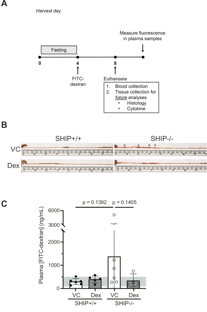
Figure 1: Dexamethasone reduces gross intestinal pathology and permeability in SHIP-/- mice. SHIP+/+ and SHIP-/- mice were orally gavaged daily with either 0.5% β-cyclodextrin as a vehicle control (VC) or dexamethasone (Dex; 3 mg/kg) from 6 to 8 weeks of age. (A) Recommended experimental timeline. (B) Gross morphology of the cecum and distal ileum was assessed, and representative images are shown (n = 4-6 mice per group). Asterisks denote areas of patchy redness and thickening observed on gross examination. The images are intended to serve as representative examples of overall pathology and are not meant for quantitative analysis. (C) To assess intestinal permeability, mice were fasted for 4 h and gavaged with 80 mg/mL FITC-dextran. After an additional 4 h of fasting, blood was collected via cardiac puncture, and plasma FITC-dextran concentrations were measured. Data are presented as mean±SD. The gray bar represents the range of plasma FITC-dextran concentrations typically observed in healthy C57BL/6 mice four hours post-gavage25. Statistical significance was determined using the Kruskal-Wallis test followed by Dunn's multiple comparisons test. Please click here to view a larger version of this figure.
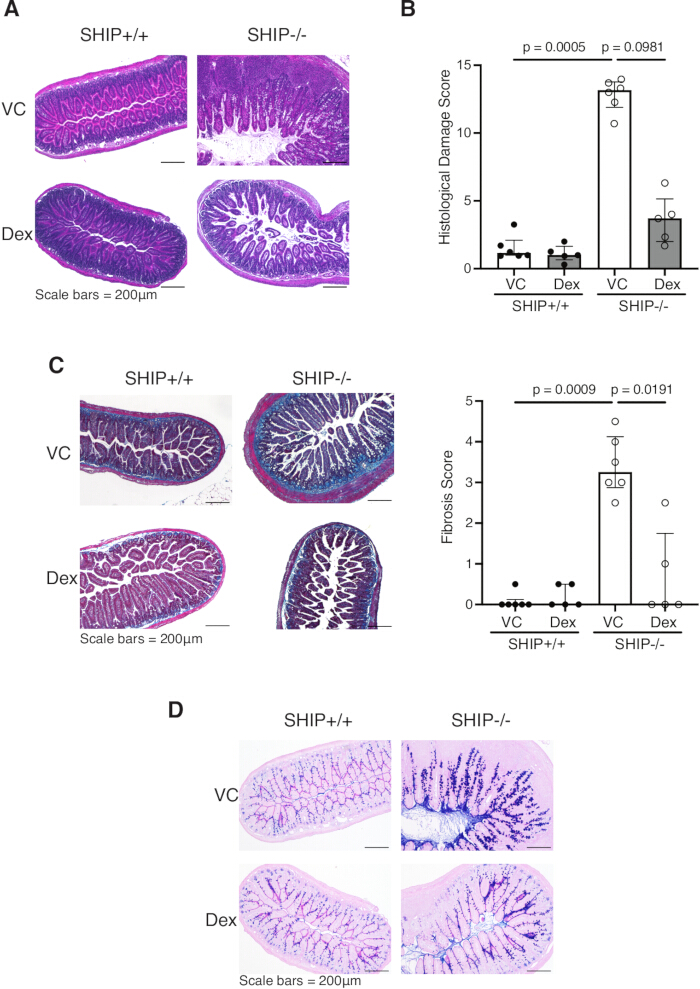
Figure 2: Dexamethasone reduces histological damage scores, collagen, muscle thickness, and goblet cell hyperplasia and hypertrophy in the distal ileum of SHIP-/- mice. SHIP+/+ and SHIP-/- mice were orally gavaged daily with either 0.5% β-cyclodextrin as a vehicle control (VC) or dexamethasone (Dex; 3 mg/kg) from 6 to 8 weeks of age. (A) H&E-stained ileal cross sections from mice were photographed at 10x magnification; scale bars = 200 µm. (B) Histological damage scores for SHIP+/+ and SHIP-/- mice expressed as median ± IQR. (C) Fixed ileal cross sections were stained with Masson's trichrome for fibrosis; collagen is stained blue. Images were taken at 10x magnification; scale bars = 200 µm. Fibrosis scores for SHIP+/+ and SHIP-/- mice expressed as median ± IQR. (D) Fixed ileal cross sections were stained with Alcian blue/PAS. Images were photographed at 10× magnification; scale bars = 200 µm. All sections are representative of n = 6 mice per group (except SHIP+/+ Dex and SHIP-/- Dex, n = 5). Statistical significance for the histological damage score was determined using the Kruskal-Wallis test followed by Dunn's multiple comparisons test. Please click here to view a larger version of this figure.
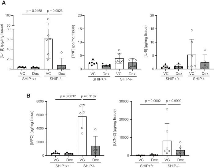
Figure 3: Dexamethasone reduces IL-1β concentrations in full-thickness ileal homogenates from SHIP-/- mice. SHIP+/+ and SHIP-/- mice were orally gavaged daily with either 0.5% β-cyclodextrin as a vehicle control (VC) or dexamethasone (Dex; 3 mg/kg) from 6 to 8 weeks of age. (A) IL-1β, TNF, and IL-6 concentrations were measured in full-thickness ileal homogenates. (B) Myeloperoxidase (MPO) and lipocalin-2 (LCN-2) concentrations were measured in full-thickness ileal tissue homogenates. Data are presented as mean ± SD. Statistical analyses were conducted using the Kruskal-Wallis test followed by Dunn's multiple comparisons test. Please click here to view a larger version of this figure.
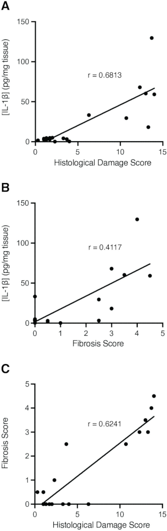
Figure 4: IL-1β concentrations positively correlate with histological damage and fibrosis and fibrosis correlates with histological damage score in SHIP-/- mice. Spearman correlation analysis was performed to assess relationships among histological damage score, fibrosis score, and IL-1β concentrations in full-thickness ileal tissue from SHIP-/- mice. Significant positive correlations were observed between (A) histological damage score and IL-1β concentrations (r = 0.6813), (B) fibrosis score and IL-1β concentrations (r = 0.4117), and (C) histological damage score and fibrosis score (r = 0.6241). Each data point represents an individual mouse. Please click here to view a larger version of this figure.
| Damage component | Score | |
| Loss of crypt architecture | 0 = none | |
| 1 = <25% loss | ||
| 2 = 25-50% loss | ||
| 3 = 50-75% loss | ||
| 4 = >75% loss | ||
| Immune cell infiltration | 0 = none | |
| 1 = occasional immune cell in lamina propria | ||
| 2 = increased immune cells in lamina propria | ||
| 3 = confluent immune cells in lamina propria and breaching mucosa | ||
| 4 = immune cell infiltration throughout the section | ||
| Goblet cell hyperplasia & hypertrophy | 0 = none | |
| 1 = <50% increase in goblet cell numbers and size | ||
| 2 = >50% increase in goblet cell numbers and size | ||
| Ulceration | 0 = none | |
| 1 = intermediate ulceration | ||
| 2 = substantial ulceration | ||
| Edema | 0 = none | |
| 1 = <50% of section | ||
| 2 = >50% of section | ||
| Muscle thickening | 0 = none | |
| 1 = intermediate thickening | ||
| 2 = substantial thickening | ||
Table 1: Histological damage scores. Histological damage of ileal cross sections from SHIP+/+ and SHIP-/- mice stained with H&E are scored based on the key features of ileitis that characterize the SHIP-/- mouse model, including loss of architecture (0-4), immune cell infiltration (0-4), goblet cell hyperplasia and hypertrophy (0-2), ulceration (0-2), edema (0-2), and muscle thickening (0-2).
| Fibrosis Component | Score | ||
| Excessive collagen | 0 = Normal architecture; no excess collagen | ||
| 1 = Mild increase in collagen; limited to submucosa or subserosa | |||
| 2 = Moderate collagen expansion into submucosa and partial muscularis propria, with disorganized bundles | |||
| 3 = Extensive, dense collagen replacing normal structure, involving submucosa and muscularis (transmural) | |||
| Fibromuscular hyperplasia/muscularization/muscular obliteration of the submucosa | 0 = None, normal muscle submucosa content | ||
| 1 = Mild increase in muscle content | |||
| 2 = Large amount of muscle in submucosa, affects the morphology but normal structure | |||
| 3= Muscle has obliterated the entire submucosal space | |||
Table 2: Fibrosis Scoring. Fibrosis in ileal cross sections from SHIP+/+ and SHIP-/- mice stained with Masson's trichrome was scored based on key features of intestinal fibrosis observed in the SHIP-/- model, including excessive collagen deposition (0-3) and fibromuscular hyperplasia or muscular obliteration of the submucosa (0-3). Collagen is assessed for extent and architectural disruption, while muscular changes are evaluated based on the degree of muscle infiltration and distortion of submucosal structure.
Discussion
This protocol outlines procedures for assessing ileitis and intestinal fibrosis in SHIP-/- mice, a murine model of CD-like intestinal inflammation with accompanying fibrotic pathology. The SHIP-/- mouse model recapitulates features of CD that are not commonly seen in other mouse models of intestinal inflammation, specifically the ileal localization, patchy and transmural inflammation, and fibrotic pathology19,26. Combined with the ease of breeding and short time to disease manifestation, the SHIP-/- model is well positioned as a powerful tool for translational research in CD19.
In this study, statistical significance for the FITC-dextran assay and histological scoring was assessed using the Kruskal-Wallis test, as these datasets did not meet the normality assumptions required for a one-way ANOVA, and histological scores represent discontinuous, non-parametric variables. While our analyses did not yield statistically significant differences between dexamethasone-treated and vehicle-treated groups, we observed clear trends. Dexamethasone reduced intestinal permeability and histopathology of SHIP-/- mice, and these effects may have reached significance with larger sample sizes.
Several technical challenges may arise when performing this protocol. With respect to tissue handling, SHIP-/- ilea require gentle manipulation during dissection as they are prone to tearing. This is particularly important when preparing tissues for gross imaging. Tissue fixation is another critical factor for histological analysis. Formalin-fixed tissue is suitable for both H&E and Masson's trichrome staining but is incompatible with Alcian Blue/PAS staining. In contrast, Carnoy's-fixed tissue is appropriate for H&E and Alcian Blue/PAS staining but not for Masson's trichrome. Carnoy's fixation leads to intense and diffuse dark blue staining, which obscures collagen and muscle layer visualization. For cytokine analysis, it is essential to process samples on ice as cytokines are highly sensitive to degradation. It is critical to aliquot clarified tissue homogenates immediately after processing. Repeated freeze-thaw cycles can substantially compromise cytokine integrity and undermine any potential differences between genotypes and/or treatment groups. Lastly, to ensure accurate quantification, we recommend performing multiple dilutions of each sample to confirm that the readings fall within the linear range of the assay's standard curve and to minimize potential interference from the complex tissue matrix. Addressing these technical considerations is essential to generate reproducible and interpretable results, but biological variability inherent to SHIP-/- mice must also be carefully managed.
There are several critical aspects of the SHIP-/- mice that should also be taken into consideration and carefully controlled to ensure reproducibility and minimize confounding variables. The gut microbiome has a profound impact on disease development and therapeutic responses in murine IBD models27,28. Our prior work has shown that intestinal inflammation in SHIP-/- mice is microbiota-dependent and can be ameliorated with antibiotics29. Therefore, littermate controls should be used to reduce microbiome-driven variability, and treatment groups must be balanced for cage origin30. In addition, careful sex matching is essential. Though no sex-based differences in penetrance or disease severity have been observed in SHIP-/- mice, other ileitis models such as SAMP1/YitFc, exhibit earlier onset and increased disease severity in females14,31. As sex hormones can also modulate microbial composition32, matching for sex is critical to minimize potential confounders in treatment studies. Moreover, individual treatments being assessed may have differential efficacy in male and female mice. Though our current findings do not reveal sex-related differences in disease or therapeutic response to dexamethasone, sex matching should be performed in all treatment studies.
In addition to the microbiome and sex considerations outlined above, it is important to include SHIP+/+ mice in treatment studies to evaluate the effects of the vehicle and therapeutic agents in the absence of intestinal inflammation. This ensures that neither the drug nor its delivery method causes or contributes to pathology in the healthy intestine, thereby improving interpretation of therapeutic efficacy and safety.
The timing of the treatment intervention is also essential and should be modified according to the experimental goals, as there are significant differences in both inflammation and fibrosis in mice at 4, 8, and 12 weeks of age19,26. SHIP-/- mice develop intestinal inflammation as early as 4 weeks of age, with all mice presenting with inflammation by 6 weeks of age, and markers of fibrotic pathology can be measured as early as 6 weeks of age and are well-established by 8 weeks of age19,26. Thus, treatments targeting ileal inflammation should begin at 6 weeks of age. Anti-fibrotic interventions can be initiated prophylactically at 6 weeks or therapeutically between 8-10 weeks. However, the onset and severity of disease may vary with the microbial environment, which differs between facilities. Investigators must first determine disease kinetics in their colony before implementing treatment protocols. Additionally, SHIP-/- mice can present up to a 17% reduction in weight by 4-5 weeks, with a further 5% by 8-10 weeks33. Animals exhibiting profound weight loss should be excluded from studies, as this reflects a severe manifestation of disease and may undermine treatment outcomes related to intestinal pathology.
An important consideration when analyzing disease progression in the SHIP-/- model is the selection of appropriate inflammatory markers. Based on our extensive experience with the SHIP-/- mouse, cytokine profiling is typically focused on specific mediators, such as IL-1β, that is known to be upregulated during disease progression. However, for initial characterization of inflammatory responses or when evaluating novel therapeutic strategies, a multiplex cytokine analysis is advisable. This unbiased approach enables broader profiling of the inflammatory milieu and may reveal unexpected targets relevant to disease pathogenesis or treatment response.
The SHIP-/- mouse model offers practical advantages that allow reproducibility and efficiency. SHIP-/- mice develop spontaneous and consistent ileal inflammation and fibrosis without the need for exogenous triggers. This eliminates variability associated with reagent preparation and non-standardized dosing protocols when inducing intestinal inflammation. Disease onset is rapid, with inflammation evident by 4-6 weeks of age and fibrosis by 8 weeks of age, making the model both time- and cost-efficient. Additionally, SHIP-/- mice are easy to breed and maintain, genotyping is straightforward, and no specialized breeding strategies are required. This simplifies colony management, reduces technical workload, and lowers the risk of introducing unintended genetic variability19,21,26. Disease can be reliably assessed using widely available techniques in most research settings, including the FITC-dextran assay, H&E and Masson's trichrome staining, and ELISA. These combined features support efficient study design, lower financial and technical barriers, and promote consistent data generation between laboratories.
While the SHIP-/- model presents unique advantages, the model also has limitations. As with all preclinical models, it cannot fully replicate the complex, multifactorial nature of human IBD, which is influenced by genetic, immune, and environmental factors34. The DSS and TNBS models remain widely popular due to their simplicity and cost-effectiveness. When combined with genetic modifications, these chemical models can serve as effective tools for understanding gene-specific contributions to barrier function35. However, their main utility lies in modeling acute epithelial injury; they fall short of capturing the chronic immune-mediated pathology that characterizes CD, and their mechanism of epithelial damage induced by chemical agents that lack direct clinical relevance36. Exclusive reliance on one model may result in an incomplete and/or misleading assessment of therapeutic potential, and it is recommended to use multiple models to be assured of treatment efficacy.
Disclosures
The authors declare no conflicts of interest.
Acknowledgements
This work is supported by the Animal Care facility at BC Children's Hospital Research Institute and funding from Natural Sciences and Engineering Research Council of Canada through the Canadian Glycomics Network (GlycoNet, A Network of Centre of Excellence) and the Canadian Institutes of Health Research (MOP-133607 to LS). K.S. and W.M. are recipients of the TRIANGLE (TRaIning a New generation of researchers in Gastroenterology and LivEr program) Doctoral scholarships.
Materials
| Name | Company | Catalog Number | Comments |
|---|---|---|---|
| 1.7mL Snap Cap Microtube | Diamed | SPE155-N | |
| 1x DPBS (no calcium chloride, no magnesium chloride) | Gibco | REF 14190-144 | |
25G x  ” needles ” needles | BD | REF 305122 | For cardiac puncture |
26G x  ” needles ” needles | BD | REF 305110 | Intestinal flushing |
| Acetic acid | Sigma-Aldrich | A6283 | Histology |
| Acid citrate dextrose solution (38mM citric acid, 107 mM sodium citrate, 136 mM dextrose) | Millipore Sigma | C3821 | |
| Animal feeding needle, non-sterile, reusable, type 304SS | Millipore Sigma | CAD7900 | For gavage Straight Size: 24G, Lxdiam. 1 in. x 1.25 mm, ball |
| Aprotinin | Cayman Chemical | 9087-70-1 | Homogenization buffer |
| BD Luer-Lok Syringe sterile, single use, 5 mL | BD | REF 309646 | Intestinal flushing |
| BD OptEIA Mouse IL-6 ELISA Set | BD Biosciences | 555240 | |
| BD OptEIA Mouse TNF ELISA Set II | BD Biosciences | 558534 | |
| BD® Luer Slip Tip Syringe sterile, single use, 1 mL | BD | REF 309659 | |
| Biopsy Foam Pads Square, 30.2 x 25.4 x 2 mm H. | Simport Scientific | REF M476-1 | Histology |
| Buffered Formalde-Fresh (Low Odor 10% Formalin) | Fisher Chemical | SF93-4 | Histology |
| Clear Flat-Bottom Immuno Nonsterile 96-Well Plates | Thermo Scientific | 439454 | |
| Eppendorf 5415R Refrigerated Centrifuge | Marshall Scientific | EP-5415R | |
| Ethyl Alcohol Anhydrous | Greenfield Global | P016EAAN | Histology |
| Fisherbrand TRU-FLOW Tissue Cassette | Fisher Scientific | 15-200-403 | Histology |
| FITC-dextran, 3-5kDa (1g) | Millipore Sigma | FD4-1G CAS Number: 60842-46-8 | |
| Leupeptin | Millipore Sigma | 103476-89-7 | Homogenization buffer |
| Mouse IL-1 beta/IL-1F2 DuoSet ELISA | R&D Systems | DY401 | |
| Multiscreen 96-well plate, solid bottom | Millipore Sigma | MSSWNFX40 | White, non-sterile, non-treated |
| OmniPur Chloroform | Millipore Sigma | 3150 | Histology |
| Polytron PT-MR2100 homogenizer | Kinematica AG | 632081 | |
| Varioskan Lux Multimode Microplate Reader | Thermo Scientific | VL0L00D0 |
References
- Khor, B., Gardet, A., Xavier, R. J. Genetics and pathogenesis of inflammatory bowel disease. Nature. 474 (7351), 307-317 (2011).
- Ng, S. C., et al. Worldwide incidence and prevalence of inflammatory bowel disease in the 21st century: a systematic review of population-based studies. Lancet. 390 (10114), 2769-2778 (2017).
- Wang, R., Li, Z., Liu, S., Zhang, D. Global, regional and national burden of inflammatory bowel disease in 204 countries and territories from 1990 to 2019: a systematic analysis based on the Global Burden of Disease Study 2019. BMJ Open. 13 (1), e065186(2023).
- Xavier, R. J., Podolsky, D. K. Unravelling the pathogenesis of inflammatory bowel disease. Nature. 448 (7152), 427-434 (2007).
- Guan, Q. A comprehensive review and update on the pathogenesis of inflammatory bowel disease. J Immunol Res. 2019, 7247238(2019).
- Chang, J. T. Pathophysiology of inflammatory bowel diseases. N Engl J Med. 383 (27), 2652-2664 (2020).
- Graham, D. B., Xavier, R. J. Pathway paradigms revealed from the genetics of inflammatory bowel disease. Nature. 578 (7796), 527-539 (2020).
- Li, J., et al. Pathogenesis of fibrostenosing Crohn's disease. Transl Res. 209, 39-54 (2019).
- Rieder, F., Mukherjee, P. K., Massey, W. J., Wang, Y., Fiocchi, C. Fibrosis in IBD: from pathogenesis to therapeutic targets. Gut. 73 (5), 854-866 (2024).
- D'Alessio, S., et al. Revisiting fibrosis in inflammatory bowel disease: the gut thickens. Nat Rev Gastroenterol Hepatol. 19 (3), 169-184 (2022).
- Louis, E., et al. Behaviour of Crohn's disease according to the Vienna classification: changing pattern over the course of the disease. Gut. 49 (6), 777-782 (2001).
- Iliopoulou, L., Kollias, G. Harnessing murine models of Crohn's disease ileitis to advance concepts of pathophysiology and treatment. Mucosal Immunol. 15 (1), 10-26 (2022).
- Rivera-Nieves, J., et al. Emergence of perianal fistulizing disease in the SAMP1/YitFc mouse, a spontaneous model of chronic ileitis. Gastroenterology. 124 (4), 972-982 (2003).
- Pizarro, T. T., et al. SAMP1/YitFc mouse strain: a spontaneous model of Crohn's disease-like ileitis. Inflamm Bowel Dis. 17 (12), 2566-2584 (2011).
- Kontoyiannis, D., Pasparakis, M., Pizarro, T. T., Cominelli, F., Kollias, G. Impaired on/off regulation of TNF biosynthesis in mice lacking TNF AU-rich elements: implications for joint and gut-associated immunopathologies. Immunity. 10 (3), 387-398 (1999).
- Steiner, C. A., et al. The TNFΔARE mouse as a model of intestinal fibrosis. Am J Pathol. 193 (8), 1013-1028 (2023).
- Li, J., et al. Mouse models of intestinal fibrosis. Methods Mol Biol. 2299, 385-403 (2021).
- Pauls, S. D., Marshall, A. J. Regulation of immune cell signaling by SHIP1: a phosphatase, scaffold protein, and potential therapeutic target. Eur J Immunol. 47 (6), 932-945 (2017).
- McLarren, K. W., et al. SHIP-deficient mice develop spontaneous intestinal inflammation and arginase-dependent fibrosis. Am J Pathol. 179 (1), 180-188 (2011).
- Kerr, W. G., Park, M. Y., Maubert, M., Engelman, R. W. SHIP deficiency causes Crohn's disease-like ileitis. Gut. 60 (2), 177-188 (2011).
- Ngoh, E. N., et al. Activity of SHIP, which prevents expression of interleukin 1β, is reduced in patients with Crohn's disease. Gastroenterology. 150 (2), 465-476 (2016).
- Fernandes, S., et al. SHIP1 deficiency in inflammatory bowel disease is associated with severe Crohn's disease and peripheral T cell reduction. Front Immunol. 9, 9(2018).
- Ma, W. J., et al. Bespoke plant glycoconjugates for gut microbiota-mediated drug targeting. Science. 388 (6706), 1410-1416 (2025).
- Park, M. Y., et al. Impaired T-cell survival promotes mucosal inflammatory disease in SHIP1-deficient mice. Mucosal Immunol. 7 (6), 1429-1439 (2014).
- Tsai, K., et al. Highly sensitive, flow cytometry-based measurement of intestinal permeability in models of experimental colitis. Cell Mol Gastroenterol Hepatol. 15 (2), 425-438 (2023).
- Lo, Y., Sauve, J. P., Menzies, S. C., Steiner, T. S., Sly, L. M. Phosphatidylinositol 3-kinase p110δ drives intestinal fibrosis in SHIP deficiency. Mucosal Immunol. 12 (5), 1187-1200 (2019).
- Sellon, R. K., et al. Resident enteric bacteria are necessary for development of spontaneous colitis and immune system activation in interleukin-10-deficient mice. Infect Immun. 66 (11), 5224-5231 (1998).
- Taurog, J. D., et al. The germfree state prevents development of gut and joint inflammatory disease in HLA-B27 transgenic rats. J Exp Med. 180 (6), 2359-2364 (1994).
- Dobranowski, P. A., Tang, C., Sauvé, J. P., Menzies, S. C., Sly, L. M. Compositional changes to the ileal microbiome precede the onset of spontaneous ileitis in SHIP-deficient mice. Gut Microbes. 10 (5), 578-598 (2019).
- Robertson, S. J., et al. Comparison of co-housing and littermate methods for microbiota standardization in mouse models. Cell Rep. 27 (6), 1910-1919.e2 (2019).
- Goodman, W. A., et al. Loss of estrogen-mediated immunoprotection underlies female gender bias in experimental Crohn's-like ileitis. Mucosal Immunol. 7 (5), 1255-1265 (2014).
- Org, E., et al. Sex differences and hormonal effects on gut microbiota composition in mice. Gut Microbes. 7 (4), 313-322 (2016).
- Helgason, C. D., et al. Targeted disruption of SHIP leads to hemopoietic perturbations, lung pathology, and a shortened life span. Genes Dev. 12 (11), 1610-1620 (1998).
- Boyapati, R., Satsangi, J., Ho, G. T. Pathogenesis of Crohn's disease. F1000Prime Rep. 7, 44(2015).
- Mizoguchi, E., Low, D., Ezaki, Y., Okada, T. Recent updates on the basic mechanisms and pathogenesis of inflammatory bowel diseases in experimental animal models. Intest Res. 18 (2), 151-167 (2020).
- Koboziev, I., Karlsson, F., Zhang, S., Grisham, M. B. Pharmacological intervention studies using mouse models of the inflammatory bowel diseases: translating preclinical data into new drug therapies. Inflamm Bowel Dis. 17 (6), 1229-1245 (2011).
- Raftery, A. L., Pattaroni, C., Harris, N. L., Tsantikos, E., Hibbs, M. L. Environmental and inflammatory factors influencing concurrent gut and lung inflammation. Inflamm Res. 73 (12), 2123-2139 (2024).
- Maxwell, M. J., et al. Genetic segregation of inflammatory lung disease and autoimmune disease severity in SHIP-1-/- mice. J Immunol. 186 (12), 7164-7175 (2011).
Reprints and Permissions
