This protocol introduces a scalp acupuncture-based method for localizing the primary motor cortex (M1) hotspot for the flexor digitorum superficialis (FDS) muscle and validates its effectiveness against the conventional C3 site.
A subscription to JoVE is required to view this content. Sign in or start your free trial.
Method Article
A Novel Scalp Acupuncture-based Method to Target the Hand Motor Hotspot for Non-invasive Brain Stimulation
In This Article
Summary
Abstract
The precise localization of the primary motor cortex (M1) corresponding to specific muscles is critical for enhancing the efficacy of neuromodulation. When navigated transcranial magnetic stimulation (nTMS) or other mapping tools are unavailable, stimulation is typically applied at the C3 site of the standard 10-20 EEG system. However, emerging evidence indicates that C3 may not be the optimal site for targeting hand muscles. Therefore, the goal of this study is to propose an innovative neuroanatomy-based localization method to identify the M1 hotspot for the hand muscle and to validate its effectiveness compared with the C3 site. A step-by-step protocol was developed to identify the M1 hotspot for the flexor digitorum superficialis (FDS) muscle using an acupuncture-based approach. The identified hotspot was then compared with the conventional C3 site by evaluating baseline and post-neuromodulation motor evoked potentials (MEPs). Representative data demonstrated that the proposed M1 hotspot yielded significantly higher MEP amplitudes than the C3 site, both at the baseline and following neuromodulation. The proposed approach provides a validated and practical method for localizing the M1 hotspot for the FDS. This technique has the potential to improve the effectiveness of neuromodulation in clinical practice when advanced neuroimaging tools are not accessible.
Introduction
Precisely localizing the cortical hotspot in the primary motor cortex (M1) for neuromodulation targeting specific muscles is critical for optimizing motor rehabilitation outcomes1. Navigated transcranial magnetic stimulation (nTMS) has been adopted as a non-invasive reliable methodology for motor mapping compared to direct cortical stimulation2. However, in the absence of such tools, C3 (left hemisphere) and C4 (right hemisphere), defined by the standard 10-20 EEG system, are commonly used as M1 targeting for delivering neuromodulation3. However, studies have indicated that C3 may not represent the actual M1 hotspot4,5, and may not be the optimal site for delivering neuromodulation, as it yields lower motor evoked potential (MEP) amplitudes compared to alternative sites in the surrounding region6. In addition, high-definition transcranial electrical stimulation (HD-tES) has the potential to enhance its modulatory effects7,8,9,10; however, it requires more precise localization of the targeted stimulation area compared to conventional montages11,12. Therefore, there is a need for a method to accurately identify the M1 hotspot when advanced neuroimaging tools are not accessible.
Chinese scalp acupuncture is a contemporary acupuncture technique integrating traditional Chinese needling methods with modern neuroanatomy of representative areas of the cerebral cortex13. In contrast to traditional acupuncture, scalp acupuncture involves subcutaneous needle insertion across defined zones that align with cortical regions responsible for central nervous system functions13. Several scalp acupuncture systems have been developed and utilized for the treatment of motor impairments, including those established by Jiao, Lin, Jin, and Tang, as well as the internationally standardized China scalp-point protocol14. Among these approaches, Jiao's scalp acupuncture integrates modern neuroanatomy and neurophysiological principles with traditional Chinese acupuncture techniques, creating an innovative method for modulating central nervous system function15,16. However, whether its concepts can be applied and validated in upper limb neuromodulation studies remains unknown.
Therefore, this protocol aims to explore a new approach for localizing the M1 hotspot of the flexor digitorum superficialis (FDS) muscle using scalp acupuncture landmarks (AC) and to validate this approach by comparing corticospinal excitability with that of the conventional C3 site before and after HD-tES intervention. However, the current approach has only been validated in healthy participants with clearly identifiable scalp anatomical landmarks and for the FDS muscle specifically. Whether this method can be generalized to other upper limb muscles or applied to individuals with neurological impairments remains uncertain.
Access restricted. Please log in or start a trial to view this content.
Protocol
Participants were right-handed healthy adults with no history of neurological or musculoskeletal disorders affecting the upper limbs. The research protocol was reviewed and approved by the Committee for the Protection of Human Subjects (CPHS) at The University of Texas Health Science Center at Houston. Written informed consent was obtained from all participants prior to participation.
1. Preparation
- Ensure the participants sit upright in a comfortable position with arms resting naturally. The M1 FDS hotspot localization protocol requires a tape measure and a marker. The process to localize the hotspot for FDS should follow the contemporary scalp acupuncture strategy.
2. Localize the upper motor point (UMP)
NOTE: The Upper Motor Point (UMP) is defined as the point located 0.5 cm posterior to the midpoint of the anterior-posterior line, which extends from the glabella (the midpoint between the eyebrows) to the inion.
- Align the "0" mark of the tape measure with the glabella (Figure 1A) and ensure the tape is held straight and level across the scalp.
- Stretch the tape to the inion, which is the tip of the external occipital protuberance (Figure 1B).
- Identify the midpoint between the glabella and the inion along the longitudinal line. From this point, measure 0.5 cm posteriorly and mark the location with a visible dot.
- Align the tape between the left and right preauricular points (or the apex of the ears) (Figure 1C), ensuring the tape is level. Mark the midpoint of this transverse line with a visible dot.
- Using the midpoint marked in step 2.4 as a reference, adjust the point identified in step 2.3 laterally, if necessary, to ensure that the final UMP is centered on the transverse midpoint (Figure 1D).

Figure 1: Localizing the Upper Motor Point. (A) The midpoint between the eyebrows (Glabella). (B) The external occipital protuberance located at the midline of the occipital bone (Inion). (C) The left preauricular point, located anterior to the tragus of the ear. (D) Located 0.5 cm posterior to the intersection of the mid-sagittal line and the transverse line connecting the left and right preauricular points; this point is defined as the Upper Motor Point (UMP). Please click here to view a larger version of this figure.
3. Localize the lower motor point (LMP)
NOTE: Lower Motor Point (LMP) is defined as the intersection of the line extending from the lateral end of the eyebrow to the inion with the anterior hairline.
- Align the "0" mark of the tape measure to the lateral end of the eyebrow on the left side of the face (Figure 2A).
- Stretch the tape measure from this point to the inion, following a straight diagonal path across the scalp (Figure 2A).
- Identify and mark with a visible dot the point where this diagonal line intersects the anterior hairline. This point is defined as the LMP (Figure 2B).
4. Localize the M1 FDS hotspot (AC)
NOTE: The M1 FDS hotspot is defined as the point located at two-fifths of the distance from the Upper Point (Up) to the Lower Motor Point (LMP) along the motor area line (Figure 2C).
- Align the "0" mark of the tape measure with the Upper Motor Point (UMP) and extend the tape measure to the Lower Motor Point (LMP).
- Record the total distance between the UMP and LMP.
- Measure and mark with a visible dot the point located at 2/5 of this distance from the UMP. This point is defined as the AC hotspot.
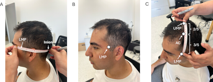
Figure 2: Localizing the Lower Motor Point and the FDS hotspot. (A) The intersection of the line extending from the lateral end of the eyebrow to the inion with the anterior hairline. (B) The Lower Motor Point (LMP). (C) The Upper Motor Point (UMP), the FDS hotspot (AC), and the LMP. Please click here to view a larger version of this figure.
5. M1 FDS Hotspot (AC) validation
NOTE: Instruct the participants to sit in a chair adjusted to a comfortable height, put their right hands on a soft pillow, and instruct them to relax during the stimulation session.
- Place bipolar surface electromyography (EMG) electrodes over the belly of the right flexor digitorum superficialis (FDS) muscle, and set the sampling frequency to 2 kHz (Figure 3A).
- Mark the C3 site based on the International 10-20 EEG system17.
- Mark the FDS hotspot (AC) using the proposed approach.
- Position the TMS coil tangentially over the scalp at the AC site, with the handle directed backward at a 45° angle (Figure 3B).
- Begin at 30% of the maximum TMS output and deliver single-pulse stimulation.
- Gradually increase the intensity to determine the resting motor threshold (RMT)18..
- Set the stimulation intensity to 120% of the RMT and record at least 20 consecutive MEPs at both the AC and C3 sites.
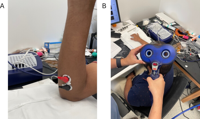
Figure 3: EMG electrode and TMS coil placement. (A) The bipolar EMG electrode placement at FDS and (B) the placement of single-pulse TMS at M1 FDS hotspot. Please click here to view a larger version of this figure.
6. HD-tES intervention
- Instruct the participants to sit comfortably in a chair with their hands resting on a table or armrest.
- Fit the EEG cap according to the standard protocol17, ensuring proper alignment with scalp landmarks (nasion and inion).
- Position one electrode over the target site (AC or C3) and place four reference electrodes evenly around the target area to ensure stable current distribution (Figure 4). Apply conductive gel to all electrode sites to reduce impedance.
- Check electrode impedance using the stimulator's built-in impedance meter, and ensure that values are below 2 kΩ.
- Set the stimulation parameters to 2 mA intensity and 21 Hz frequency for a duration of 20 min.
- Turn on the HD-tES and gradually ramp the intensity to 2 mA over 30 s.
- Monitor the participant's reactions throughout the stimulation, checking for signs of discomfort, skin irritation, or unusual sensations.
- After 20 min, turn off the device, carefully remove the electrodes, and clean the scalp areas.
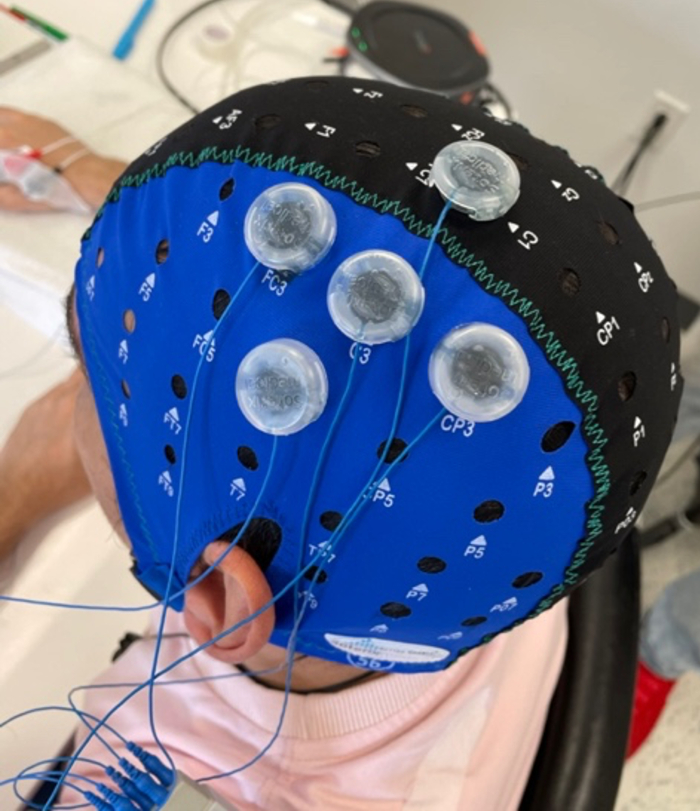
Figure 4: The setup of HD-tES. The center electrode is targeting at C3 or AC with 4 reference electrodes located at the surrounding channels. Please click here to view a larger version of this figure.
Access restricted. Please log in or start a trial to view this content.
Results
The results revealed three key findings: i) The proposed method (AC), based on neuroanatomy-guided scalp acupuncture, successfully identified the M1 hotspot for the FDS muscle, as it overlapped with the TMS-guided hotspot. ii) The identified M1 FDS hotspot did not overlap with the conventionally targeted C3 site; moreover, the AC site exhibited higher baseline MEP amplitudes compared to C3. iii) HD-tACS intervention at the AC site significantly increased MEP amplitudes, whereas no significant change was observed followin...
Access restricted. Please log in or start a trial to view this content.
Discussion
In this study, an innovative approach was developed to localize the M1 hotspot for the FDS muscle based on scalp acupuncture landmarks. The proposed AC site showed strong potential as the M1 hotspot for the FDS muscle, with higher baseline and post-modulation MEP amplitudes compared to the conventionally used C3 site.
The corticospinal tract is one of the most critical descending pathways originating from the motor cortex for voluntary motor behavior20,
Access restricted. Please log in or start a trial to view this content.
Disclosures
The authors declare that they have no known competing financial interests or personal relationships that could have appeared to influence the work reported in this paper.
Acknowledgements
The authors are grateful to Dr. Yingchun Zhang, Professor in the Department of Biomedical Engineering at the University of Miami, for providing valuable guidance and expertise in conducting the EMG analysis.
Access restricted. Please log in or start a trial to view this content.
Materials
| Name | Company | Catalog Number | Comments |
|---|---|---|---|
| Electrode Gel | Soterix Medical Inc., New York, USA | https://soterixmedical.com/research/hd-tdcs/accessories/hd-gel | |
| EMG | TMSi, Oldenzaal, The Netherlands | https://www.tmsi.artinis.com/product-overview | |
| High-definition transcranial electrical stimulation (HD-tES) | Soterix Medical Inc., New York, USA | https://soterixmedical.com/research/hd-tdcs/4x1 | |
| TMS (Magstim Bistim² 200) | Magstim Company Ltd., Whitland, Wales, UK | 3231-00 |
References
- Rossini, P. M., et al. Non-invasive electrical and magnetic stimulation of the brain, spinal cord, roots and peripheral nerves: basic principles and procedures for routine clinical and research application. An updated report from an IFCN committee. Clin Neurophysiol. 126 (6), 1071-1107 (2015).
- Jeltema, H. R., et al. Comparing navigated transcranial magnetic stimulation mapping and "gold standard" direct cortical stimulation mapping in neurosurgery: a systematic review. Neurosurg Rev. 44 (4), 1903-1920 (2021).
- Tang, X., et al. Transcranial direct current stimulation for upper extremity motor dysfunction in poststroke patients: a systematic review and meta-analysis. Clin Rehabil. 38 (6), 749-769 (2024).
- Silva, L. M., Silva, K. M. S., Lira-Bandeira, W. G., Costa-Ribeiro, A. C., Araujo-Neto, S. A. Localizing the primary motor cortex of the hand by the 10-5 and 10-20 systems for neurostimulation: an MRI study. Clin EEG Neurosci. 52 (6), 427-435 (2021).
- Sparing, R., Buelte, D., Meister, I. G., Paus, T., Fink, G. R. Transcranial magnetic stimulation and the challenge of coil placement: a comparison of conventional and stereotaxic neuronavigational strategies. Hum Brain Mapp. 29 (1), 82-96 (2008).
- Kim, H., Wright, D. L., Rhee, J., Kim, T. C3 in the 10-20 system may not be the best target for the motor hand area. Brain Res. 1807, 148311(2023).
- Roy, A., Baxter, B., He, B. High-definition transcranial direct current stimulation induces both acute and persistent changes in broadband cortical synchronization: a simultaneous tDCS-EEG study. IEEE Trans Biomed Eng. 61 (7), 1967-1978 (2014).
- Meng, H., et al. High-definition transcranial alternating current stimulation (HD-tACS) has regional neuromodulation effects in the forearm and hand muscles. Brain Stimul. 18 (1), 94-96 (2025).
- Masina, F., et al. Neurophysiological and behavioural effects of conventional and high-definition tDCS. Sci Rep. 11 (1), 7659(2021).
- Parlikar, R., et al. High-definition transcranial direct current stimulation (HD-tDCS): a systematic review on the treatment of neuropsychiatric disorders. Asian J Psychiatr. 56, 102542(2021).
- Alam, M., Truong, D. Q., Khadka, N., Bikson, M. Spatial and polarity precision of concentric high-definition transcranial direct current stimulation (HD-tDCS). Phys Med Biol. 61 (12), 4506-4521 (2016).
- Villamar, M. F., et al. Technique and considerations in the use of 4×1 ring high-definition transcranial direct current stimulation (HD-tDCS). J Vis Exp. (77), e50309(2013).
- Hao, J. J., Hao, L. L. Review of clinical applications of scalp acupuncture for paralysis: an excerpt from Chinese scalp acupuncture. Glob Adv Health Med. 1 (1), 102-121 (2012).
- Lee, G. E., Lee, P. T., Ran, N., Zhou, J. Scalp acupuncture for children with cerebral palsy: a protocol for a systematic review. Medicine (Baltimore). 98 (48), e18062(2019).
- Wang, J., et al. Acupuncture treatment on the motor area of the scalp for motor dysfunction in children with cerebral palsy: study protocol for a multicenter randomized controlled trial. Trials. 21 (1), 29(2020).
- Wu, Y., et al. Potential scalp acupuncture and brain stimulation targets for common neurological disorders: evidence from neuroimaging studies. Chin Med. 20 (1), 58(2025).
- American Electroencephalographic Society. Guideline thirteen: guidelines for standard electrode position nomenclature. J Clin Neurophysiol. 11 (1), 111-113 (1994).
- Westin, G. G., Bassi, B. D., Lisanby, S. H., Luber, B. Determination of motor threshold using visual observation overestimates transcranial magnetic stimulation dosage: safety implications. Clin Neurophysiol. 125 (1), 142-147 (2014).
- Meng, H., Houston, M., Francisco, G. E., Zhang, Y., Li, S. Scalp acupuncture guidance for identifying the optimal site for transcranial electrical stimulation of the hand. Exp Brain Res. 242 (9), 2083-2091 (2024).
- Lemon, R. N. Descending pathways in motor control. Annu Rev Neurosci. 31, 195-218 (2008).
- Liu, R., Moe, A. A. K., Liu, W., Zoghi, M., Jaberzadeh, S. Does acupuncture at motor-related acupoints affect corticospinal excitability? a systematic review and meta-analysis. J Integr Med. 23 (2), 113-125 (2025).
- Song, W., Amer, A., Ryan, D., Martin, J. H. Combined motor cortex and spinal cord neuromodulation promotes corticospinal system functional and structural plasticity and motor function after injury. Exp Neurol. 277, 46-57 (2016).
- Krucoff, M. O., Rahimpour, S., Slutzky, M. W., Edgerton, V. R., Turner, D. A. Enhancing nervous system recovery through neurobiologics, neural interface training, and neurorehabilitation. Front Neurosci. 10, 584(2016).
- Søndergaard, R. E., Martino, D., Kiss, Z. H. T., Condliffe, E. G. TMS motor mapping methodology and reliability: a structured review. Front Neurosci. 15, 709368(2021).
- Wang, T. Acupuncture for brain. , Springer. Cham. (2021).
- Meng, H., et al. Efficacy of high-definition transcranial alternating current stimulation (HD-tACS) at the M1 hotspot versus C3 site in modulating corticospinal tract excitability. Biomedicines. 12 (11), 2635(2024).
Access restricted. Please log in or start a trial to view this content.
Reprints and Permissions
