This protocol provides a step-by-step guide for performing transforaminal full-endoscopic foraminotomy under local anesthesia. This motion-preserving technique is presented as a minimally invasive surgical alternative to fusion extension for treating L5/S1 foraminal stenosis in patients with adjacent segment disease.
Method Article
Transforaminal Full-Endoscopic Lumbar Foraminotomy Under Local Anesthesia for L5/S1 Adjacent Segment Foraminal Stenosis
In This Article
Summary
Abstract
Foraminal stenosis in patients with L5/S1 adjacent segment disease (ASD) presents a significant clinical challenge, as conventional treatment often requires fusion extension surgery. This approach sacrifices spinal mobility and is associated with considerable surgical morbidity. This video article describes a motion-preserving surgical alternative, namely, transforaminal full-endoscopic foraminotomy performed under local anesthesia, detailing a step-by-step protocol to navigate the challenges of these revision cases. The technique utilizes a posterolateral corridor, avoiding prior surgical scarring. Key procedural steps include meticulous preoperative trajectory planning, foraminoplasty via resection of the superior articular process (SAP), and partial resection of the inferior articular process to fully expose the ligamentum flavum (LF). A specific "detach" technique is then demonstrated, involving undercutting the ventral rim of the S1 SAP to release the LF before its removal. Representative results demonstrate successful osseous decompression confirmed by postoperative computed tomography. The procedure resulted in immediate improvement in radicular pain and motor weakness, with the patient's visual analog scale score decreasing from 9/10 to 1/10 at the one-month follow-up. This minimally invasive technique provides effective neural decompression and facilitates rapid recovery, offering a valuable alternative to more extensive surgery for this challenging patient population.
Introduction
Lumbar spinal stenosis is a common condition with a significant impact on quality of life, and foraminal stenosis is a frequent subtype causing radicular symptoms due to nerve root compression1,2. A particularly challenging scenario arises in patients with Adjacent Segment Disease (ASD), a long-term complication following lumbar fusion surgery. ASD frequently manifests as new-onset foraminal stenosis at levels adjacent to the fused segments, with a reported incidence of up to 30% within five years post-surgery3.
Traditionally, revision surgeries for symptomatic ASD have involved open decompression with fusion extension4,5. However, this approach is associated with considerable surgical morbidity, increased blood loss, and prolonged recovery, especially in elderly patients with comorbidities6. Consequently, minimally invasive spine surgery (MISS) techniques have gained traction. Among these, full-endoscopic spine surgery (FESS) offers the potential for adequate neural decompression with minimal tissue disruption, reduced postoperative pain, and faster recovery, often with shorter operative times7,8. The transforaminal endoscopic approach, in particular, provides direct access to the foramen, making it suitable for treating foraminal stenosis9. Despite its benefits, addressing L5/S1 foraminal stenosis via a transforaminal endoscopic approach presents unique anatomical challenges, primarily due to the presence of a high iliac crest, a narrow foramen, and in cases with ASD, altered anatomy or scar tissue from previous surgeries1,10. Performing such procedures under local anesthesia further enhances patient safety by allowing for intraoperative patient feedback, and is particularly beneficial for patients who are poor candidates for general anesthesia11,12.
This article provides a comprehensive, step-by-step demonstration of our updated transforaminal full-endoscopic lumbar foraminotomy technique. This method is specifically indicated for patients with unilateral radiculopathy due to L5/S1 foraminal stenosis adjacent to a prior lumbar fusion, who do not present with significant segmental instability. Our protocol highlights key surgical maneuvers, including precise superior articular process (SAP) resection and use of the "detach technique" for safe and complete ligamentum flavum removal, to achieve thorough neural decompression in these challenging revision scenarios13.
Protocol
All procedures described herein were performed after obtaining written informed consent from the patient and with the approval of the Institutional Review Board of Tokushima University Hospital. Preoperative images of the lumbar spine, including plain radiographs, computed tomography (CT) scans, and magnetic resonance images, were reviewed for each patient to confirm the diagnosis of L5/S1 foraminal stenosis adjacent to prior lumbar fusion and to meticulously plan the surgical trajectory, with a focus on the iliac crest and any anatomical variations due to previous surgery.
Inclusion criteria
Patients were included in this study if they met the following criteria: (1) Symptomatic unilateral radiculopathy corresponding to the L5 nerve root; (2) Radiological evidence of L5/S1 foraminal stenosis confirmed by magnetic resonance imaging (MRI) and CT; (3) A history of previous lumbar fusion at a superior level (L4/5 or higher), consistent with ASD; (4) Failure to respond to at least 3 months of conservative management, including medication, physical therapy, and/or nerve root blocks; (5) Provision of written informed consent.
Exclusion criteria
Patients were excluded from this study if they presented with any of the following: (1) Evidence of gross segmental instability (e.g., degenerative spondylolisthesis > Grade I) at the L5/S1 level on dynamic radiographs; (2) Predominant symptoms caused by central canal stenosis rather than foraminal stenosis; (3) Active spinal infection, tumor, or significant systemic disease; (4) Severe scoliosis or other spinal deformities that would preclude a safe transforaminal approach; (5) Bilateral foraminal stenosis requiring treatment of both sides in the same session.
1. Preoperative preparation
- Trajectory planning and patient preparation
- Meticulously plan the skin entry point and needle trajectory using axial CT or MRI before surgery.
- First, determine the optimal disc puncture site (target point "a") on the disc annulus, typically on a line connecting the medial borders of the superior and inferior pedicles on an anteroposterior (AP) view and at the level of the superior endplate of the caudal vertebra on a lateral view.
- Next, draw a line tangential from this target point ("a") passing the SAP; the intersection of this line with the skin surface defines the skin puncture point ("b"). Measure the distance from the midline (spinous process) to point "b" (Figure 1). This detailed "preoperative drawing" is critical for L5/S1 transforaminal full-endoscopic spine surgery (TF-FESS) due to the narrow operative corridor, which is often constrained by a high iliac crest or osteophytes, particularly in ASD. The trajectory aims for the SAP to ensure a safe bony landmark for initial docking.
NOTE: During this planning phase, carefully evaluate the preoperative CT to assess the influence of existing implants. Metal artifacts from pedicle screws can obscure anatomical landmarks, making trajectory planning difficult. If a significant artifact is present, consider using CT with metal artifact reduction software or cross-reference with MRI to accurately visualize the safe corridor and avoid interference with implants.
- Perform a discography if necessary to confirm the patient's ability to maintain a prone position and to ensure the disc can be accessed without issue.
- Administer appropriate intravenous antibiotic prophylaxis approximately 30 min before the planned skin incision, according to institutional guidelines, to minimize the risk of surgical site infection.
- Meticulously plan the skin entry point and needle trajectory using axial CT or MRI before surgery.
- Conscious sedation and monitoring
- Once the patient is in the prone position on the operating table, administer half an ampule of hydroxyzine hydrochloride and half an ampule of pentazocine intravenously for anxiolysis and analgesia. Continuously monitor vital signs (including blood pressure for hypertension and heart rate for vagal reflex-induced bradycardia) and patient responsiveness. Ensure nicardipine and atropine are readily available in the operating room for immediate use if needed.
- After completing surgical disinfection and draping, administer the remaining half ampule of hydroxyzine hydrochloride and half ampule of pentazocine intravenously just before the start of the surgical procedure.
- Operating room setup
- Position a C-arm fluoroscope to allow for clear AP and lateral imaging of the L5/S1 segment throughout the procedure. Ensure the X-ray beam is parallel to the target disc space and that the vertebral endplates and SAP are clearly visualized.
- Set up the endoscopic tower, including the light source, camera system, monitor, irrigation pump system, and radiofrequency ablation system. Confirm that all systems are functioning correctly. Prepare a sterile surgical table with all necessary instruments and disposables.
2. Surgical technique
- Patient positioning and initial marking
- Place the patient in a prone position on a radiolucent operating table with appropriate padding (e.g., thoracic and pelvic support pillows) to ensure free abdominal movement and minimize epidural venous pressure.
- Adjust the operating table to achieve adequate hip and knee flexion, which can help widen the L5/S1 interlaminar space and facilitate the transforaminal approach, particularly if a high iliac crest is present.
- Using C-arm fluoroscopy, obtain true AP and lateral views of the L5/S1 segment. Ensure vertebral endplates are parallel on the lateral view and spinous processes are midline on the AP view for accurate localization.
- Confirm the pre-planned trajectory with C-arm fluoroscopy and mark the final skin entry point. This point is typically located 5-8 cm lateral to the midline at the level of the L5/S1 disc space or aimed towards the S1 SAP. Next, mark the iliac crest (ala) on the skin under fluoroscopic guidance to provide a clear surface landmark for this critical bony structure (Figure 2).
NOTE: Intraoperative fluoroscopy is essential for verifying that the pre-planned trajectory is accurate in the actual patient position. Adjust the entry point based on the real-time visualization of patient anatomy (e.g., iliac crest height, lumbar lordosis). In ASD cases, pay close attention to fluoroscopically identified bony landmarks to navigate safely around surgical implants and scar tissue.
- Local anesthesia and skin incision
- Administer initial local anesthesia at the marked skin entry point using a 23 G, 60 mm needle (following institutionally approved protocols). Infiltrate approximately 3-5 mL of 1% lidocaine solution into the skin and subcutaneous tissue to create a distinct skin wheal.
- Under intermittent AP and lateral fluoroscopic guidance, advance the same 23 G needle along the planned trajectory towards the S1 SAP. Incrementally infiltrate approximately 7 mL of 1 % lidocaine solution into the paraspinal muscles and soft tissues along this pathway.
- Switch to a 18 G, 200 mm needle for targeted deep infiltration of anesthesia. Under continued intermittent fluoroscopic guidance, advance the needle to the following specific anatomical targets:
- Inject 2 mL of 1% lidocaine into the L5/S1 facet joint capsule.
- Inject 2 mL into the tip of the S1 SAP, and 2 mL into the middle portion of the S1 SAP.
- Inject 2 mL onto the S1 superior endplate, if safely accessible.
- Advance the needle tip to the surface of the L5/S1 annulus fibrosus. Confirm satisfactory needle positioning with an AP fluoroscopic view, then inject approximately 2 mL onto the annular surface.
- Further advance the needle into the L5/S1 disc space, and inject an additional 2 mL.
- Next, inject 1-2 mL of indigo carmine solution into the disc space for staining if disc material identification is anticipated to be critical11.
NOTE: Throughout the deep infiltration process, maintain verbal communication with the patient to monitor for any radicular pain. Aspirate before each injection to prevent intravascular administration. The "walking" technique can be employed to navigate the needle along bony surfaces to the target locations. - Make an 8 mm incision at the fully anesthetized skin entry point using a #11 or #15 scalpel blade, extending through the fascia.
- Placement of the working cannula
- Introduce a guidewire (optional) or directly insert the initial blunt-tipped serial dilator through the 8 mm skin incision along the anesthetized trajectory.
- Under intermittent AP and lateral C-arm fluoroscopic guidance, sequentially advance the serial dilators with gentle rotating movements towards the lateral aspect of the S1 SAP.
NOTE: During dilators insertion, gently "scrape" or "walk" the tips of the dilators along the bony surface of the SAP. This maneuver, as emphasized in similar endoscopic techniques11,12, helps to bluntly dissect and clear overlying soft tissues from the SAP, ensuring clear visualization of bone upon insertion of the endoscope. It also helps to confirm solid bony contact. - Confirm that the tip of each dilator is accurately positioned on the bony surface of the S1 SAP, ideally at the junction of the SAP and pedicle. Adhere strictly to the "bone is my friend" principle: ensure the final dilator rests securely on bone.
- Advance the beveled working cannula (8 mm inner diameter, 165 mm length) over the final dilator until it is firmly docked on the lateral aspect of the S1 SAP.
NOTE: To protect the exiting L5 nerve root, initially orient the cannula tip slightly caudally during insertion. Once past the presumed level of the nerve root, carefully rotate the cannula to its final position on the SAP. Orient the bevel of the cannula (e.g., medially or craniomedially) to facilitate the initial endoscopic view and subsequent bone resection. - Confirm the final cannula position using AP and lateral fluoroscopy. On the AP view, dock the cannula on the lateral aspect of the SAP. On the lateral view, position the cannula tip at the posterior aspect of the foramen, directly overlying the SAP. Ensure the cannula is not inserted too deeply into the foramen or spinal canal at this stage.
- Carefully remove the final dilator, leaving the working cannula in place as the operative portal.
- Insertion and initial orientation of the endoscope
- Gently insert the 25° angled endoscope through the docked working cannula.
- Establish continuous saline irrigation and suction through the endoscope's integrated channels. Set the irrigation pump to maintain a pressure of 60-80 mmHg to ensure a clear visual field and hemostasis.
NOTE: Ensure optimal visualization. Adjust the irrigation flow rate as needed. Clear bubbles or debris by temporarily increasing outflow or gently repositioning the cannula. - Perform initial endoscopic visualization to orient to the surgical field. Identify the primary landmark: the lateral aspect of the S1 SAP where the cannula is docked.
NOTE: In ASD cases, postsurgical changes might obscure typical landmarks. Exercise greater caution during dissection. Meticulously identify the bony surface of the SAP and confirm positive bony contact before proceeding with any tissue removal, adhering to the "bone is my friend" principle. - Carefully remove any overlying soft tissue from the SAP surface using endoscopic rongeurs or a radiofrequency bipolar probe (30 W) to achieve a clear, unobstructed view of the bone.
NOTE: If blue dye was injected into the disc during local anesthesia (as per section 2.2), look for any stained disc material that might be visible deep to the SAP or within the foramen, which can help in later orientation towards the disc pathology if a discectomy component is planned. - Once the lateral aspect of the S1 SAP is clearly exposed, systematically explore its surface to identify the junction with the S1 pedicle (caudally) and the path towards the L5/S1 foramen (cranially and ventrally) to create a roadmap for foraminoplasty.
- Foraminoplasty: Resection of the SAP
- With the lateral aspect of the S1 SAP clearly visualized, commence the foraminoplasty using a high-speed endoscopic drill set to approximately 65,000 rpm equipped with a 3.5 mm diamond-tipped bur.
- Start drilling at the caudal base of the S1 SAP, at its junction with the S1 pedicle. This starting point ensures bone removal begins from a safe bony landmark, away from the exiting nerve root (ENR).
- Systematically resect the ventral and cranial portions of the S1 SAP. Drill in a caudal-to-cranial direction, gradually shaving the SAP from its base towards its tip (Figure 3). Unroof the L5/S1 foramen dorsally and laterally to expose the underlying Ligament flavum (LF) and decompress the exiting L5 nerve root.
NOTE: The L5 ENR is located in the cranial and ventral part of the foramen; direct the drilling trajectory away from this location. In ASD, the SAP is often hypertrophied. Be prepared to resect a larger volume of bone than in primary cases to achieve adequate decompression. Perform meticulous, layer-by-layer bone removal. - Continue drilling until approximately 80%-90% of the hypertrophied portion of the SAP is resected and the foraminal LF and the shoulder of the L5 ENR are sufficiently exposed. Use preoperative CT scans and the intraoperative endoscopic view to guide the extent of resection.
- After adequate resection of the S1 SAP, the thickened LF remains partially obscured by the L5 inferior articular process (IAP). Next, resect a portion of the L5 IAP to achieve full exposure of the LF.
- Exposure of the LF via L5 IAP resection
- Identify the L5 IAP ventrally at the cranial aspect of the surgical field.
- Using the high-speed endoscopic drill, carefully resect a portion of the ventral aspect of the L5 IAP.
NOTE: Do not perform a complete facetectomy. The goal is to remove just enough bone from the L5 IAP to fully visualize the entire foraminal extent of the underlying LF, ensuring the subsequent detach technique can be performed safely under direct visualization. - Continue this partial resection until the LF is completely exposed within the foramen (Figure 4).
- Detach technique: Releasing the LF from the S1 SAP
- With the LF now fully exposed, clearly identify its caudal attachment to the remaining ventral rim of the S1 SAP.
- Perform the "detach technique": press the cranial half of a spherical bur tip against the bony edge of the S1 SAP at the ligament's insertion point. Employ a rotational shaving motion to resect this bone, while simultaneously pulling the drill proximally to direct the resection away from the thecal sac and safely release the LF (Figure 5).
- Continue this undercutting process until the thickened LF is observed to be completely mobile and floating, fully separated from the S1 SAP. Ensure complete detachment before proceeding to removal.
- Observe the detached LF for pulsation. At this stage, with the ligament fully mobilized, the annular surface of the L5/S1 disc and the exiting nerve root are typically visualized for the first time (Figure 5).
- LF Removal and final decompression
- Grasp the free-floating, detached LF with a 3.5 mm Kerrison punch or endoscopic rongeurs. Carefully remove the ligament from the foramen (Figure 6).
NOTE: In ASD, the LF is often significantly thickened and may be calcified. Grasp it gently but firmly, and avoid aggressive pulling to prevent dural tears or nerve root irritation. - After removal of the LF, visualize the L5 exiting nerve root (ENR) and the lateral aspect of the thecal sac (Figure 6). Inspect the entire visible course of the nerve root for any residual compression from bony spurs or ligamentous remnants.
- Release the nerve root and confirm decompression. Using a blunt, angled probe, meticulously trace the full circumference of the L5 nerve root, from its axilla to its shoulder.
- If any remaining ligamentous adhesions or bony spurs are detected, carefully resect them with endoscopic rongeurs or the high-speed drill, respectively. Confirm the final release by passing the probe smoothly along the entire nerve path without resistance, ensuring it is fully mobilized and free from any tethering or points of compression.
- Observe the decompressed nerve root. A relaxed appearance and visible pulsation are strong indicators of successful decompression.
NOTE: In cases of chronic compression or significant epidural fibrosis, the nerve root may not show immediate, vigorous pulsation. In these instances, visual confirmation of ample space and free mobility of the nerve are the primary indicators of an adequate decompression.
- Grasp the free-floating, detached LF with a 3.5 mm Kerrison punch or endoscopic rongeurs. Carefully remove the ligament from the foramen (Figure 6).
- Final decompression check, hemostasis, and closure
- Perform a final, thorough inspection of the surgical field. Confirm that all compressive elements (e.g., hypertrophied bone, LF, extruded disc fragments) have been removed from the L5 ENR and the lateral thecal sac.
- Achieve meticulous hemostasis under continuous irrigation. Use a radiofrequency bipolar probe to coagulate any minor bleeding points. If oozing persists, apply a flowable hemostatic matrix directly to the bleeding site.
- Once hemostasis is confirmed and the surgical field is clear, slowly withdraw the endoscope and working cannula.
- Close the 8 mm skin incision with one or two dermal sutures.
NOTE: A surgical drain is not required for this procedure. - Apply a sterile dressing to the wound.
3. Postoperative care
- Following the procedure, assist the patient in transferring from the operating table to a stretcher for transport to their hospital room
NOTE: Patients under local anesthesia can often move with minimal assistance - Upon return to the ward, fit the patient with a soft lumbar corset. Encourage and assist the patient in standing and ambulating as tolerated.
NOTE: Many patients experience significant improvement or complete resolution of their preoperative leg pain immediately after the surgery. - Manage minimal postoperative pain at the incision site with oral non-steroidal anti-inflammatory drugs or other oral analgesics as needed.
- Implement a postoperative rehabilitation program, which typically involves approximately 2 weeks of Pilates-based exercises13. Discharge the patient after completing this initial rehabilitation period, provided they are comfortable, neurologically stable, and ambulating safely.
- Provide discharge instructions covering wound care, continuation of exercises, and a gradual return to normal activities, with restrictions on heavy lifting or strenuous activities for a specified period.
- Schedule a follow-up appointment within one month postoperatively for a wound check and clinical assessment. Arrange additional follow-ups as clinically indicated.
Results
A patient with a history of L4/L5 fusion surgery performed 16 years earlier presented with ASD at the L5/S1 level. The chief complaint was chronic pain in the right lower extremity in the sciatic nerve distribution. Physical examination revealed motor weakness of the right extensor hallucis longus, graded as 4/5 on manual muscle testing. The straight leg raise test was positive at 80° on the right. The preoperative visual analog scale score (VAS score) for pain was 8 to 9/10. Preoperative imaging studies confirmed right-sided L5/S1 foraminal stenosis with a herniated nucleus pulposus compressing the exiting L5 nerve root (Figure 7).
The patient underwent a transforaminal full-endoscopic lumbar foraminotomy on the right at L5/S1 under local anesthesia, following the detailed protocol. The surgical procedure involved meticulous foraminoplasty via resection of the hypertrophied SAP (as demonstrated in Figure 3), exposure and detachment of the ligamentum flavum using the 'detach' technique (Figure 4 and Figure 5), and complete removal of the compressive ligamentous and herniated disc material. Final inspection confirmed a fully decompressed and pulsating L5 nerve root (Figure 6).
Postoperative CT scans, when compared with preoperative images, confirmed adequate osseous decompression of the foramen (Figure 8). In this case, the postoperative course was excellent. Immediately following surgery, the motor strength in the extensor hallucis longus improved to 5-/5 with no new neurological deficits. The patient was discharged on postoperative day 5. At the one-month follow-up, there was a significant reduction in both pain and numbness. The VAS score for pain improved from 8 to 9/10 preoperatively to 1/10 during daily activities with a corset. Furthermore, functional outcomes measured by the Japanese Orthopaedic Association Back Pain Evaluation Questionnaire (JOABPEQ) demonstrated remarkable improvement at the one-month follow-up. Scores improved across all five domains: pain-related disorders (from 43 to 8), lumbar spine dysfunction (from 92 to 20), gait disturbance (from 7 to 2), social life dysfunction (from 32 to 7), and psychological disturbance (from 46 to 12). The surgical wound healed cleanly without any signs of infection.

Figure 1: Determination of the skin entry point and needle trajectory. The path is planned based on the target point (a) and the superior articular process. Please click here to view a larger version of this figure.
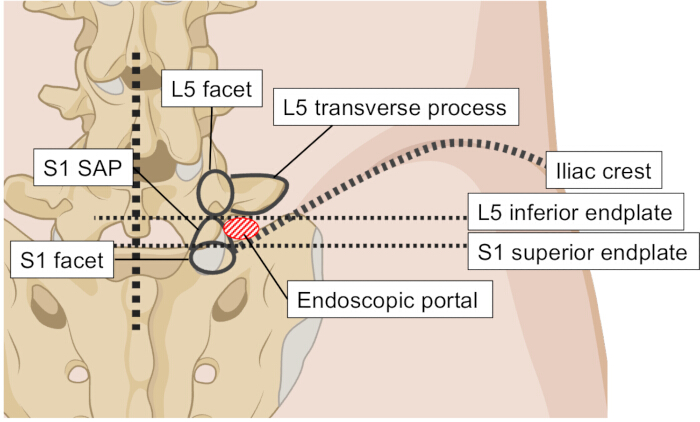
Figure 2: Preoperative skin marking. The skin entry point is marked under direct fluoroscopic visualization, ensuring the trajectory avoids the iliac crest. SAP, superior articular process. Please click here to view a larger version of this figure.
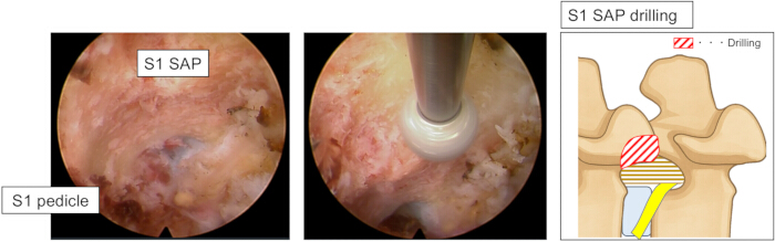
Figure 3: Endoscopic view: Foraminoplasty of the S1 SAP. The left panel shows the initial endoscopic orientation after placement of the cannula, with the lateral aspect of the S1 SAP identified as the primary bony landmark. The right panel demonstrates the commencement of the foraminoplasty, with a high-speed endoscopic drill resecting the caudal base of the S1 SAP at its junction with the S1 pedicle. SAP, superior articular process. Please click here to view a larger version of this figure.
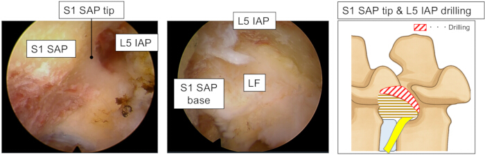
Figure 4: Endoscopic view: Exposure of the LF via L5 IAP resection. The left panel shows the SAP tip and its articulation with the L5 IAP. The right panel demonstrates the results after partial resection of the L5 IAP with a high-speed drill, which has now fully exposed the underlying foraminal LF. IAP, inferior articular process; LF, ligamentum flavum; SAP, superior articular process. Please click here to view a larger version of this figure.
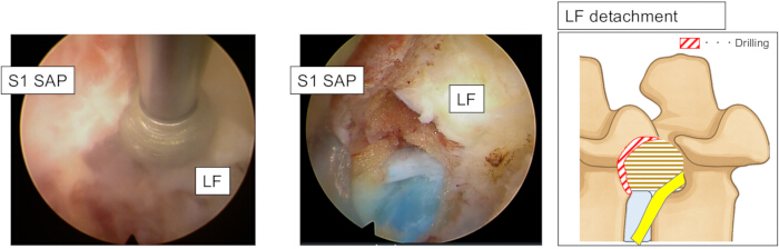
Figure 5: Endoscopic view: Release of the LF from the S1 SAP using the "detach" technique. The left panel demonstrates the detach technique, where the ventral rim of the remaining S1 SAP is undercut with a drill to release the caudal attachment of the LF. The right panel shows the LF in a "floating" state, completely mobilized after being detached from the S1 SAP. LF, ligamentum flavum; SAP, superior articular process. Please click here to view a larger version of this figure.

Figure 6: Endoscopic view: Final decompression. The left panel shows the removal of the detached floating LF using an endoscopic Kerrison punch. The right panel shows the final view after all compressive elements have been removed, revealing a fully decompressed, relaxed, and pulsating L5 ENR, which indicates successful decompression. ENR, exiting nerve root; IAP, inferior articular process; LF, ligamentum flavum. Please click here to view a larger version of this figure.
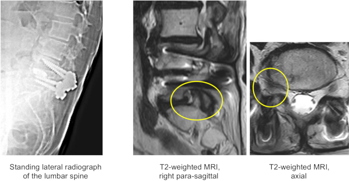
Figure 7: Radiological findings in a patient with adjacent segment disease following L4/5 PLIF. The left panel is a standing lateral radiograph of the lumbar spine that reveals instrumentation at L4/5 and degenerative changes at the adjacent level. The middle panel is a right para-sagittal T2-weighted MRI scan showing disc degeneration and foraminal narrowing at L5/S1. The right panel is an axial T2-weighted MRI scan showing nerve root compression within the right L5/S1 foramen. PLIF, posterior lumbar interbody fusion; MRI, magnetic resonance imaging. Please click here to view a larger version of this figure.

Figure 8: Pre- and postoperative CT images demonstrating successful osseous decompression. Left panels: Preoperative sagittal and axial CT images show severe right-sided L5/S1 foraminal stenosis. The hypertrophied superior articular process (SAP, yellow arrow) is compressing the neural foramen (dotted outline). Right panels: Corresponding postoperative images confirm adequate decompression, showing significant enlargement of the foramen (dotted outline) after resection of the ventral-cranial portion of the SAP (white arrow). Please click here to view a larger version of this figure.
Discussion
Transforaminal full-endoscopic foraminotomy under local anesthesia for L5/S1 foraminal stenosis adjacent to previous fusion is a valuable motion-preserving technique. It directly addresses the neural compression while avoiding the morbidity of fusion extension or extensive posterior revision surgery. This discussion will elaborate on the advantages of this method, key technical considerations for its success, and its limitations and future directions.
Advantages of the transforaminal endoscopic approach in ASD
ASD, particularly foraminal stenosis at a level adjacent to a previous lumbar fusion, presents a significant clinical challenge4,14. Conventional management for symptomatic ASD often involves extending the fusion to include the affected segment , which necessitates sacrificing the motion of an additional segment and is associated with increased surgical morbidity5,15. Revision surgery via a traditional posterior approach is also technically difficult due to postsurgical scarring, altered anatomical landmarks, and potential adherence of neural structures, which can increase the risk of complications, such as dural tears or nerve root injury15.
In contrast, the TF-FESS technique offers distinct advantages. The transforaminal approach accesses the foramen via a posterolateral corridor that typically remains undisturbed by prior posterior fusion surgery. Accessing the target through native tissue planes significantly reduces the risks associated with dissecting through scar tissue and has been shown to reduce operative time and blood loss compared with traditional posterior revision16. Furthermore, this technique directly addresses the stenosis without requiring additional fixation, thus preserving motion at the affected segment and potentially mitigating or delaying further degeneration of adjacent segments17.
Performing this procedure under local anesthesia with conscious sedation further enhances its usability and efficiency, particularly for the elderly or those with comorbidities who may be poor candidates for general anesthesia11,12. This approach not only minimizes physiological insult but also provides a crucial safety feature: real-time patient feedback, which helps prevent neural irritation1,18. The minimally invasive nature of the procedure contributes to shorter hospital stays, reduced postoperative pain, and a faster return to daily activities, potentially lowering overall healthcare costs when compared to revision fusion surgery19.
Key technical considerations and troubleshooting
The success of this technique hinges on several critical protocol steps. First, meticulous preoperative trajectory planning is paramount to navigate the narrow corridor, which is often obstructed by a high iliac crest. Second, strict adherence to the "bone is my friend" principle ensures the safety of the exiting nerve root by maintaining contact with the SAP as a reliable landmark. Finally, the "detach technique" for ligamentum flavum removal is a cornerstone of achieving complete decompression, as it allows the thickened ligament to be fully mobilized before removal, minimizing dural traction.
Common intraoperative challenges can be effectively managed. A high iliac crest can be addressed by adjusting patient positioning or planning a slightly steeper trajectory. Intraoperative bleeding is typically controlled by increasing irrigation pressure and using a radiofrequency probe. In revision cases with unclear landmarks, relying on fluoroscopy and carefully exposing the bony anatomy of the SAP is the safest approach.
Limitations and scope of the technique
Despite its advantages, the described method has important limitations and a specific scope. It is crucial to acknowledge that ASD presents in various forms, including central stenosis, degenerative spondylolisthesis, or frank instability. The technique detailed herein is specifically indicated for radiculopathy caused by foraminal stenosis without significant instability. Patients with other forms of ASD may require different treatment strategies, such as interlaminar decompression for central stenosis or fusion extension for instability.
Furthermore, this protocol focuses on caudal ASD at the L5/S1 level. While the principles of endoscopic foraminotomy can be applied to cranial adjacent segments, the surgical trajectory and anatomical challenges differ, and were not the focus of this article. A significant limitation of this case report is the lack of long-term follow-up and dynamic radiographic evaluation. As such, the potential for delayed instability at the operated segment after this motion-preserving decompression remains a valid concern that requires further investigation. Finally, as with all advanced endoscopic procedures, a steep learning curve must be considered to minimize operative risks such as dural tears or incomplete decompression20.
Future directions
The limitations of this study highlight clear directions for future research. A prospective, long-term study including dynamic radiographs is essential to evaluate the durability of clinical outcomes and the incidence of postoperative instability. To establish the definitive role of this technique, a randomized controlled trial comparing endoscopic foraminotomy with revision fusion surgery for foraminal stenosis in ASD is warranted. Such a trial should assess clinical outcomes, radiographic changes at the index and adjacent levels, and cost-effectiveness. Additionally, future studies could focus on adapting and evaluating this technique for cranial-level ASD and investigating the use of advanced technologies like intraoperative navigation to improve safety and accuracy.
Conclusion
Transforaminal full-endoscopic foraminotomy under local anesthesia for L5/S1 foraminal stenosis adjacent to previous fusion is a valuable technique that directly addresses the neural compression while avoiding the morbidity of fusion extension or extensive posterior revision surgery. Its minimally invasive nature, coupled with the benefit of local anesthesia, makes it an attractive option for a challenging patient population.
Disclosures
The authors have no conflicts of interest to declare.
Acknowledgements
This research received no specific grant from any funding agency in the public, commercial, or not-for-profit sectors.
Materials
| Name | Company | Catalog Number | Comments |
|---|---|---|---|
| 10K Arthroscope tube set | Zimmer Biomet | LC-10K0-100-00 | Irrigation system console |
| 10K Irrigation console | Zimmer Biomet | LC-10K0-000-00 | Irrigation system console |
| 1-Coarse Diamond Bur SPL (3.5) | Nakanishi | PDS-1CD330-35 | High-speed drill |
| 21G PTCD needle, 200 mm | Hakko Shoji | U5099-MM | Needle for local anesthesia |
| 23G Catelan needle | NIPRO | 2022 | Needle for local anesthesia |
| 32-inch 4K LCD monitor | Sony | LMD-X3200MD | Monitor |
| Aquarius NET Server | TeraRecon | N/A | CT reconstruction and analysis software |
| Atarax-P | Pfizer | N/A | Hydroxyzine hydrochloride |
| Centricity Universal Viewer Zero Footprint Client | GE Healthcare | Version 6.0 SP11.2.2 | MRI visualization and analysis software |
| CLICKLINE grasping forceps | Karl Storz | 28163FSI | Intervertebral disc rongeur |
| Curved rongeur, WL 360 mm, φ2.5 mm | RIWOspine | 89240.1044 | Soft tissue rongeur |
| Dilator, Ø7.0 mm, effective length 225 mm (2 holes) | Elliquence | 11-2311 | Dilator |
| ENDOCAM Logic 4K camera controller | Richard Wolf GmbH | 55253011 86-103 | Camera |
| ENDOCAM Logic 4K camera head | Richard Wolf GmbH | 85525942 86-104 | Camera |
| Fiber light cable (φ3.5 mm/3.0 mm) | Richard Wolf GmbH | 806635301 86-124 | Camera |
| Flexible probe (Φ2.5 mm, WL 350 mm) | RIWOspine | 892501925 | Flexible probe |
| Floseal Hemostatic Matrix | Baxter | ADS201844 5mL Kit | Hemostatic agent |
| Guide wire | Nihon MDM | 28163GWT | Dilator |
| Kerrison punch handle | Elliquence | 12-1947 | Kerrison |
| Kerrison punch, angled type, Ø3.5 mm, effective length 360 mm | Elliquence | 12-1938 | Kerrison |
| Kerrison punch, angled type, Ø4.0 mm, effective length 360 mm | Elliquence | 12-1940 | Kerrison |
| LED light source unit for endoscopy (LED 1.2 set) | Richard Wolf GmbH | 51610011 86-164 | Camera |
| Primado2 Drill System | Nakanishi | P200-CU-100 | High-speed drill |
| Primado2 Foot Controler | Nakanishi | FC-73 | High-speed drill |
| Primado2 Slim motor handpiece | Nakanishi | P200-SMH-HS | High-speed drill |
| PTC needle type B, 18G × 200 mm | Hakkou Shoji | 22411830 | Dilator |
| Rongeur, WL 360 mm, φ3.0 mm | RIWOspine | 89240.1003 | Soft tissue rongeur |
| Sosegon | Maruishi Pharma | N/A | Pentazocine |
| Step dilator set (5 pieces) | Elliquence | CSD-5 | Dilator |
| Super Slim attachment 200 | Nakanishi | P200-RA330-L | High-speed drill |
| Surgi-Max Air | Elliquence | IEC4-SP | Bipolar device |
| Trigger-Flex | Elliquence | DTF-40 | Bipolar device |
| VERTEBRIS lumbar, 25°, 6.9 mm, WL 207 mm | RIWOspine GmbH | 89210.1254 | Rigid endoscope |
| Working channel, duckbill type, Ø8.0 mm, length 165 mm | Richard Wolf GmbH | 11-2910 | Duckbill cannula |
| Working channel, elevator type, Ø8.0 mm, length 165 mm | Richard Wolf GmbH | 11-2916 | Oblique cannula |
References
- Sairyo, K., et al. State-of-the-art transforaminal percutaneous endoscopic lumbar surgery under local anesthesia: Discectomy, foraminoplasty, and ventral facetectomy. J Orthop Sci. 23 (2), 229-236 (2018).
- Ahn, Y., Lee, S. H., Park, W. M., Lee, H. Y. Posterolateral percutaneous endoscopic lumbar foraminotomy for L5-S1 foraminal or lateral exit zone stenosis. J Neurosurg. 99 (3 Suppl), 320-323 (2003).
- Cho, K. S., et al. Risk factors and surgical treatment for symptomatic adjacent segment degeneration after lumbar spine fusion. J Korean Neurosurg Soc. 46 (5), 425-430 (2009).
- Park, P., Garton, H. J., Gala, V. C., Hoff, J. T., McGillicuddy, J. E. Adjacent segment disease after lumbar or lumbosacral fusion: review of the literature. Spine. 29 (17), 1938-1944 (2004).
- Harrop, J. S., Youssef, J. A., Maltenfort, M., et al. Lumbar adjacent segment degeneration and disease after arthrodesis: a systematic review. J Neurosurg Spine. 10 (5), 354-362 (2009).
- Bashti, M., et al. Surgical management of thoracolumbar adjacent segment disease: Techniques and outcomes in 107 patients undergoing surgical intervention. Int J Spine Surg. 18 (3), 295-303 (2024).
- Ahn, Y., Kim, H. S., Lee, J. H., Kim, J. S., Jang, I. T. Radiographic assessment on magnetic resonance imaging after percutaneous endoscopic lumbar foraminotomy. Neurol Med Chir (Tokyo). 57 (12), 649-657 (2017).
- Song, Q. P., et al. Full-endoscopic foraminotomy with a novel large endoscopic trephine for severe degenerative lumbar foraminal stenosis at L5-S1 level: An advanced surgical technique. Orthop Surg. 13 (2), 659-668 (2021).
- Sairyo, K., Chikawa, T., Nagamachi, A. Strengths and merits of transforaminal full-endoscopic lumbar spine surgery under the local anesthesia: A review article. J Minim Invasive Spine Surg Tech. 9 (Suppl 2), S143-S151 (2024).
- Matsumura, T., Chikawa, T., Nishidono, K., Sairyo, K. Step-by-step explanation of full-endoscopic lumbar foraminotomy at the L5/S1 level under local anesthesia: An editorial technical note. EC Orthop. 14 (7), 47-53 (2023).
- Ahn, Y., Song, S. -K. Transforaminal endoscopic lumbar foraminotomy for octogenarian patients. Front Surg. 11, 1324843(2024).
- Zheng, B., et al. Anesthesia methods for full-endoscopic lumbar discectomy. Front Med. 10, 1193311(2023).
- Yamashita, K., et al. Percutaneous full endoscopic lumbar foraminoplasty for adjacent level foraminal stenosis following vertebral intersegmental fusion in an awake and aware patient under local anesthesia: A case report. J Med Invest. 64 (3-4), 291-295 (2017).
- Zhang, C., Berven, S. H., Fortin, M., Weber, M. H. Adjacent segment degeneration versus disease after lumbar spine fusion for degenerative pathology: A systematic review with meta-analysis of the literature. Clin Spine Surg. 29 (1), 21-29 (2016).
- Okuda, S., et al. Surgical complications of posterior lumbar interbody fusion with total facetectomy in 251 patients. J Neurosurg Spine. 4 (4), 304-309 (2006).
- Guo, J., et al. Comparative advantages of transforaminal percutaneous endoscopic lumbar decompression versus traditional revision surgery for degenerative adjacent segment degeneration following lumbar fusion: A retrospective propensity score matching study. BMC Surg. 24 (1), 177(2024).
- Hashimoto, K., Aizawa, T., Kanno, H., Itoi, E. Adjacent segment degeneration after fusion spinal surgery: A systematic review. Int Orthop. 43 (4), 987-993 (2019).
- Gore, S., Yeung, A. The "inside out" transforaminal technique to treat lumbar spinal pain in an awake and aware patient under local anesthesia: Results and a review of the literature. Int J Spine Surg. 8, 28(2014).
- Findlay, M. C., Hamrick, F. A., Kim, R. B., Twitchell, S., Mahan, M. A. Hospital cost differences between open and endoscopic lumbar procedures. J Neurosurg Spine. 40 (1), 77-83 (2023).
- Hsu, H. T., Chang, S. J., Yang, S. S., Chai, C. L. Learning curve of full-endoscopic lumbar discectomy. Eur Spine J. 22 (4), 727-733 (2013).
Reprints and Permissions
