Here, we present the stent-bridging pancreaticogastrostomy (PG) technique, which uses a stent to bridge the remnant pancreas to the stomach during open pancreaticoduodenectomy. The stent-bridging PG aims to simplify the anastomosis, achieve complete diversion of pancreatic juice, and minimize manipulation of the pancreatic remnant.
Method Article
Application of Simplified Stent-bridging Pancreaticogastrostomy in Open Pancreaticoduodenectomy
* These authors contributed equally
In This Article
Summary
Abstract
The standardized technique for pancreatic anastomosis in pancreaticoduodenectomy (PD) has not achieved consistent acceptance due to persistent technical complexity and the incidence of postoperative pancreatic fistula (POPF). This paper presents a case in which a simplified stent-bridging pancreaticogastrostomy (PG) is employed. The patient is placed in the supine position, and a longitudinal midline abdominal incision is made. Following a subtotal stomach-preserving PD, reconstruction is performed. An appropriately sized polyethylene catheter is inserted to bridge the Wirsung duct and the stomach, enabling drainage of pancreatic juice into the gastric cavity. The entire pancreatic stump is continuously sutured. The Wirsung duct and the polyethylene catheter are looped and ligated. Two discontinuous figure-of-eight sutures are prearranged without knotting, and an incision is made in the posterior gastric wall. The distal end of the polyethylene catheter is inserted into the gastric cavity to provide internal drainage. Two seromuscular purse-string sutures are tightened to approximate the pancreatic remnant to the gastric lumen. The gastric omentum with adjacent preperitoneal fat is positioned over the anastomosis site. The procedural steps of the stent-bridging PG are described in detail to demonstrate technical feasibility. By diverting pancreatic fluid and limiting manipulation of the remaining pancreatic tissue, the technique is intended to reduce mechanical stress at the anastomosis. Further experience is required to clarify optimal indications and long-term outcomes.
Introduction
Pancreaticoduodenectomy (PD) has shown progressive outcomes1; nevertheless, it continues to pose certain challenges due to its inherent complexity2. Clinically relevant postoperative pancreatic fistula (POPF) may lead to intra-abdominal infection, hemorrhage, and potentially life-threatening outcomes3. The surgeons have increasingly recognized the importance of an evidence-based reconstructive approach to improve patient outcomes and advance clinical standards, thereby minimizing the risk of POPF. Among various pancreatic anastomoses in pancreaticojejunostomy (PJ) or pancreaticogastrostomy (PG)4, studies failed to demonstrate a significant difference in outcomes5. The single- or double-layer, invaginating PG anastomosis, where the remnant pancreatic stump is inserted into the gastric cavity with an external stent, is the preferred variant in PG. However, this method entails risks of stent slipping out or occlusion4. The use of internal stents entails a risk of migration in up to 17% of patients5, and biodegradable internal stents are rather unsuccessfully6.
The potential mechanisms underlying POPF mainly comprise minor leakage of sutures, poor blood supply at the anastomosis site, anastomotic tension, and anastomotic delay healing7. Also, suture material induces changes in the pancreas similar to those of acute pancreatitis, which supports the rationale for using thinner and fewer sutures8. In this video case presentation, we present the stent-bridging PG, which utilizes an appropriately sized polyethylene catheter to bridge the Wirsung duct and the stomach, aiming to simplify reconstruction and facilitate controlled pancreatic juice diversion. For soft pancreas with small duct diameters, a modified peridural catheter is utilized9. However, the clinical evidence regarding the stent is not yet entirely clear. The anastomoses over internal or externalized stents are at the surgeon's discretion. The issue of long-term follow-up for internal stents remains to be addressed, and further clinical validation in a randomized setting is necessary.
Protocol
Ethics approval is not required for this procedure as part of routine clinical care. Informed consent is obtained from the individual participant in the study. The patient is a 70-year-old female diagnosed with a pancreatic head malignancy, presenting with progressive jaundice, which remained painless for 3 months. Contrast-enhanced computed tomography (CT) scan reveals a pancreatic head malignancy involving the descending section of the adjacent duodenum and portal vein - superior mesenteric vein.
1. Preoperative preparation
- Carefully evaluate preoperative computed tomography (CT) or magnetic resonance imaging (MRI) images to determine the exact location of the tumor and its spatial relationship with the surrounding tissues. Figure 1 illustrates the appearance of the lesion on a contrast-enhanced CT scan.
- Implement perioperative management in accordance with the standardized principles of the Enhanced Recovery After Surgery (ERAS) protocol, including counseling, prehabilitation, and nutritional support.
- Prescribe a soft diet on the day preceding surgery and ensure fasting from midnight before the operation.
- After induction of general anesthesia using standard procedures, insert a central venous catheter for fluid administration and a peripheral arterial catheter for continuous blood pressure monitoring.
2. Surgical technique
- Operation setting: Place the patient in a supine position. Make a longitudinal incision along the midline of the abdomen.
- Exploration and dissection phase: After standard subtotal stomach-preserving PD, perform the reconstruction phase.
- Reconstruction phase
- Perform the hepaticojejunostomy approximately 3–5 cm distal to the blind end of the jejunum in a retrocolic fashion.
- Perform the gastrojejunostomy approximately 65–70 cm distal to the hepaticojejunostomy, using an antecolic approach.
- Subsequently, perform a Braun jejunojejunostomy, which involves a side-to-side anastomosis between the afferent and efferent jejunal loops, 20 cm distal to the gastrojejunostomy site to prevent bile reflux gastritis.
- Finally, perform the novel PG anastomosis technique, referred to as "the stent-bridging PG" method. (Figure 2)
- Pancreas mobilization and pancreatic duct stenting: Carefully isolate the pancreatic stump approximately 2 cm to facilitate the creation of the PG anastomosis, ligate and divide small veins between the pancreas and the splenic vein, and lift the stomach to expose the posterior gastric wall. Select a polyethylene catheter of an appropriate size (usually 6–10 French); for a small Wirsung duct of 0.1 cm, choose a modified peridural catheter. Cut the multiple drainage side holes according to the remaining length of the pancreas, trim the distal end into an inclined shape, and insert the distal end into the Wirsung duct (Figure 2A).
- Suture the whole pancreatic stump continuously, approximately 1 cm from the cut edge, to achieve hemostasis and secure the polyethylene catheter. Further, loop around and ligate the Wirsung duct and the polyethylene catheter with nonabsorbable suture (4/0) (Figure 2B).
- Prearrange two discontinuous figure-of-eight sutures (3/0) without knotting at the posterior gastric wall just above the pancreatic remnant (Figure 2C). Create a full-layer hole on the posterior wall of the stomach, with its diameter approximately matching that of the Wirsung duct, to achieve an effective bypass (Figure 2D).
- Insert only the distal end of the polyethylene catheter into the gastric cavity as internal drainage (Figure 2E). Hereafter, gently retract the stomach inferiorly in close apposition to the pancreatic remnant for a relatively short sinus.
- Appropriately tighten the two discontinuous figure-of-eight sutures in case of stent occlusion, thus completing the stent-bridging PG procedure (Figure 2F).
- Cover the remaining ends of the gastric-duodenal artery and anastomotic site with the falciform ligament. Place two drainage tubes in the proximity of the pancreatic and biliary anastomoses (Figure 2G).
- Localize the anastomosis site with the gastric omentum with adjacent preperitoneal fat in case of pancreatic leakage (Figure 2H).
Results
After surgery, prophylactic octreotide is not administered. A nasojejunal tube is placed for a liquid diet from postoperative day 1. The operation time is 6 h with a blood loss of 100 mL. The patient's postoperative recovery is uneventful, with no evidence of POPF observed, as defined by the International Study Group on Pancreatic Fistula (ISGPF) criteria10 during the follow-up period. The postoperative CT scan confirms optimal positioning and safe fixation of the stent, and no evidence of fluid accumulation around the anastomosis, peripancreatic exudation, or pancreatic duct dilatation (Figure 3). The nasojejunal tube and the intra-abdominal drains are removed on postoperative day 5, and the patient is discharged on postoperative day 8. Pathology reveals a 3 cm pancreatic poorly to moderately differentiated adenocarcinoma, R0 resection, and 22 negative lymph nodes with 1 malignancy.
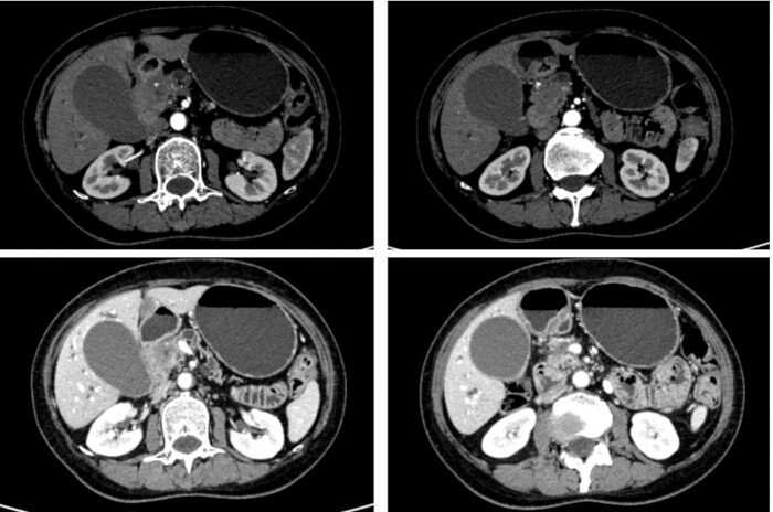
Figure 1. Preoperative contrast-enhanced CT scan. Please click here to view a larger version of this figure.
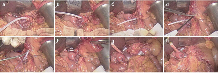
Figure 2. Stent-bridging pancreaticogastrostomy. (A) Insert the matching polyethylene catheter into the Wisung duct as a stent. (B) Perform continuous suturing on the entire pancreatic stump. (C) Prearrange two discontinuous figure-of-eight sutures (3/0) without knotting at the incision site just above the pancreatic remnant. (D) Create a small posterior gastrotomy where the catheter can be inserted without undue tension using electrocoagulation. (E) Insert only the matching polyethylene catheter into the gastric cavity. (F) Tighten the two discontinuous figure-of-eight sutures around the incision for fixation, and retract the stomach cavity to the pancreatic stump. (G) Wrap the gastric-duodenal artery stump and the pancreatic stump with the falciform ligament and the round ligament. Place two easy-flow drainage tubes near the anastomoses. (H) Localize the anastomosis site using the omentum tissue. Please click here to view a larger version of this figure.
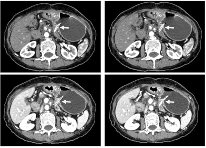
Figure 3. Postoperative contrast-enhanced CT scan. The white arrow indicates optimal positioning and stable fixation of the stent. There is an absence of fluid accumulation around the anastomosis, peripancreatic exudation, or pancreatic duct dilatation. Please click here to view a larger version of this figure.
Discussion
As one of the most technically demanding surgeries in abdominal surgery, PD or LPD has been predominantly performed at high-volume tertiary centers1. Adhering to the basic principles of anastomosis reconstruction and being able to perform anastomosis surgeries that are safe, reliable, repeatable, and associated with a low incidence of clinically significant POPF remains crucial for achieving excellent surgical outcomes5. Unlike gastrointestinal anastomosis, pancreatic anastomoses are diverse and remain the focus of future research in PD11.
The critical procedures include suturing the entire pancreatic stump, securing the stent, ensuring close attachment of the pancreatic stump to the stomach, and localizing the pancreatic stump. The small branches of the pancreatic duct at the pancreatic remnant can lead to pancreatic leakage7. Thus, it is essential to continuously suture the pancreatic stump to reduce leakage. Given that stent displacement is the most prevalent complication associated with internal stents5, it is of great importance to firmly secure the stent. We employ three steps to fix the stent: ligating the Wirsung duct together with the catheter, suturing the entire pancreatic stump, and performing two interrupted figure-of-eight sutures on the gastric wall incision. When initially dissecting the pancreas and isolating the pancreatic duct, it is recommended to keep it adequately long to facilitate the binding with the catheter. The surgeon needs to be cautious and tie the knot appropriately to avoid obstructing the pancreatic duct. The greater omentum flap and a portion of the preperitoneal fat should be used to localize the anastomotic site. It has been suggested that POPF may lead to more severe complications due to reduced or delayed formation of abdominal adhesions12. Wrapping the residual ends of the hepatic artery and the gastroduodenal artery helps prevent invasive intraabdominal bleeding after pancreatic resection by isolating the exposed arteries from pancreatic fistulas and infections13.
In a multicenter RCT, PG has been proven more effective than PJ in reducing the incidence of POPF14. From a pathophysiological perspective, the increased pressure in the lumen of the jejunal loop, combined with the detrimental effects of activated pancreatic enzymes in the presence of intestinal enterokinase and bile in PJ, may gradually lead to fatal POPF caused by self-digestion15. PG may effectively achieve complete diversion of pancreatic fluid, prevent the accumulation of pancreatic leakage and the subsequent activation of pancreatic enzymes within the jejunum cavity, thus interrupting the pathological physiological cascade reaction16. There is also a hypothesis suggesting that postoperative pancreatic fistula might be caused by accidental damage to the pancreatic duct, akin to acute pancreatitis, or the fragile pancreatic tissue during the process of suturing or knotting17. Thus, the thinner and fewer sutures are recommended8. This recommendation is particularly applicable when dealing with soft, fragile, high-fat-content, and small-duct-diameter pancreatic tissues. Compared with duct-to-mucosa or invaginating end-to-side PG18, the stent-bridging PG reduces tangential tension and shear forces by only catheter placement in the gastric cavity and fewer sutures. Moreover, a smaller posterior gastrotomy is created, allowing the catheter to be inserted without undue tension. The short distance between the pancreatic remnant and the stomach enables the gastric wall to function as a serosal covering for the pancreatic remnant. The thick gastric wall has desirable suture retention strength and rich blood supply. It may also reduce the potential risk of suture cutting caused by a single- or double-layer full-thickness purse string suture of the pancreatic stump.
Surgeons are seeking simpler and safe methods of pancreatic anastomosis to facilitate the surgery and promote adoption from duct-to-mucosa to the bundled method11,18. Existing risk models have shown limited predictive accuracy, potentially leading to unreliable estimations regarding the impact of the anastomosis technique and surgical duration19. The stent-bridging PG can shorten the pancreatic reconstruction time without excessive manipulation of the stomach and the pancreatic remnant9. Due to the complexity of these procedures, debates surrounding surgeon experience have hindered the broader adoption and clinical trial dissemination of such techniques, largely due to steep learning curves and suboptimal early results20,21. A definitive conclusion regarding the number of operations required to overcome the learning curve has not yet been reached22. As this surgery is still in its early stages, many issues remain unclear, such as the long-term side effects of the internal stent. Further evaluation and validation of the above therapeutic effects and safety in a randomized setting are still necessary.
Disclosures
The authors have no conflicts of interest to disclose.
Acknowledgements
Funding: the Central High-level TCM Hospital Clinical Research Capability Improvement Project (HLCMHPP2023124).
Materials
| Name | Company | Catalog Number | Comments |
|---|---|---|---|
| 4K Optical 3D fluorescence laparoscopy System (Stellar) | OptoMedic | Stellar | Video recording equipment |
| Coated VICRYL TM PLUS | ETHICON | VCP772D | Absorbable surgical suture |
| Coated VICRYL TM PLUS | ETHICON | VCP752D | Absorbable surgical suture |
| Osmo Action 5 Pro | DJI | 10120634062917 | Video recording equipment |
| PDS* Plus Antibacterial With Irgacare MP 2360 uglm VIOLET MONOFILAMENT | ETHICON | PDP304H | Absorbable surgical suture |
| Silicone rubber catheter | Chensheng | 20100048 | Pancreatic ductal tube |
| Surgical instruments | SHINVA | Surgical instruments |
Reprints and Permissions
