This protocol describes a method for integrating continuous renal replacement therapy (CRRT) into exsitu normothermic liver perfusion by connecting it to the perfusion reservoir. This configuration supports hemodynamic stability, helps prevent excessive intravascular pressures, and may reduce the risk of tissue edema, in particular during extended perfusion.
Method Article
Integrating Continuous Renal Replacement Therapy into Ex-situ Normothermic Liver Machine Perfusion
* These authors contributed equally
In This Article
Summary
Abstract
Ex situ normothermic machine perfusion (NMP) of the liver has emerged as a dynamic preservation strategy, enabling the maintenance of metabolic activity and assessment of grafts prior to transplantation. Prolonged perfusions, however, are limited by the accumulation of metabolic waste, electrolyte imbalances, and inflammatory mediators that can compromise graft function. Continuous renal replacement therapies (CRRT), especially continuous venovenous hemodiafiltration (CVVHDF), offer a potential means to support solute clearance and homeostatic regulation during extended perfusion periods.
In this protocol, we describe a reproducible and safe method for integrating CVVHDF into an ex situ liver NMP system by connecting the filtration circuit independently from the main organ perfusion circuit. This configuration supports hemodynamic stability, helps prevent excessive intravascular pressures, and may reduce the risk of tissue edema. The physical connection, priming, and monitoring of the CRRT circuit are explained in detail, along with instructions on how to utilize a Hoffman clamp to simulate physiological venous pressure. When compared to direct in-line circuit integration, representative results suggest that the out-of-circuit approach is associated with reduced graft edema, while maintaining stable circuit pressure and solute clearance. This approach can be easily integrated into other similar perfusion devices in order to improve solute handling while maintaining stable perfusion dynamics.
Introduction
Ex situ normothermic machine perfusion (NMP) is a dynamic preservation strategy that aims to maintain the metabolic activity of the organ, providing oxygen and metabolic substrates at physiological temperatures1,2,3. Under these conditions, it is possible to assess injury in and to some degree the functionality of organs prior to their transplantation. As well, ex situ NMP offers a potential platform for the reconditioning, treatment, and evaluation of organs, for both transplant and non-transplant purposes3,4.
Ex situ organ perfusion duration is conditioned by different pathophysiological processes arising consequent to the perfusion itself. Most perfusions in both preclinical and clinical studies reported to date have been performed for relatively limited durations of time, commonly 6-24 h in standard protocols, with selected reports extending to or exceeding days to weeks under experimental conditions5,6,7,8. Adverse processes and events that arise during ex situ NMP include the accumulation of metabolic waste substances, alteration of the perfusate electrolyte composition, production of intermediates and inflammatory mediators, such as damage-associated molecular patterns (DAMPs), and edema/weight gain related to fluid shifts, which can alter graft viability9. Consequently, incorporation of extracorporeal blood purification (EBP) methods has been evaluated as an important means to regulate and remove these different products10. Of all those available, the most widely used are continuous renal replacement therapies (CRRT).
CRRT comprises a set of techniques typically used in patients with renal failure to filter blood and clear different components, thereby regulating acidosis and electrolyte imbalances and eliminating waste products11,12. These include hemodialysis (HD), hemofiltration (HF), and hemodiafiltration (HDF)13. HD relies on diffusion to remove small (<15 kDa) solutes, such as urea or β2-microglobulin, and is effective in correcting electrolyte imbalances14,15,16,17. HD has limitations, however, such as low efficacy in the removal of larger or protein-bound toxins and induction of immune activity due to membrane contact17,18,19. HF relies on convective mechanisms to remove larger molecules, including pro- and anti-inflammatory cytokines, though it can also remove essential plasma components, such as vitamins, trace elements, and micronutrients20,21,22,23. HDF combines both diffusion and convection mechanisms to achieve broader clearance of medium-sized toxins and inflammatory mediators, thereby allowing for more effective modulation of the inflammatory milieu24,25,26,27,28.
CRRT relies on carefully controlled gradients to facilitate the exchange of solutes and fluids across membranes. In the case of HD, diffusion is driven by concentration differences, while HF and HDF require transmembrane pressure gradients to enable convective transport29. When these systems are integrated into ex situ perfusion circuits, controlling pressure dynamics becomes especially relevant. Organ perfusion systems depend on a precise balance of pressures to maintain adequate flow, tissue oxygenation, vascular integrity, and organ metabolic function30. The addition of a CRRT system introduces pressure changes that can alter the delicate balance within the organ perfusion circuit.
Herein, we describe a practical and reproducible strategy to connect CRRT during ex situ liver NMP independently from the main perfusion circuit, by interfacing both access and return lines with the perfusion reservoir rather than the vascular loop. Compared with direct "in-circuit" integration (i.e., connecting to a vascular perfusion circuit), this out-of-circuit configuration hydraulically decouples CRRT from organ vasculature, supporting more stable pressure and flow regulation and helping to mitigate pressure-induced edema formation caused by the in-line configuration. This approach helps to preserve hemodynamic stability and solute handling during extended organ perfusions. In addition, the reservoir-based connection facilitates operational steps, such as priming and filter exchange, without interrupting the main NMP circuit. This protocol is readily adaptable to perfusion platforms that provide reservoir access, and its principles can be applied across CRRT modalities with minor configuration adjustments.
Protocol
Animal care and experimental protocols were approved by the institutional and regional ethics committees (Division of Agriculture, Livestock, and Food of the Community of Madrid and the Animal Welfare Body [OEBA] of the Autonomous University of Madrid). All animals were housed and procedures conducted at the Experimental Surgery Service of Hospital Universitario La Paz, in accordance with current national and European regulations, accredited by the International Standards Organization (ISO9001:2015) and authorized by the regional authority (ES280790001941). Seventeen livers were obtained from healthy porcine donors and subjected to a minimal-injury model.
1. Minimal-injury model
- Induce relative in vivo warm ischemia (13 ± 5 min) in order to recover donor blood for use on the machine, followed by cold ischemia (77 ± 16 min).
- For each experiment, initiate CRRT using CVVHDF with the following operational parameters: blood flow rate (BFR) 100 mL/min, dialysate flow rate (DFR) 250 mL/h, and replacement fluid flow delivered post-filter (PBP) 100 mL/h. Set net ultrafiltration (UF) to 20 mL/h to account for fluid inputs (e.g., heparin, parenteral nutrition, antibiotics) and outputs (e.g., bile), aiming for near-neutral fluid balance.
- Ensure that the hemofilter employs a polyethersulfone high-flux membrane with an effective surface area of approximately 0.3 m2 and a molecular-weight cut-off of about 40 kDa.
- Anticoagulate the entire ex situ perfusion circuit, including the CRRT system, with unfractionated heparin (UFH), administered directly into the perfusion reservoir as an initial bolus of 5,000 IU followed by continuous infusion at 1,500 IU/h diluted in normal saline to prevent clotting and ensure circuit patency.
- Maintain the NMP circuit at 37.0 ± 0.2 °C using an integrated heat exchanger within the perfusion device. Ensure that the CRRT machine utilizes an inline fluid warmer so that return flow enters the reservoir at 37 °C, mitigating cooling from replacement/dialysate solutions. Monitor temperature continuously both in the perfusate and by the CRRT internal sensors.
2. Physical and functional connection of CRRT to the ex situ machine perfusion system
- Identify connection points.
- Identify an appropriate outflow port on the perfusion system reservoir to serve as the access (draw) point for the CRRT circuit.
- Identify an appropriate inflow port on the perfusion system reservoir to serve as the return (reinfusion) point.
- Install sterile tubing ensuring contact with the reservoir perfusate, if not already present.
NOTE: Both access and return ports should ideally be outside the pressure-regulated circuit of the ex situ perfusion system, to avoid disturbing vascular resistance, impairing flow stability, and ultimately damaging the graft. The blood reservoir is the preferred site for connection.
- Purge and secure connection lines.
- Attach sterile male Luer-lock connectors to both the access and return ports of the perfusion system, if not already present.
- Prime the outflow line from the perfusion system with the perfusate using a syringe to eliminate air.
- Clamp the outflow line to prevent backflow until final connection.
- Connect the CRRT system to the perfusion system.
- Connect the access line to the purged outflow port of the perfusion system using the Luer-lock connector.
- Place a Hoffman clamp or equivalent on the CRRT return line near its connection to the perfusion system to simulate physiological venous pressure (Figure 1).
NOTE: The Hoffman clamp serves to provide sufficient downstream resistance, preventing alarms or back pressure interruptions during CRRT. Ensure the clamp is not fully closed and return flow is continuous and visible once treatment begins. - Connect the return line to the inflow port of the perfusion system using the Luer-lock connector.
- Initiate perfusate purification treatment.
- Unclamp both the access line and the outflow port of the perfusion system before starting the CRRT treatment.
- Confirm that the system begins treatment and that blood flow is visible through both lines.
- WARNING: All system components, including the perfusate and organ, should be treated as potentially infectious biological material. Always use appropriate personal protective equipment (PPE), including gloves, face shields, and lab coats, during setup, operation, and waste disposal.
- Monitor CRRT circuit functionality.
- Record system temperature and pressure values hourly, including access pressure, return pressure, prefilter pressure, transmembrane pressure (TMP), and effluent pressure.
- Observe blood flow through the tubing and ensure no air bubbles are present.
- Confirm dialysate and replacement fluid flows are occurring as programmed.
CAUTION: Sudden deviations from normal values may indicate clotting, filter blockage, line kinking, or inadequate venous resistance at the return site. Typical reference values and their deviations and justifications during CRRT can be found in Table 1 and Table 2, respectively.
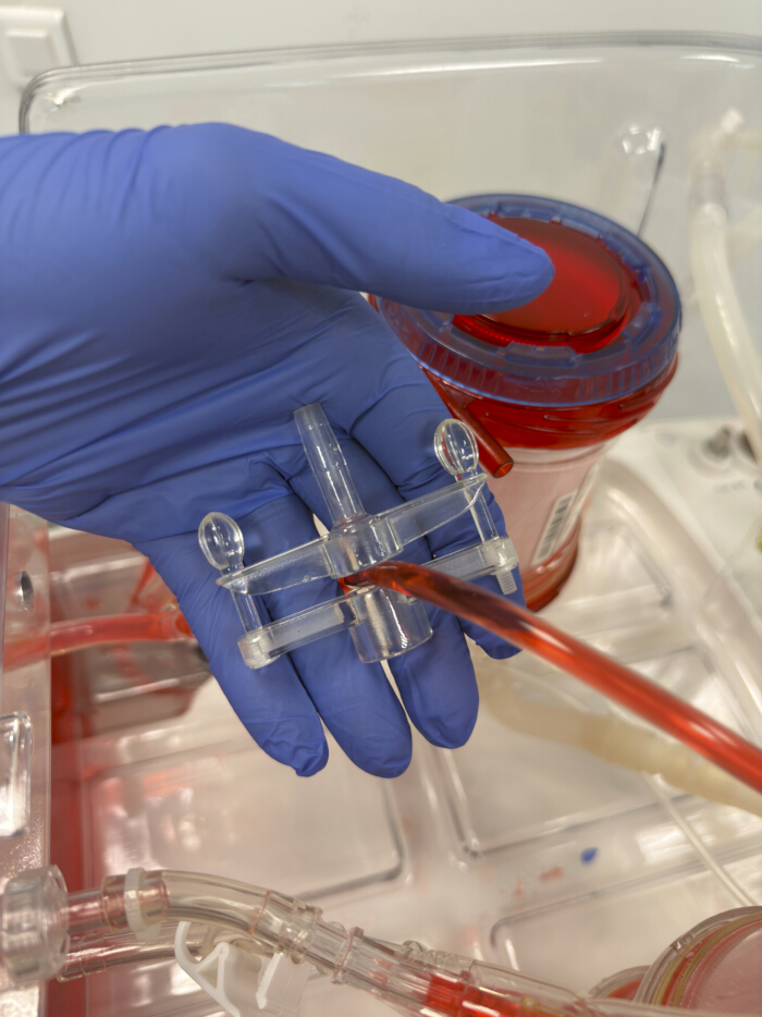
Figure 1: Hoffman clamp positioned on tubing to regulate venous return pressure. The clamp is installed on the return line before connection to the reservoir of the normothermic perfusion system. This configuration enables fine regulation of flow resistance and simulates physiological venous pressure, which is crucial in preventing negative return pressures and false alarms in the CRRT machine. Please click here to view a larger version of this figure.
| Parameter | Expected Range (mmHg) | Interpretation |
| Access pressure | –50 to –200 | Negative pressure due to blood draw from reservoir. |
| Return pressure | < 200 | Reflects mild resistance in the return line. |
| Pre-filter pressure | 100–300 | Indicates blood entry to filter under standard flow. |
| Transmembrane pressure (TMP) | < 300 | Marker of filter performance and clot risk. |
| Effluent pressure | < 300 | Variable depending on bag fill and resistance. |
Table 1: Reference pressure ranges for continuous renal replacement therapy during ex situ perfusion. This table summarizes the expected pressure ranges (in mmHg) for key monitoring points in the extracorporeal circuit, including access pressure, return pressure, pre-filter pressure, transmembrane pressure, and effluent pressure. These reference values serve as baseline indicators of circuit stability, hydraulic efficiency, and filter performance when CRRT is integrated into an ex situ perfusion system. Deviation for these ranges may suggest flow, resistance, occlusion, or filter clotting requiring circuit assessment. Abbreviations: CRRT = continuous renal replacement therapy; TMP = transmembrane pressure.
| Parameter | Abnormal Finding (mmHg) | Possible Cause | Recommended Action |
| Access pressure | > –50 | Poor reservoir contact, line occlusion, kinked or clamped line, air in line or low blood flow | Check reservoir fluid level and position, remove kinks, ensure line is fully submerged and unclamped, increase blood flow |
| Return pressure | > 200 | Return line submerged too deep, line occlusion, clamp too tight, kinking, or clotting | Adjust return line depth, loosen clamp, reposition line, or check for kinks or clots, consider filter change |
| Pre-filter pressure | > 300 | Flow obstruction, excessive viscosity, filter clotting, or clamps left on | Verify circuit patency, reduce flow, check for clots, remove clamps. |
| Transmembrane pressure (TMP) | > 300 | Filter clotting, high resistance, filtrate line or bag clamped, air in filter | Change filter, check for clots or kinks, unclamp lines, remove air, re-prime if needed. |
| < -30 | Low blood flow from reservoir, or filtrate pump slower than treatment pump. | Check reservoir level or increase flow | |
| Effluent pressure | > 300 | Effluent bag full, line occlusion, or bag not hanging freely | Change effluent bag, check for kinks, ensure bag is hanging freely |
| Sudden drop in any pressure | Leak or disconnection | Inspect entire circuit immediately. |
Table 2: Troubleshooting guide for continuous renal replacement therapy during ex situ perfusion. This table provides a systematic guide for identifying and resolving abnormal pressure readings or performance deviations in the CRRT circuit. For each pressure parameter, common abnormal findings are listed with their possible causes and recommended corrective actions. This troubleshooting reference supports rapid detection of circuit malfunctions, maintenance of adequate filtration performance, and prevention of pressure-related complications during ex situ organ perfusion.
3. Waste disposal
- Collect the effluent generated by the CRRT circuit in sealed waste collection bags directly connected to the effluent lines of the machine. Once filled, securely seal and dispose of these bags as liquid biohazard waste, in accordance with institutional and local biosafety regulations.
- Discard the used perfusate, together with any residual pharmacological agents or additives, into designated clinical waste containers intended for liquid and sharp biohazard materials.
- Upon completion of perfusion, drain filters and tubing into liquid waste bags to remove any residual perfusate before disposal. Place all components that have been in contact with biological material in approved clinical waste containers for regulated medical waste and process them according to institutional procedures.
- Place the perfused organ in sealed biological waste bags and discard through certified biohazard waste disposal channels, following applicable institutional and regional biosafety protocols.
WARNING: All system components, including the perfusate and organ, should be treated as potentially infectious biological material. Always use appropriate personal protective equipment (PPE), including gloves, face shields, and lab coats, during setup, operation, and waste disposal.
Results
The aim of this protocol is to enable the integration of CRRT during ex situ liver NMP in a manner that preserves perfusion stability. CRRT operates under negative pressure and flow conditions that, when directly connected to a perfusion circuit within the system, can disrupt circuit hemodynamics (Figure 2A). To overcome this, the protocol introduces a configuration in which the CRRT circuit interfaces with the graft/perfusate reservoir, maintaining functional independence between the two circuits (Figure 2B). CRRT was introduced between the second and fourth hour of perfusion to allow the system to stabilize initially and to better isolate the effects attributable to extracorporeal filtration.
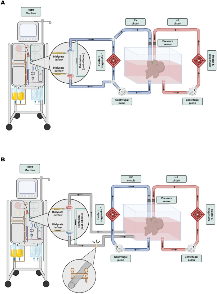
Figure 2: Representation of in-circuit and out-of-circuit CRRT integration in ex situ normothermic liver machine perfusion. (A) In the in-circuit configuration, the CRRT access line is connected directly to the venous outflow of the organ, and the return line rejoins just before the cannula that connects the perfusion machine to the portal vein. This setup can increase portal venous pressure and promote periportal edema. (B) In the out-of-circuit configuration, both the access and return lines of the CRRT are connected to the perfusion reservoir, hydraulically decoupling CRRT from the main circuit. This preserves hemodynamic stability and helps minimize pressure-induced periportal edema. The Hoffman clamp on the return line allows fine adjustment of downstream resistance1. Depending on the perfusion system, the portal vein perfusion circuit may or may not include an oxygenator, the inclusion of which is irrelevant for the connection of the CRRT device. Created with Biorender.com Please click here to view a larger version of this figure.
Successful integration of CRRT into ex situ liver NMP is demonstrated by stable perfusion pressures, consistent flow, and minimal graft weight gain throughout the perfusion period.
When CRRT is integrated during ex situ liver NMP directly in the high-volume, low-pressure portal perfusion circuit (n = 9), derivation of flow through the former induces hemodynamic alterations that require application of higher pressures to maintain constant, adequate flow to the graft. Under baseline conditions, flow through the circuit was maintained using a pressure-driven control algorithm, and physiological portal flow rates were achieved with a steady low pressure of 2.0 ± 0.5 mmHg. However, after the start of CRRT, derivation of some of the perfusate through the CRRT circuit caused portal flow to decline, and the switch had to be made from pressure- to flow-controlled perfusion, to maintain a minimum portal flow rate. Portal vein circuit pressure rose to 6.1 ± 1.4 mmHg within 2 h after the start of CRRT and 7.9 ± 1.3 mmHg after 16 h (p < 0.001 for both comparisons relative to baseline) (Figure 3A). Given that it was difficult using this configuration to accurately control actual pressure in the graft portal vein, the sustained circuit pressure elevation was accompanied by significant graft weight gain, reaching 53.3 ± 31.2% at 24 h (Figure 3C), primarily as a consequence of pressure-induced periportal edema formation.
In contrast, the out-of-circuit CRRT configuration, with CRRT connected to the perfusion reservoir (n = 8), maintained portal venous pressure more stable (Figure 3A). Portal flow remained stable throughout the perfusion period, with no relevant fluctuations observed between early and late timepoints (Figure 3B). Furthermore, the lower portal pressures achieved in this configuration were associated with a significantly reduced change in graft weight (53.3 ± 31.2 vs. 15.5 ± 13.1%, p=0.03), indicating effective prevention of periportal edema formation compared to the in-circuit setup.
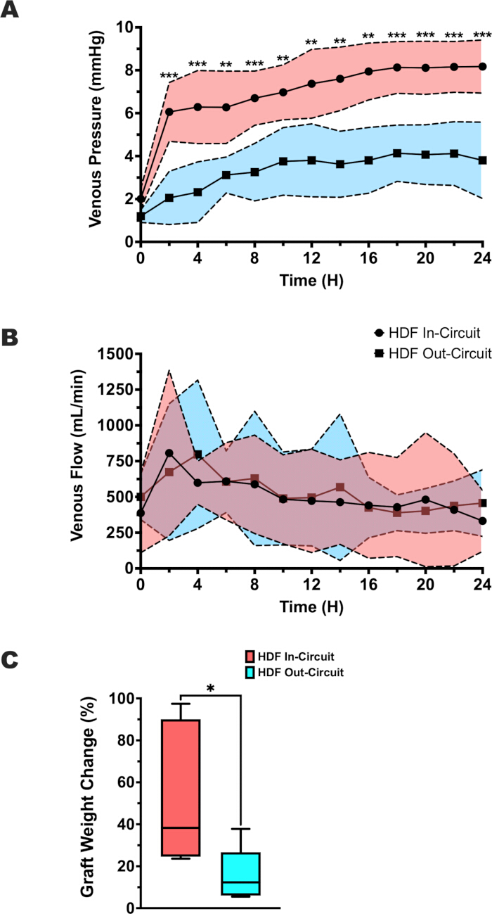
Figure 3: Portal venous pressure, flow, and graft weight change during normothermic liver perfusion with in-circuit and out-of-circuit CRRT configurations. (A) Portal venous pressure (mmHg) was measured continuously over a 24 h perfusion period. The in-circuit CRRT group (red shading, circles, n = 9) exhibited a significant and sustained increase in pressure following CRRT initiation, whereas the out-of-circuit group (blue shading, squares, n = 8) maintained stable pressure profiles within physiological limits. (B) Portal venous flow (mL/min) was recorded concurrently. Both groups demonstrated comparable flow stability over time, with no relevant fluctuations, reflecting the perfusion machine's flow regulation system. (C) Relative graft weight change (%) at the end of the 24 h perfusion period. The out-of-circuit group exhibited significantly lower weight gain compared with the in-circuit group, indicating reduced edema formation under stable hemodynamic conditions. Data are presented as (A,B) mean ± SD or as (C) box-and-whisker plots; Statistical comparisons were performed using one-way ANOVA test for portal venous pressure, Kruskal-Wallis test for portal venous flow, and Unpaired t test for graft weight change. Statistical significance is indicated as *p < 0.05; **p < 0.01; ***p < 0.001. Please click here to view a larger version of this figure.
All these results demonstrate that the reservoir-based CRRT integration avoids the negative consequences of direct in-circuit connection by maintaining perfusion dynamics.
To corroborate the solute-clearance efficiency of the new out-of-circuit configuration, serial measurements of biochemical and electrolyte parameters were performed throughout the 24 h perfusion (Figure 4). Both setups demonstrated a consistent decline in solute concentrations over time, indicating preserved filtration efficiency across configurations. In the in-circuit group, creatinine decreased from 0.91 ± 0.12 to 0.13 ± 0.03 mg/dL by 24 h, while in the out-of-circuit group it fell from 0.93 ± 0.06 to 0.15 ± 0.04 mg/dL (P > 0.99; Figure 4A). Similarly, BUN reached peak values 10.45 ± 4.33 mg/dL and 9 ± 2.18 mg/dL in the in-circuit and out-of-circuit groups, respectively, before declining to 4.44 ± 0.88 and 3.58 ± 1.34 mg/dL at 24 h, with no significant differences between configurations (p > 0.99; Figure 4B).
Electrolyte concentrations remained within physiological ranges over time. Sodium and bicarbonate levels (Figure 4C,E) were maintained between 135 and 145 mmol/L and 23-30 mmol/L, respectively, in both groups, reflecting appropriate regulation of osmotic and acid-base balance. Potassium concentrations (Figure 4D) transiently decreased during the first 4 h of perfusion, reaching < 3 mmol/L in both configurations (p < 0.001 compared with baseline), followed by subsequent stabilization as equilibrium was achieved between perfusate and replacement solutions. Minor early differences were not sustained at later time points.
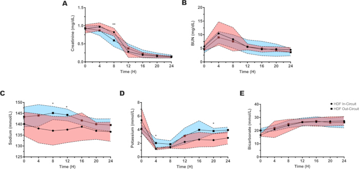
Figure 4: Solute clearance and electrolyte stability during normothermic liver perfusion with in-circuit and out-of-circuit CRRT configurations. (A) Creatinine (mg/dL) levels measured over 24 h perfusion period. Both configurations exhibited a progressive and comparable decline in concentration, indicating preserved small-solute clearance efficiency across setups. (B) BUN (mg/dL) evolution over time. BUN reached peak values early after CRRT initiation before declining steadily in both groups, with no significant differences between in-circuit (red shading, circles, n = 9) and out-of-circuit (blue shading, squares, n = 8) configurations. (C-E) Electrolyte dynamics during perfusion: (C) sodium, (D) potassium, and (E) bicarbonate. Sodium and bicarbonate concentrations remained within physiological limits throughout the 24 h perfusion, reflecting stable osmotic and acid-base regulation. Potassium levels showed a transient decrease during the first 4 h due to reperfusion, followed by a gradual normalization toward physiological values facilitated by the incorporation of CRRT. (A-E) Data are presented as mean ± SD. Statistical comparisons were performed using one-way ANOVA test for creatinine, sodium and bicarbonate levels; Kruskal-Wallis test for BUN; and Brown-Forsythe and Welch ANOVA test for potassium levels. Statistical significance is indicated as *p < 0.05; **p < 0.01; ***p < 0.001. Please click here to view a larger version of this figure.
Together, these findings indicate that solute clearance and electrolyte regulation were fully preserved when CRRT was integrated using the out-of-circuit configuration. The reservoir-based connection achieved equivalent clearance performance to the conventional in-circuit setup, while maintaining stable perfusion conditions throughout prolonged ex situ liver perfusion.
Discussion
This protocol presents a detailed and reproducible method for integrating a CRRT device during ex situ liver NMP by connecting the filtration circuit to the perfusion reservoir. The key advantage of this approach is the preservation of perfusion circuit hemodynamics and prevention of the deleterious pressure alterations typically observed with direct in-circuit CRRT integration. Critical steps include the correct placement of CRRT lines within the reservoir to prevent air entrapment, the use of a Hoffman clamp to simulate physiological venous pressure, and continuous monitoring of pressures and flows in both the CRRT and perfusion circuits.
In these experiments, CVVHDF was employed using a high-flux polyethersulfone hemofilter. HDF typically utilizes high-flux membranes to achieve the convective volumes required for middle-molecule clearance (~0.5-35 kDa), while medium cut-off (MCO) and, in selected applications, high cut-off (HCO) membranes may be used to extend solute removal toward larger molecular weights, balancing efficacy with the risk of albumin loss16. Although specific flow and UF parameters were optimized for this study, these can be adjusted according to the intrinsic characteristics of the perfusion, including perfusate viscosity, hematocrit, organ size, and the targeted filtration balance. Values typically range from 80 to 150 mL/min for BFR, 200 to 800 mL/h for DFR, and 100 to 800 mL/h for replacement flow (pre- or post-filter), with UF rates maintained near neutrality (± 25-50 mL/h adjustments, as needed). Because the circuit interfaces with the reservoir rather than the vascular pathway, variations in flow settings within these physiological ranges exert minimal influence on perfusion hemodynamics. When perfusion demands require higher blood or dialysate flow, incremental adjustments of the Hoffman clamp maintain venous-return pressure and prevent over-perfusion or edema formation, preserving stable perfusion dynamics throughout extended experiments.
Although several studies have explored the use of CRRT and other filtration-based methods during ex situ organ perfusion, only a handful have incorporated comprehensive CRRT machines7,8,9,31,32,33,34,35,36,37,38,39. These reports rarely provide detailed information regarding the technical aspects of circuit connection, nor do they systematically evaluate the hemodynamic or metabolic consequences of integrating a CRRT into the perfusion system. Most published protocols rely instead on custom-built filtration systems implemented without real-time monitoring or integrated safety features. While technically feasible, this lack of standardization and control introduces significant risks, especially in the context of isolated organ perfusion, where even minor deviations can have immediate and severe consequences for the graft40.
The use of homemade or non-automated devices for continuous EBP has considerable limits and may present hazards, regardless of the specific modality used. The lack of precise control mechanisms for fluid management, pressure regulation, and temperature stability are all major issues. Without monitoring and feedback, maintaining fluid balance becomes particularly challenging, as there is a heightened risk of sudden pressure fluctuations within the circuit. Uncontrolled ultrafiltration or convective transport may result in excessive or insufficient fluid removal, predisposing to hemodynamic instability, tissue edema, vascular and endothelial injury, hypovolemia with reduced organ perfusion and hypoxia, or even fluid overload41,42,43,44. The absence of real-time monitoring of transmembrane pressure and circuit pressures further increases the risk of undetected circuit instability, filter obstruction, or clotting, which can abruptly interrupt therapy and lead to the accumulation of metabolic waste products and fluid removal, ultimately compromising organ viability45,46. Temperature regulation is another crucial feature often overlooked in improvised devices. Using room-temperature solutions without active thermal management might cause occasional hypothermia, modifying their metabolic activity and impacting the function and preservation of the perfused organ47,48,49.
The approach for integrating CRRT during ex situ NMP presented herein lowers the risk of uncontrolled pressure fluctuations and fluid imbalances by utilizing the safety features and real-time monitoring capabilities of CRRT machines. An additional practical advantage of the reservoir-based configuration is the feasibility of circuit or filter replacement during extended perfusions. In clinical CRRT, filters and tubing sets typically have a recommended use time of ≤ 72-80 h to prevent progressive mechanical wear. In this experimental setup, because the CRRT circuit is hydraulically decoupled from the organ perfusion loop, replacement of the filter and tubing can be performed without interrupting perfusion or altering flow dynamics. Once the programmed operating time of the consumables is reached, the used components are discarded and replaced with new sterile lines following standard priming and system-start procedures. The entire process generally requires ≤ 20 min, during which the NMP reservoir and organ remain continuously perfused. Consequently, circuit changes can be executed safely and reproducibly, with negligible impact on graft viability and perfusion stability during long-term experiments. A potential limitation of this protocol is that it was validated using minimally injured porcine livers under experimental conditions; its performance with marginal or injured grafts remains to be tested. Additionally, while this setup is optimized for HDF, adaptations may be necessary when employing other EBP therapies, such as adsorption systems or plasma exchange, which could have different pressure and flow requirements.
The protocol's modular design enables easy adaptation to different ex situ organ perfusion platforms or filtration modalities, making it suitable for use in organs other than the liver, such as the heart, lung, pancreas, intestines, and even the kidney. By providing a clear, safe, and standardized approach, this method enables the reliable integration of CRRT into various experimental and translational settings, facilitating the broader application of EBPs during ex situ organ NMP.
Disclosures
The authors have no other conflicts of interest to declare. The funding bodies had no role in study design, data collection and analysis, interpretation of results, or manuscript preparation.
Acknowledgements
The authors thank Guangdong Shunde Innovative Design Institute for lending the perfusion system and for their research funding support, as well as NorrDia Spain Medical Device S.L., for providing a CRRT system for liver perfusion experiments. Jordi Vengohechea, Joaquim Albiol, Amelia Judith Hessheimer, and Constantino Fondevila have received research funding from Guangdong Shunde Innovative Design Institute. This study was also supported by funds from the Instituto de Salud Carlos III (ISCIII), PI18/00894 and PI23/00364, and co-funded by the European Union. Jordi Vengohechea was supported by a predoctoral fellowship from the University of Barcelona (PREDOCS-UB 2020).
Materials
| Name | Company | Catalog Number | Comments |
|---|---|---|---|
| 20 mL syringe Luer slip | Nipro Medical Spain SL | SY3-10ESC-GEC | |
| Accusol 35 K+ 2 (Potassium 2 mmol/L) | NIKKISO Europe GmbH | PPE02 | Replacement fluid for CRRT |
| AqualineS Bloodline | NIKKISO Europe GmbH | AQUASET03LV | Pediatric bloodline set for CRRT |
| Aquamax Filter HF03 | NIKKISO Europe GmbH | AQUASET03LV | Pediatric hemofilter (up to 30 kg donors) |
| AquariusTM System | NIKKISO Europe GmbH | Continuous renal replacement therapy machine | |
| Ex situ liver perfusion system | Guangdong Devocean Medical Instrument Co., Ltd. | Devocean-Liver&Kidney 3000 | Main organ perfusion platform composed by two centrifugal pumps with pulsatile/continual pressure perfusion, control panel (sensors for pressure, flow and temperature) and heater unit |
| Fresenius isotonic saline solution 500 mL | Fresenius Kabi | 138900 | |
| Hoffman clamp | Guangdong Devocean Medical Instrument Co., Ltd. | Custom made | Regulation of return line pressure |
| Male Luer-lock connectors | Provided by the Hospital | N/A | For the connection between the perfusion system tubing and the bloodline of the CRRT machine |
| Sodium Heparin 25.000 UI | Laboratorios Farmacéuticos Rovi S.A | 641639.6 | |
| Sterile tubing set | Guangdong Devocean Medical Instrument Co., Ltd. | Custom made | For reservoir access and return connections |
| Universal hose clamp | Achim SchuLz-Lauterbach Vertrieb medizinischer Producte GmbH | KL 20 |
References
- Jing, L., Yao, L., Zhao, M., Peng, L., Liu, M. Organ preservation: from the past to the future. Acta Pharmacol Sin. 39 (5), 845-857 (2018).
- Ozgur, O. S., et al. Current practice and novel approaches in organ preservation. Front Transplant. 2 (1), 1156845(2023).
- Ramos, P., et al. Abdominal organ preservation solutions in the age of machine perfusion. Transplantation. 107 (2), 326-340 (2023).
- Broere, R., Luijmes, S. H., De Jonge, J., Porte, R. J. Graft repair during machine perfusion: a current overview of strategies. Curr Opin Organ Transplant. 29 (4), 248-254 (2024).
- Lascaris, B., Hoffmann, R. F., Nijsten, M. W. N., Porte, R. J., De Meijer, V. E. Continuous renal replacement therapy during long-term normothermic machine perfusion of human donor livers for up to 7 d. Transplant Direct. 10 (2), e1568(2024).
- Huang, J., et al. Incorporating a hemodialysis filter into a commercial normothermic perfusion system to facilitate long-term preservation of human split livers. Artif Organs. 48 (9), 1008-1017 (2024).
- Clarke, G., et al. A reproducible extended ex vivo normothermic machine liver perfusion protocol utilising improved nutrition and targeted vascular flows. Commun Med. 4 (1), 214(2024).
- Cillo, U., Nalesso, F., Bertacco, A., Indraccolo, S., Gringeri, E. Normothermic perfusion of a human tumoral liver for 17 days with concomitant extracorporeal blood purification therapy: case description. J Hepatol. 81 (3), e96-e98 (2024).
- Nalesso, F., et al. The rationale for combining normothermic liver machine perfusion with continuous renal replacement therapy to maintain physiological perfusate during ex vivo organ perfusion. J Clin Med. 13 (17), 5214(2024).
- Vengohechea, J., Vaquero-Rey, A., Fondevila, C., Hessheimer, A. J. The role of renal replacement therapy in improving normothermic machine perfusion of liver grafts. Gastroenterol Hepatol. 48 (4), 502336(2025).
- Ostermann, M., et al. Nomenclature of extracorporeal blood purification therapies for acute indications: the Nomenclature Standardization Conference. Blood Purif. 53 (5), 358-372 (2024).
- Saunders, H., Rehan, A., Hashmi, M. F., Sanghavi, D. K. Continuous renal replacement therapy. StatPearls. , (2024).
- Verma, S., Palevsky, P. M. Prescribing continuous kidney replacement therapy in acute kidney injury: a narrative review. Kidney Med. 3 (5), 827-836 (2021).
- Jörres, A. Hemofiltration or hemodialysis for acute kidney injury. Crit Care. 16 (4), 147(2012).
- Kellum, J. A., Romagnani, P., Ashuntantang, G., Ronco, C., Zarbock, A., Anders, H. -J. Acute kidney injury. Nat Rev Dis Primers. 7 (1), 52(2021).
- Maduell, F. Hemodiafiltration. Hemodial Int. 9 (1), 47-55 (2005).
- Perez-Garcia, R., Alcazar-Arroyo, R., De Sequera-Ortiz, P. What is the role of expanded hemodialysis in renal replacement therapy in 2020. Nefrol Engl Ed. 41 (3), 227-236 (2021).
- Campo, S., Lacquaniti, A., Trombetta, D., Smeriglio, A., Monardo, P. Immune system dysfunction and inflammation in hemodialysis patients: two sides of the same coin. J Clin Med. 11 (13), 3759(2022).
- Losappio, V., et al. Molecular mechanisms of premature aging in hemodialysis: the complex interplay between innate and adaptive immune dysfunction. Int J Mol Sci. 21 (10), 3422(2020).
- Finkel, K. W., Waguespack, D. R. Renal replacement therapies. Onco-Nephrol. , 290-298 (2020).
- Fishman, G., Singer, P. Metabolic and nutritional aspects in continuous renal replacement therapy. J Intensive Med. 3 (3), 228-238 (2023).
- Harbord, N., Winchester, J. F., Charen, E., Sheth, N., Bhansali, A. Extracorporeal therapies in acute intoxication and poisoning. Crit Care Nephrol. , 588-594 (2019).
- Tolwani, A. J., Connor, M. J., Palevsky, P. M. Renal replacement therapy for acute kidney injury. Chronic Kidney Dis Dial Transplant. , 739-753 (2019).
- Ağbaş, A., et al. Hemodiafiltration is associated with reduced inflammation, oxidative stress and improved endothelial risk profile compared to high-flux hemodialysis in children. PLoS One. 13 (6), e0198320(2018).
- Canaud, B., Blankestijn, P. J., Grooteman, M. P. C., Davenport, A. Why and how high-volume hemodiafiltration may reduce cardiovascular mortality in stage 5 chronic kidney disease dialysis patients? A comprehensive literature review. Semin Dial. 35 (2), 117-128 (2022).
- Canaud, B., Davenport, A. The rationale and clinical potential of online hemodiafiltration as renal replacement therapy. Semin Dial. 35 (5), 380-384 (2022).
- Guimarães, M. G. M., et al. Hemodiafiltration versus hemodialysis in end-stage kidney disease: a systematic review and meta-analysis. Kidney Med. 6 (6), 100829(2024).
- Maduell, F., Rodríguez-Espinosa, D., Broseta, J. J. Latest trends in hemodiafiltration. J Clin Med. 13 (4), 1110(2024).
- Saunders, H., Rehan, A., Hashmi, M. F., Sanghavi, D. K. Continuous renal replacement therapy. StatPearls. , (2025).
- Machine perfusion for donor organ repair: from vision to everyday clinical practice. Organ Repair Regen. , 43-73 (2021).
- Abraham, N., et al. Orthotopic transplantation of the full-length porcine intestine after normothermic machine perfusion. Transplant Direct. 8 (11), e1390(2022).
- Banan, B., et al. Normothermic extracorporeal liver perfusion for donation after cardiac death livers. Surgery. 158 (6), 1642-1650 (2015).
- Banan, B., Watson, R., Xu, M., Lin, Y., Chapman, W. Development of a normothermic extracorporeal liver perfusion system toward improving viability and function of human extended criteria donor livers. Liver Transpl. 22 (7), 979-993 (2016).
- Bitsch, R. G., Düx, M., Helmberger, T., Lubienski, A. Effects of vascular perfusion on coagulation size in radiofrequency ablation of ex vivo perfused bovine livers. Invest Radiol. 41 (4), 422-427 (2006).
- Clavien, P. -A., et al. Transplantation of a human liver following 3 days of ex situ normothermic preservation. Nat Biotechnol. 40 (11), 1610-1616 (2022).
- De Haan, M. J. A., et al. A cell-free nutrient-supplemented perfusate allows four-day ex vivo metabolic preservation of human kidneys. Nat Commun. 15 (1), 3818(2024).
- Eshmuminov, D., et al. An integrated perfusion machine preserves injured human livers for 1 week. Nat Biotechnol. 38 (2), 189-198 (2020).
- Kappler, B., et al. Attenuated cardiac function degradation in ex vivo pig hearts. Int J Artif Organs. 43 (3), 173-179 (2020).
- Schuler, M. J., et al. Observations and findings during the development of a subnormothermic/normothermic long-term ex vivo liver perfusion machine. Artif Organs. 47 (2), 317-329 (2023).
- Clark, W., Villa, G., Ronco, C. Continuous renal replacement therapy machine technology. Crit Care Nephrol. , 853-860 (2019).
- Berger, D., Takala, J. Hypotension and hypovolemia during hemodialysis: is the usual suspect innocent. Crit Care. 20 (1), 140(2016).
- Canaud, B., Chazot, C., Koomans, J., Collins, A. Fluid and hemodynamic management in hemodialysis patients: challenges and opportunities. Braz J Nephrol. 41 (4), 550-559 (2019).
- Inal, M. T., Memiş, D., Karamanlıoglu, B., Sut, N. Effects of polygeline and hydroxyethyl starch solutions on liver functions assessed with LIMON in hypovolemic patients. J Crit Care. 25 (2), 361.e1-361.e5 (2010).
- Prowle, J., Mehta, R. Fluid balance management during continuous renal replacement therapy. Semin Dial. 34 (6), 440-448 (2021).
- Li, P., et al. Effect of dynamic circuit pressures monitoring on the lifespan of extracorporeal circuit and the efficiency of solute removal during continuous renal replacement therapy. Front Med. 8 (1), 621921(2021).
- Tsujimoto, Y., Fujii, T. How to prolong filter life during continuous renal replacement therapy. Crit Care. 26 (1), 62(2022).
- Akhoundi, A., et al. Incidence of adverse events during continuous renal replacement therapy. Blood Purif. 39 (4), 333-339 (2015).
- Bell, M., et al. Modern CRRT systems are associated with lower risk of hypothermia. Sci Rep. 14 (1), 74977(2024).
- Morsch, C. M. F., et al. Hypothermia related to continuous renal replacement therapy: incidence and associated factors. Rev Bras Ter Intensiva. 33 (1), 111-118 (2021).
Reprints and Permissions
This article has been published
Video Coming Soon