This protocol describes a scalable method for the expansion of Adoptive Cell Therapies (ACT) products, namely Chimeric Antigen Receptor (CAR)-T cells and Cytokine-Induced Killer (CIK) cells, using Gas-permeable Rapid expansion (G-Rex) devices, demonstrating efficient and robust growth in a clinically relevant setting.
Method Article
Efficient and Rapid Generation of CAR-T and Cytokine-Induced Killer Cells in GMP-scalable Devices
* These authors contributed equally
In This Article
Summary
Abstract
Adoptive cell therapies (ACT) have progressed rapidly in recent years, playing a pivotal role in cancer immunotherapy and significantly influencing treatment outcomes. A critical step in cell therapy development, at both preclinical and clinical stages, is the manufacturing process. Conventional static culture methods require frequent cell manipulation and are often limited by reduced gas exchange and nutrient supply, which can compromise cell yield and phenotype. Here, we present a step-by-step, reproducible, and scalable protocol for the ex vivo expansion of two ACT products, namely Chimeric Antigen Receptor (CAR)-T and Cytokine-Induced Killer (CIK) cells, using Gas-permeable Rapid expansion (G-Rex) devices. G-Rex devices are specifically designed to enhance nutrient exchange and support high-density cultures with minimal user intervention. Among various ACT approaches, CAR-T cells have demonstrated remarkable success in treating hematological malignancies, facilitating a rapid advance from experimental research into clinical applications. In addition to CAR-T cells, CIK cells have also been widely applied in clinical trials due to their characteristic phenotype and the absence of treatment-related adverse events and Graft-versus-Host Disease (GvHD). This protocol outlines the key technical steps for both cell types, starting with the isolation and seeding of peripheral blood mononuclear cells (PBMCs). Briefly, PBMCs are stimulated with anti-CD3/CD28 antibodies, followed by lentiviral transduction to expand CAR-T cells, or with Interferon-γ (IFN-γ), anti-CD3 antibody, and Interleukin 2 (IL-2) to obtain CIK cells. Flow cytometry is employed to monitor the viability and phenotype of the cells, and functional assays are performed to confirm the therapeutic potential of the cell product and to validate the scalability of the approach. Overall, this protocol provides a practical solution for producing large numbers of effector cells in a preclinical setting, facilitating clinical application with several immune cell populations. Finally, it offers a scalable solution for high-yield effector cell manufacturing.
Introduction
Adoptive cell therapy (ACT) has become an important pillar of advanced therapies for cancer treatment. This approach is based on administering ex vivo expanded cellular products to achieve anti-tumor efficacy1. To date, many cell types have been used in ACT, ranging from antigen-aspecific approaches, e.g., natural killer (NK)2, cytokine-induced killer (CIK)3,4, natural killer T (NKT)5, or γδ T cells6, to highly specific and personalized ones, such as tumor-infiltrating lymphocytes (TILs)7 and engineered cell-based therapies, e.g., chimeric antigen receptor (CAR)1,8 and T cell receptor (TCR) cells9,10.
CAR-T cells are genetically modified T cells generated by ex vivo viral transduction to express a synthetic immune receptor designed to recognize a specific molecular target. The CAR structure consists of an antigen-specific single-chain variable Fragment (scFv) antibody combined with intracellular signaling domains, including the TCRζ chain and costimulatory molecules. Upon antigen engagement, the intracellular domain delivers activation signals that promote T cell proliferation and antitumor activity11.
CIK cells are a heterogeneous population of activated T lymphocytes that acquire expression of CD56 during ex vivo expansion from peripheral blood mononuclear cells (PBMCs) or umbilical cord blood (UCB)12. This expansion involves stimulation with Interferon-γ (IFN-γ), anti-CD3 monoclonal antibody, and Interleukin-2 (IL-2), followed by culture in IL-2 for 14 days. CIK cells functionally resemble both NK and T cells, exerting antitumor activity through both major histocompatibility complex (MHC)-independent and dependent mechanisms13,14,15,16. These properties allow CIK cells to be used in both allogeneic and autologous therapeutic settings with minimal graft-versus-host disease (GvHD) risk17.
In clinical practice, both CAR-T and CIK cell therapies require the administration of large numbers of cells. CAR-T cells are infused at 50-200 million doses per patient18. Similarly, CIK cells are typically given at doses of up to 10 billion cells per infusion, with patients often receiving multiple infusions depending on the treatment protocol19. Traditional cell culture methods using flasks or plates are inefficient, time-consuming, and require frequent passaging, making the large-scale expansion unfeasible and increasing the risk of contamination. Automated or semi-automated systems using bag cultures or bioreactors allow for large-scale expansion but have limitations, i.e., shear stress or the need for density adjustments and frequent medium perfusion20,21, and the handling of one batch at a time, leading to high costs in both academic and industrial contexts. In this challenging situation, an alternative is represented by the gas-permeable rapid expansion (G-Rex) device, a static culture method featuring a silicon membrane at the bottom of the well, which ensures efficient oxygen exchange and increased height of the vessels, allowing for high-volume medium capacity. Thus, the system supports cell growth without medium replacement or density adjustment22,23. G-Rex-M systems have demonstrated scalability up to 5000 mL and are compatible with automated harvest systems, providing a contamination-free and closed culture platform that is easily translatable from academic to clinical settings for the development of advanced therapy medicinal products (ATMPs)24.
In the outlined procedure, we leveraged the advantages and characteristics of G-Rex devices to optimize the expansion protocol of both CAR-T and CIK cells. We demonstrated that both cell types can be efficiently expanded and scaled up, maintaining a consistent phenotype and functional profile across different culture systems. A schematic overview of the expansion protocol for both CIK and CAR-T cells is shown in Figure 1.
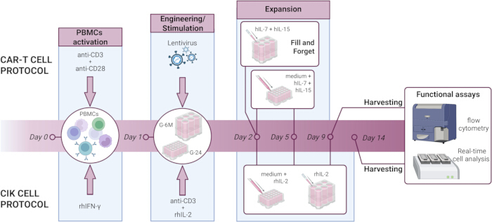
Figure 1: Overview of CAR-T and CIK cell expansion protocols using G-24 or G-6M culture devices. CAR-T and CIK cells are expanded from isolated PBMCs starting on Day 0. For the expansion of CAR-T cells, PBMCs are stimulated on Day 1 with anti-CD3 and anti-CD28 antibodies, genetically engineered on Day 2, and subsequently cultured with hIL-7 and hIL-15 supplementation every 3-4 days, with final harvest for functional assays on Day 9. In the G-6M format, cytokine supplementation is limited to Day 2, with no additional cytokine feeding required throughout the culture period. For the expansion of CIK cells, PBMCs are stimulated on Day 1 with rhIFN-γ and on Day 2 with anti-CD3 antibody and rhIL-2, which is then provided every 3-4 days. In G-24, a medium change is required concurrently with the cytokine supplement. CIK cells are harvested on Day 14 for downstream functional assays. Please click here to view a larger version of this figure.
Protocol
Anonymized buffy coats from healthy donors previously tested for the absence of pathogen contamination (e.g., HIV, HCV, HBV, syphilis) were obtained from the Blood Bank of Padova Hospital prior to written informed consent. Blood samples were manipulated within certified biosafety cabinets, and personnel wore appropriate Personal Protective Equipment (PPE).
NOTE: All procedures involving lentiviral vectors were conducted under BioSafety Level 2 (BSL-2) conditions, as per institutional guidelines. Lentiviral vector handling requires additional precautions, including double gloving. Biohazardous waste (including viral materials, transduced cells, and human-derived samples) was autoclaved before disposal, while chemical waste (including fluorescent dyes and reagents) required segregation according to institutional hazardous waste protocols. Note that specific biosafety requirements may vary between institutions and should be verified prior to protocol implementation.
1. PBMCs isolation
- Isolate PBMCs from healthy donor buffy coats or peripheral blood samples by density gradient centrifugation. Dilute the buffy coat with an equal volume of sterile 1x Phosphate Buffer Saline (PBS) and carefully layer the diluted blood over the same volume of Lymphoprep in a 50 mL conical tube. Dilution with 1x PBS is not necessary for peripheral blood. Centrifuge at 400 x g for 20 min at room temperature without brake.
- Following centrifugation, collect the PBMCs layer at the plasma-Lymphoprep interface and wash it with sterile 1x PBS. Centrifuge and resuspend PBMCs in hematopoietic cell culture medium (CM) supplemented with 1% penicillin/streptomycin (hereafter cCM). Assess cell count and viability using the Trypan Blue exclusion assay.
2. CAR-T cell expansion from PBMCs in G-24 culture vessels
- PBMCs activation (Day 0)
- Resuspend 2 x 10⁶ freshly isolated PBMCs in 2 mL of cCM supplemented with 5% fetal bovine serum (FBS) (hereafter cCM-F), resulting in a final seeding density of 1 x 10⁶ PBMCs/cm2/mL, and transfer into a well of a 24-well G-Rex (G-24) culture vessel. Seed as many wells as the number of different transduction conditions to perform at Day 1 (Step 2.2- Day 1).
- Add 4 µl of anti-CD3 (pure-functional grade, final concentration: 0.2 µg/mL) and 20 µl anti-CD28 (pure-functional grade, final concentration: 1 µg/mL) antibodies directly to each well to activate T cells. Culture cells in a humidified incubator maintained at 37 °C, 5% CO2.
- T cell transduction with lentiviral vectors (Day 1)
- Following overnight stimulation, harvest and count the activated T cells using the Trypan Blue exclusion assay to assess viability. Calculate 1 x 10⁶ of viable activated cells for each well of G-24 (well area of 2 cm2), corresponding to a seeding density of 0.5 x 10⁶ cells/cm². Transfer cells in a tube and centrifuge at 300 x g for 5 min.
- Prepare the transduction media for each well by combining the appropriate lentiviral vector volume, based on the concentration (TU/mL) of the lentiviral vector preparation (here, CD19-CAR-BBζ lentivirus obtained from St. Jude Children's Research Hospital25; multiplicity of infection (MOI) 20), with 5 µl of the transduction enhancer protamine sulfate (8 mg/mL stock solution is obtained by dissolving protamine sulfate in 1x PBS and sterile filtered using 0.2 µm filter; final concentration of 50 µg/mL). Use CM medium supplemented with 5% FBS without the addition of penicillin/streptomycin to bring to a final volume of 0.8 mL/well (0.4 mL/cm²). For untransduced (UTD) control prepare the described transduction media avoiding the addition of lentiviral vector.
- Resuspend pelleted cells (step 2.2.1) in 0.8 mL/well of transduction media and plate them in a new G-24 well. Incubate overnight at 37 °C, 5% CO2.
NOTE: If transduction of multiple wells with the same lentiviral vector is planned, prepare a master mix of the transduction medium containing the virus and protamine sulfate at the desired concentration. Mix thoroughly to ensure homogeneity. Then, aliquot 0.8 mL of this master mix into each tube of pelleted cells to guarantee consistent transduction conditions across all samples.
- CAR-T cells culture and expansion (Day 2 to Day 9)
- On Day 2, increase the culture volume to a final volume of 8 mL/well by adding 7.2 mL of fresh cCM-F, supplemented with human Interleukin-7 (hIL-7) and human Interleukin-15 (hIL-15) at a final concentration of 10 ng/mL each (volume of cytokines calculated considering a total volume of 8 mL per well).
- On Day 5, refresh the culture media by carefully removing 6 mL of conditioned medium from each well without disturbing the cells at the bottom of the well and replacing it with 6 mL of fresh cCM-F supplemented with hIL-7 and hIL-15 (final concentration: 10 ng/mL calculated considering a total volume of 8 mL per well). If working with multiple wells, prepare a master mix of cCM-F media supplemented with hIL-7 and hIL-15 cytokines to ensure an even distribution of the cytokines.
- On Day 9, the CAR-T cells are efficiently expanded and ready for downstream applications. To harvest the CAR-T cells, gently remove and discard 5 mL of conditioned media. Using a 1000 µL pipette, resuspend the CAR-T cells in the remaining 3 mL of media and move them to a 15 mL tube. Count CAR-T cells and check their viability using the Trypan Blue exclusion assay. Typically, about 3 x 10⁷ CAR-T cells (3.24 ± 1.67 x 10⁷ cells) are obtained from each G-24 well (Supplementary Table 1).
3. CIK expansion in G-24 culture vessels
- PBMCs activation (Day 0)
- Seed 1 x 10⁶ freshly isolated PBMCs for each well of a G-24 culture vessel, which corresponds to a density of 0.5 x 10⁶/cm2, in 8 mL of cCM supplemented with recombinant human Interferon-γ (rhIFN-γ, final concentration: 1000 IU/mL). Incubate the cells at 37 °C, 5% CO2.
- CIK cell stimulation and expansion (Day 1 to Day 14)
- On day 1, 22-24 h after rhIFNγ stimulation, add directly to each well 4 µL of anti-CD3 antibody (final concentration: 50 ng/mL) and 4 µL of recombinant human Interleukin-2 (rhIL-2, final concentration: 500 IU/mL). Incubate the cells at 37 °C, 5% CO2.
- Every 3 to 4 days (on day 5, 9, and 12 of cell culture), replace 6 mL of conditioned medium with fresh cCM and rhIL-2 (final concentration: 500 IU/mL), calculating the final concentration of rhIL-2 relative to the full 8 mL volume.
- On day 14, harvest CIK cells by discarding 5 mL of conditioned media and resuspending them in the remaining 3 mL of conditioned media. Transfer cells into a 15 mL tube and assess total cell number and cell viability using the Trypan Blue exclusion assay. Typically, about 8 x 10⁷ bulk CIK cells (8.6 ± 4.58 x 10⁷ cells) are obtained from each well (Supplementary Table 2).
NOTE: During medium changes, carefully aspirate the supernatant, taking care not to disturb the cell layer population or contact the gas-permeable membrane. Damage to the membrane may impair gas exchange and negatively affect cell viability.
4. Large-scale CAR-T and CIK cells' manufacturing
NOTE: The CAR-T and CIK cell expansion protocols described in Sections 2 and 3 can be easily scaled up to larger G-Rex culture devices, namely the G-6M format. When transitioning from the G-24 format (surface area: 2 cm2) to the G-6M device (surface area: 10 cm2), all concentrations and densities are maintained constant, while all volumes and absolute quantities are proportionally increased by a 5-fold factor to match the increased surface area. This means that cell seeding density, media supplementation concentrations, and cytokine concentrations remain unchanged, but the total volumes of media, number of cells seeded, and absolute amounts of cytokines added will be scaled up proportionally. This scalability feature enables a smooth transition from small-scale research protocols to preclinical or early clinical manufacturing without requiring significant protocol modifications, thereby maintaining consistent effector cell characteristics.
- CAR-T cells expansion in G-6M format devices
- Perform PBMCs' activation on Day 0 and lentiviral transduction on Day 1, as described in Section 1, maintaining all concentrations and densities while scaling up volumes and absolute quantities by a 5-fold factor.
- On Day 2, bring the culture volume to a total of 100 mL using cCM-F medium. Calculate and add hIL-7 and hIL-15 to achieve the final concentration of 10 ng/mL accordingly.
NOTE: Unlike the G-24 format, the taller design of the G-6M device allows for a greater medium volume, enabling a steady nutrient and cytokine supply throughout the culture period without the need for media replacement ("Fill and Forget" approach). Therefore, no additional cytokines are required on Day 5. - To harvest CAR-T cells on Day 9, discard about 90 mL of medium from each well using a serological pipette and resuspend the cells in the remaining 10 mL. Transfer cells to a 15 mL tube and use them for subsequent assays.
- CIK cells expansion in G-6M format devices
- Perform PBMCs' seeding on Day 0 following the protocol described in Section 3 and scale up all volumes and reagents by a 5-fold factor to match the larger device format (G-6M).
- On Day 1, adjust the culture volume to a total of 100 mL with cCM media. Add a final concentration of 500 IU/mL rhIL-2 and 50 ng/mL anti-CD3 to each well.
- Supplement 500 IU/mL rhIL-2 every 3-4 days (day 5, 9, and 13) to each well without medium change.
- Harvest CIK cells on Day 14 by removing about 90 mL of medium from each well with a serological pipette. Resuspend the cells in the remaining 10 mL, move them to a 15 mL tube, and use them for subsequent assays.
5. Effector cell phenotype characterization by flow cytometry
NOTE: At the end of the expansion protocol, surface marker analysis by flow cytometry is performed on CAR-T and CIK cells to evaluate their phenotype, including distinct differentiation states, i.e., Naïve (CD45RA+CD62L+), Central Memory (CM, CD45RA-CD62L+), Effector Memory (EM, CD45RA-CD62L-), and Effector Memory CD45RA-positive (EMRA, CD45RA+CD62L-; Supplementary Figure 1A-B).
- Collect 3 x 10⁵ cells for each staining tube. Wash cells in 1x PBS with 1% FBS (PBS-F), centrifuge at 300 x g for 5 min, and resuspend the cell pellet in 50 µL of antibody mix solution prepared in staining buffer (1 part Brilliant stain buffer and 1 part PBS-F) according to the desired staining panel (Table 1 and Table 2).
- Incubate the cells for 20 min at 4 °C protected from light, then wash the stained cells and centrifuge. If a second staining step is required, perform the staining with the specific antibody mix, as described before (step 5.1).
- Lastly, wash cells and stain with a viability dye, here Fixable Viability Stain 780 (FVS780), in 50 µL of 1x PBS, incubating for 15 min at 4 °C protected from light. A wash step is not required following viability staining. After incubation, add 200 µL of 1x PBS to each sample, and proceed with flow cytometric acquisition. Detailed gating strategy is shown in Supplementary Figure 1A-B.
| Target | Clone | Fluorochrome | Concentration/Dilution | Purpose | |
| First staining step | rhCD19-His | / | / | 2 µg/mL | CAR molecule detection |
| Incubation at 4 °C for 20 min | |||||
| Washing step | |||||
| Second staining step | His | GG11-8F3.5.1 | PE | 1/50 | CAR molecule detection |
| CD3 | UCHT1 | BV510 | 1/40 | Lineage | |
| CD4 | SK3 | BV650 | 1/40 | Lineage | |
| CD8 | RPA-T8 | BV421 | 1/20 | Lineage | |
| CD45RA | HI100 | PerCP-Cy5.5 | 1/83 | Maturation | |
| CD62L | DREG-56 | FITC | 1/10 | Homing, activation | |
| Incubation at 4 °C for 20 min | |||||
| Washing step | |||||
| Third staining step | FVS780 | / | / | 1/5000 | Live/Dead cell discrimination |
| Incubation at 4 °C for 15 min | |||||
Table 1: Antibody panel used for CAR-T cells staining. Staining panel used to evaluate CAR expression, T cell phenotype, and differentiation status. Antibody clone, concentration, and conjugated fluorochrome for each target of interest are reported.
| Target | Clone | Fluorochrome | Dilution | Purpose | |
| First staining step | CD56 | HCD56 | PE | 1/40 | Lineage |
| NKG2D | 1D11 | APC | 1/83 | Activation | |
| CD3 | UCHT1 | BV510 | 1/40 | Lineage | |
| CD4 | SK3 | BV650 | 1/40 | Lineage | |
| CD8 | RPA-T8 | BV421 | 1/20 | Lineage | |
| CD45RA | HI100 | PerCP-Cy5.5 | 1/83 | Maturation | |
| CD62L | DREG-56 | FITC | 1/10 | Homing, activation | |
| Incubation at 4 °C for 20 min | |||||
| Washing step | |||||
| Second staining step | FVS780 | / | / | 1/5000 | Live/Dead cell discrimination |
| Incubation at 4 °C for 15 min | |||||
Table 2: Antibody panel used for CIK cells staining. Staining panel used to evaluate CIK subpopulations, phenotype, and differentiation status. Antibody clone, concentration, and conjugated fluorochrome for each target of interest are reported.
6. Evaluation of the specific cytotoxic activity of CAR-T and CIK cells
NOTE: The cytotoxic activity of CAR-T and CIK cells is measured by co-culturing them with target cells at the desired effector-to-target (E:T) cell ratio. The type of assay depends on the type of target cells, whether they are derived from solid tumors or hematological malignancies. In this protocol, we describe a fluorescent cell dye-based flow cytometry assay for suspension cells and an impedance-based Real-Time Cell Analysis (RTCA) assay for adherent cells.
- Fluorescent cell dye-based flow cytometry assay
NOTE: Antigen-specific cytotoxicity against hematological malignancies can be evaluated using a fluorescent cell dye-based flow cytometry assay. Before co-culture, target cells are labelled with a fluorescent dye to enable their identification. Following co-culture with effector cells, target cell death is assessed by staining with a viability dye. Flow cytometric analysis allows for the quantification of antigen-specific killing, based on the proportion of non-viable fluorescent dye-positive cells.- For each co-culture condition, collect 2 x 10⁵ target cells and resuspend them in 400 µL of complete culture media to achieve a final concentration of 5 x 10⁵ cells/mL. Add fluorescent dye at a final concentration of 5 µM (here Cell Trace Violet dye, CTV). Incubate for 30 min at 37 °C, protected from light. Following incubation, wash the cells once with complete medium and resuspend the pellet at a final concentration of 4 x 10⁵ cells/mL.
- Dispense 500 µL of the stained target cell suspension (here 2 x 10⁵ of Raji cells as a CD19+ suspension target cell model) into clean 5 mL tubes according to the experimental setup. Confirm successful staining by flow cytometry by identifying target cells as cells positive to the fluorescent dye (SSC-A versus CTV). Around 98% of fluorescent dye-positive cells is recommended.
- In parallel, count and prepare effector cells (here 5 x 10⁶ of CD19 CAR-T cells) for each co-culture condition. Wash the cells and resuspend them in complete culture medium at the appropriate concentration to match the desired effector-to-target (E:T) ratio. Add 500 µL of the effector cell suspension to each corresponding tube containing target cells.
- To achieve a 25:1 E:T ratio, add 5 x 10⁶ effector CD19 CAR-T cells to 2 x 10⁵ Raji target cells in each tube, and prepare as many tubes as required by the experimental design.
- Incubate the co-cultures for 6 h at 37°C in a humidified incubator with 5% CO2. Include a control condition containing only target cells to measure spontaneous cell death. This control is important for distinguishing background death from effector cell-specific cytotoxicity.
- Wash the co-cultured cells and stain them with FVS780 vitality dye diluted in 1x PBS (final dilution 1:5000). Incubate for 15 min at 4 °C, protected from light. Wash the cells and resuspend the final cell pellet in 600 µL of PBS-F buffer. Proceed with flow cytometry acquisition to evaluate effector cell-specific cytotoxicity.
- Perform the flow cytometer gating strategy as follows: identify target cells as cells positive to the fluorescent dye (SSC-A versus CTV), and then, within this population, calculate the percentage of dead target cells (SSC-A versus FVS780, Supplementary Figure 1C). Calculate Specific Lysis by subtracting spontaneous target cell death (%FVS780+ within CTV+ in the target only condition) from the effector cell-specific cytotoxicity (%FVS780+ within CTV+ in the effector-target co-culture condition).
- Real-time cell analysis (RTCA) assay
NOTE: The RTCA assay is a method for assessing the cytotoxic activity of effector cells against adherent target cells26. Target cell death is detected as a difference in the impedance signal due to the detachment of cells from the E-plate well engaged in the xCELLigence device. Perform the RTCA assay following the manufacturer's recommended protocol to ensure optimal assay performance and reproducibility.- Subculture target cells one day before the assay (Day -1) to ensure they reach 60%-80% confluency by Day 0. This step is critical to maintain the cells in active proliferation at the beginning of the experiment. It is preferred to use target cells between passages 5-20, with a minimum of 1 week (3-4 passages) post-thaw to ensure optimal cell performance.
- On Day 0, carefully add 50 µL of culture medium to each well of the E-Plate, engage the plate inside the device, and acquire the background signal. Then, transfer the plate back to the hood for the cell seeding step.
- Count target cells and transfer the required total number into a clean tube. Resuspend cells in an appropriate volume of culture medium to achieve the desired concentration for seeding into the E-Plate, and transfer 100 µL of target cells into each well (here 4 x 10⁴ of MCF-7 cells as an adherent tumor cell line model). Keep the plate inside the hood at room temperature for at least 30 min to ensure even attachment of target cells. Engage the plate inside the device at 37 °C in a CO2 incubator and start the real-time acquisition (acquisition interval of 15 min for 22 h).
NOTE: Perform target cell titration before using a new target cell line, as suggested by the manufacturer. - On Day 1 (around 20-22 h post target cell seeding), count the effector cells and resuspend them in fresh culture medium, adjusting the cell concentration to achieve the desired effector-to-target (E:T) ratio. For example, to achieve a 10:1 E:T ratio, add 4 x 10⁵ effector CIK cells to 4 x 10⁴ MCF-7 target cells for each well.
- Prepare each treatment condition in technical duplicates using a sterile 96-well U-bottom plate and equilibrate it for 15 min at 37°C. Dispense 50 µL of the effector cell suspension for each condition using a multi-channel pipette. Place the E-Plate back into the instrument and continue the real-time acquisition (acquisition interval of 15 min for 24 h). Calculate Specific Lysis using the following equation:

Results
The protocol described here provides an efficient and robust workflow for the ex vivo expansion and engineering of effector cells from isolated PBMCs, namely CAR-T and CIK cells. This method requires minimal cell manipulation and takes advantage of the gas-exchange capacity of G-Rex devices, providing superior nutrient availability and oxygenation compared to conventional static culture systems23,24. A key advantage of this protocol is its inherent scalability from small-scale 24-well devices (G-24), suitable for preclinical studies, to larger 6M devices (G-6M), compatible with translational or clinical manufacturing needs, without requiring any changes to the protocol other than proportional scaling.
Using this protocol, both CAR-T and CIK cells were successfully expanded in parallel from healthy donor PBMCs in G-24 and G-6M formats, as described in Sections 2-4. At the end of the culture period, both cell types exhibited high fold expansion and viability, with comparable proliferation between the two device formats (Figure 2, Supplementary Table 1, Supplementary Table 2), highlighting the reproducibility of the protocol across different culture scales.
To validate the scalability of the culture devices, effector cells were further characterized phenotypically and functionally. For the CD19 CAR-T cells, we quantified CAR expression and evaluated the CD4+ and CD8+ subsets, noting a predominance of CD19 CAR+ CD8+ T cells (Figure 3A-B, Supplementary Table 1). For CIK cells, we assessed the frequency of T (CD3+CD56-), NK (CD3-CD56+), and CIK (CD3+CD56+) populations (Figure 3D), as well as the expression of the activating receptor NKG2D (Figure 3E, Supplementary Table 2). Moreover, we observed overall comparable phenotypes and expression of specific population markers between the G-24 and G-6M culture formats for both CAR-T and CIK cells. The differentiation status of both CD19 CAR-T and CIK cell products was analyzed by CD62L and CD45RA staining (Figure 3C-F, Supplementary Table 1, Supplementary Table 2), revealing similar memory and effector phenotypes between cells cultured in G-24 and G-6M formats. These findings demonstrate that scaling up the process does not compromise the phenotypic integrity or differentiation status of the expanded effector cell populations.
Lastly, to evaluate functional potency, we employed target-specific cytotoxicity assays. Both CD19 CAR-T and CIK cells expanded in either device exhibited potent cytolytic activity, confirming the maintenance of functional properties across scale (Figure 4). Overall, these results support the suitability of the expanded products for downstream applications in preclinical models and potentially clinical testing.
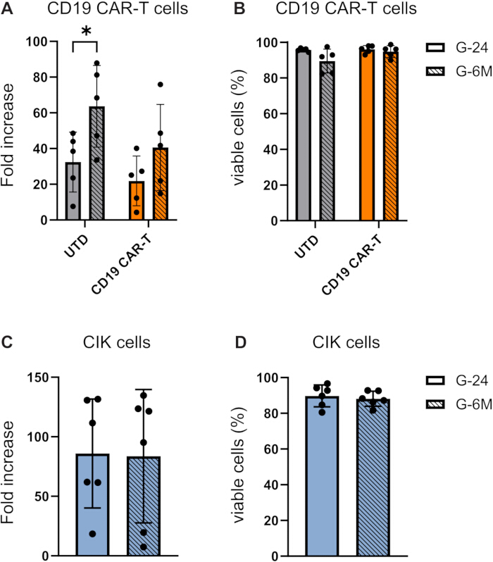
Figure 2: Proliferation and viability ofCD19 CAR-T and CIK cells. CAR-T cells (n = 5) and CIK cells (n = 6) expanded from PBMCs in G-24 and G-6M plates were collected on Day 9 and Day 14 of expansion, respectively, to perform cell count and evaluation of the (A-C) fold increase and (B-D) percentage of viable cells. Histograms represent the mean expression ± standard deviation (SD), and dots indicate single donors (see Supplementary Table 1 and Supplementary Table 2 for individual values). Data was analyzed by two-tailed multiple paired t-test (A-C) or multiple two-tailed Wilcoxon matched-pairs rank test (B-D), with Holm-Sidak correction for multiple comparisons (*p < 0.05). Please click here to view a larger version of this figure.

Figure 3: Assessment of the phenotype of CD19 CAR-T and CIK cells expanded in G-24 and G-6M devices. The expression of CAR molecules on CD19 CAR-T cells (n = 5) was analyzed by flow cytometry to assess (A) the transduction efficiency in G-24 and G-6M expansion protocols, as well as the expression of (B) CD4+/CD8+ and (C) naïve/memory subpopulations in the CD3+CAR+ subset. The phenotype of bulk CIK cells (n = 6) was analyzed by flow cytometry to investigate (D) the percentage of CIK (CD3+CD56+), NK (CD3-CD56+) and T (CD3+CD56-), as well as the expression of (E) NKG2D and (F) naïve/memory subpopulation in the CD3+CD56+ subset (G-24 n=5, G-6M n=6). Naïve (CD45RA+CD62L+), Central Memory (CM, CD45RA-CD62L+), Effector Memory (EM, CD45RA-CD62L-), and Effector Memory CD45RA-positive (EMRA, CD45RA+CD62L-). Histograms represent the mean expression ± SD, and dots indicate single donors (see Supplementary Table 1 and Supplementary Table 2 for individual values). Data was analyzed by two-tailed multiple paired t-test (B-C-D-F) or two-tailed Wilcoxon matched rank test (A-E), with Holm-Sidak correction for multiple comparisons (*p < 0.05). Please click here to view a larger version of this figure.
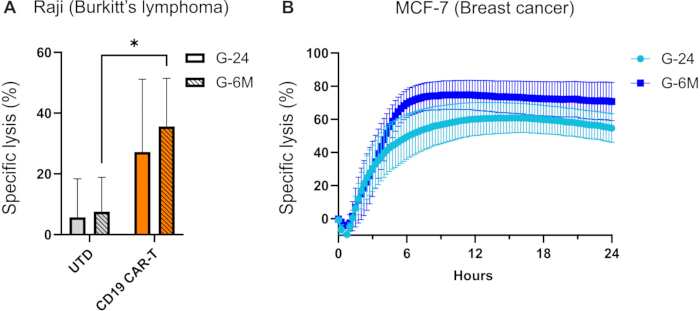
Figure 4: Evaluation of CD19 CAR-T and CIK cell cytotoxicity. (A) Specific lysis of CD19 CAR-T cells (orange, n = 5) or untransduced (UTD) cells (gray, n = 5) against the Burkitt's lymphoma Raji cell line (as a suspension cell line model) was performed by a 6 h flow cytometry-based killing assay. Results are shown as the mean specific lysis ± SD. (B) Cytotoxicity of CIK cells cultured in either G-24 (light blue, n = 7) or G-6M (dark blue, n=6) against the breast cancer MCF-7 cell line (as an adherent cell line model) was monitored every 15 min over 24 h using a real-time cell analyzer. Results are shown as the mean specific lysis ± SEM. Data was analyzed by Mann-Whitney test, Holm-Sidak correction for multiple comparisons (*p < 0.05). Please click here to view a larger version of this figure.
Supplementary Figure 1: Gating strategy used for flow cytometry analyses. Representative plots illustrating the gating strategies for (A) CAR-T cell and (B) CIK cell phenotypic characterization, and (C) fluorescent cell dye-based cytotoxicity assay. Please click here to download this File.
Supplementary Table 1: Growth, viability, and phenotype of CD19 CAR-T cell cultures from individual donors. Total cell number, fold increase, viability, and phenotypic characterization of CD19-CAR T cells harvested on Day 9 of culture in G-24 and G-6M devices are shown for each donor. Please click here to download this File.
Supplementary Table 2: Growth, viability, and phenotype of CIK cell cultures from individual donors. Total cell number, fold increase, viability, and phenotypic characterization of CIK cells harvested at Day 14 of culture in G-24 and G-6M devices are shown for each donor. Please click here to download this File.
Discussion
This protocol outlines the implementation of an efficient and scalable platform for the generation of two different ACT products, namely CAR-T and CIK cells, from PBMCs using G-Rex culture systems. We implemented proof-of-concept functional and phenotypic assays to validate the scalability of the approach. We showed that both phenotype and activity of the expanded cells are maintained during scale-up, ensuring the system suitability for research applications and direct clinical translation. Indeed, we successfully demonstrated that both CAR-T and CIK cells can be efficiently expanded with high cell yield, while maintaining a functional effector phenotype and potent antigen-specific cytotoxicity across different culture devices.
Combined with the gas-permeable membrane technology of the culture devices, this expansion protocol leads to faster expansion kinetics, reduces manual handling, and allows consistent high yields across donors. These features make the protocol highly reproducible and less labor-intensive compared to standard culture systems20,21, which often involves multiple media exchanges and increased risk of cell loss and contamination.
Moreover, this protocol enables seamless transition across manufacturing scales: from small-scale open systems (G-24, suitable for preclinical research and screening, for example, of CAR constructs, cytokine combinations, or donor variability) to larger-scale open systems (G-6M, for pre-GMP validation), and ultimately to larger-scale closed systems (for GMP production). As described in this study, the progression from small-scale to larger-scale open systems requires only proportional adjustments of volumes and cell input relative to device size, which represents a significant practical advantage. Likewise, the only implementation needed for the transition from open to closed systems for GMP manufacturing, and thus to adapt the G-6M protocol to large-scale GMP production, is utilizing appropriate GMP-grade reagents and closed system devices (G-10M-CS with a surface area of 10 cm2, corresponding to a single G-6M well, or G-100M-CS with a surface area of 100 cm², corresponding to the G-100M open counterpart) without any further modification of the protocol itself. These closed devices differ from their open counterparts in the integration of sterile tubing connections for automated media handling, which work in conjunction with a liquid handling pump (GatheRex) to create a fully closed environment. This closed system approach eliminates manual media exchanges and reduces contamination risks, making it ideally suited for GMP-compliant manufacturing environments where sterility and reproducibility are paramount. Notably, scalability represents a critical feature for bridging the gap between early-stage discovery and clinical translation, particularly in academic or early biotech settings with limited resources.
Further implementation of this protocol toward full GMP compliance should consider transitioning from FBS-containing media to serum-free/xeno-free formulations, aligning with current regulatory trends from the FDA and EMA, in order to eliminate batch-to-batch variability, remove potential xenogeneic contamination risks, and enhance regulatory compliance27.
Moreover, the minimal intervention required by the proposed expansion system provides an ideal platform for implementing multi-dimensional phenotypic profiling28 by reducing procedure-induced artifacts that might confound phenotypic analysis during manufacturing procedures. Additionally, beyond CAR-T and CIK cells, the system could be highly adaptable to other lymphocyte populations and engineered immune cells, broadening its utility in immunotherapy development29,30.
Although the protocol is robust and does not require modifications, a few steps are particularly critical for success. For CIK cells expansion, the timing of cytokine addition is essential to ensure optimal differentiation and expansion. For CAR-T cells, the transduction should occur at 24 h after activation to maximize efficiency, and lentiviral aliquots should be thawed only once, as repeated freeze-thaw cycles significantly reduce transduction efficiency31.
Biological variability among primary cell preparations naturally contributes to differences in expansion rates and functional activity, reflecting donors' diversity. This inevitable and intrinsic variability can be effectively managed by performing parallel expansions from multiple donors, ensuring robust and reproducible outcomes across experiments.
In conclusion, this protocol enables rapid, scalable, and reliable expansion of functional effector lymphocytes directly from PBMCs, offering flexibility for both early-phase research and large-scale clinical translation. This system can be readily adapted to various cell types and genetic modifications, providing a versatile platform for immunotherapy development.
Acknowledgements
The CAR construct was provided under MTA by St. Jude Children's Research Hospital. This work was supported by ScaleReady G-Rex Grant 2025; University of Padova - SOMMAGGIO_BIRD24_01; AIRC Foundation under 5x1000 2019 - ID. 22759 program; Alleanza Contro il Cancro, Progetto CAR-T_2 RCR-2023-23684268. This research received "Ricerca Corrente" funding from the Italian Ministry of Health to cover publication costs. Figures were created with GraphPad Prism 9 Software and BioRender.com.
Materials
| Name | Company | Catalog Number | Comments |
|---|---|---|---|
| Brilliant Stain Buffer | BD Biosciences | 566349 | |
| CD28 Antibody, anti-human | Miltenyi Biotech | 130-093-375 | Clone 15E8 |
| CD3 Antibody, anti-human | Miltenyi Biotech | 130-093-387 | Clone OKT3 |
| CD3 BV510 | BD Biosciences | 740147 | Clone UCHT1 |
| CD314 APC (NKG2D) | Biolegend | 320808 | Clone 1D11 |
| CD4 BV650 | BD Biosciences | 563875 | Clone SK3 |
| CD45RA PerCP-Cy5.5 | BD Biosciences | 563429 | Clone HI100 |
| CD56 PE | Biolegend | 318306 | Clone HCD56 |
| CD62L FITC | BD Biosciences | 555543 | Clone FREG-56 |
| CD8 BV450 | BD Biosciences | 562428 | Clone RPA-T8 |
| CellTrace Violet Cell Proliferation Kit | ThermoFisher scientific | C34557 | |
| Dimethyl sulfoxide (DMSO) | ThermoFisher scientific | D2650 | |
| Dulbecco′s Phosphate Buffered Saline (D-PBS) | Sigma Aldrich | D5652 | |
| E-plate 16 | Agilent | 5469813001 | |
| Fetal Bovine Serum (FBS), Value | ThermoFisher Scientific | A5256701 | |
| Fixable Viability Stain 780 (FVS780) | BD Biosciences | 565388 | |
| G-Rex 24 Well Plate | Wilson Wolf | 80192M | |
| G-Rex 6M Well Plate | Wilson Wolf | 80660M | |
| HEPES Buffer Solution | Lonza | ECM0180D | |
| His Antibody | Miltenyi Biotech | 130-120-718 | |
| Human Albumin 20% | CSL Behring | ||
| Human IL-15 Recombinant Protein | PeproTech | 200-15 | |
| Human IL-7 Recombinant Protein | PeproTech | 200-07 | |
| LSRFortessa X-20 | BD Biosciences | ||
| Lymphoprep | STEMCELL Technologies | 18060 | |
| MCF-7 | ATCC | HTB-22 | Breast cancer cell line |
| Penicillin and Streptomycin | Lonza | ECB3001D | |
| Physiological solution (0.9% Sodium Chloride) | Monico | ||
| Protamine Sulfate | Sigma Aldrich | P3369 | |
| Raji | ATCC | CCL-86 | Burkitt's lymphoma cell line |
| Recombinant Human IFN-gamma | R&D Systems | 285-IF-100/CF | |
| Recombinant IL-2 (Proleukin) | Novartis | ||
| rhCD19-His | Invitrogen | A42607 | |
| Roswell Park Memorial Institute 1640 (RPMI-1640) | Euroclone | ECB9006L | |
| Stable L-Glutamine | Lonza | ECB3004D | |
| Trypan-Blue solution | Sigma Aldrich | T8154 | |
| xCELLigence Real-Time Cell Analysis | Agilent | ||
| X-VIVO 15 Serum-free Hematopoietic Cell Medium | Lonza | 02-053Q |
References
- Rosenberg, S. A., Restifo, N. P. Adoptive cell transfer as personalized immunotherapy for human cancer. Science. 348 (6230), 62-68 (2015).
- Shimasaki, N., Jain, A., Campana, D. NK cells for cancer immunotherapy. Nat Rev Drug Discov. 19 (3), 200-218 (2020).
- Cappuzzello, E., et al. How can cytokine-induced killer cells overcome CAR-T cell limits. Front Immunol. 14, 1229540(2023).
- Sharma, A., Schmidt-Wolf, I. G. H. 30 years of CIK cell therapy: Recapitulating the key breakthroughs and future perspective. J Exp Clin Cancer Res. 40, 388(2021).
- Bayatipoor, H., et al. Role of NKT cells in cancer immunotherapy-from bench to bed. Med Oncol. 40, 29(2022).
- Hayday, A., Dechanet-Merville, J., Rossjohn, J., Silva-Santos, B. Cancer immunotherapy by γδ T cells. Science. 386, 6717(2024).
- Rohaan, M. W., van den Berg, J. H., Kvistborg, P., Haanen, J. B. A. G. Adoptive transfer of tumor-infiltrating lymphocytes in melanoma: A viable treatment option. J Immunother Cancer. 6, 102(2018).
- June, C. H., O'Connor, R. S., Kawalekar, O. U., Ghassemi, S., Milone, M. C. CAR T cell immunotherapy for human cancer. Science. 359 (6382), 1361-1365 (2018).
- Fesnak, A. D., June, C. H., Levine, B. L. Engineered T cells: The promise and challenges of cancer immunotherapy. Nat Rev Cancer. 16, 566-581 (2016).
- Zhang, J., Wang, L. The emerging world of TCR-T cell trials against cancer: A systematic review. Technol Cancer Res Treat. 18, 1533033819831068(2019).
- Sadelain, M., Brentjens, R., Rivière, I. The basic principles of chimeric antigen receptor design. Cancer Discov. 3 (4), 388-398 (2013).
- Cappuzzello, E., Sommaggio, R., Zanovello, P., Rosato, A. Cytokines for the induction of antitumor effectors: The paradigm of cytokine-induced killer (CIK) cells. Cytokine Growth Factor Rev. 36, 99-105 (2017).
- Palmerini, P., et al. A serum-free protocol for the ex vivo expansion of cytokine-induced killer cells using gas-permeable static culture flasks. Cytotherapy. 22 (9), 511-518 (2020).
- Pietà, A. D., et al. Innovative therapeutic strategy for B-cell malignancies that combines obinutuzumab and cytokine-induced killer cells. J Immunother Cancer. 9, e002475(2021).
- Pievani, A., et al. Dual-functional capability of CD3+CD56+ CIK cells, a T-cell subset that acquires NK function and retains TCR-mediated specific cytotoxicity. Blood. 118 (12), 3301-3310 (2011).
- Cappuzzello, E., Tosi, A., Zanovello, P., Sommaggio, R., Rosato, A. Retargeting cytokine-induced killer cell activity by CD16 engagement with clinical-grade antibodies. OncoImmunology. 5, e1199311(2016).
- Rambaldi, B., Rizzuto, G., Rambaldi, A., Introna, M. Genetically modified and unmodified cellular approaches to enhance graft versus leukemia effect, without increasing graft versus host disease: The use of allogeneic cytokine-induced killer cells. Front Immunol. 15, 1459175(2024).
- Rotte, A., et al. Dose-response correlation for CAR-T cells: A systematic review of clinical studies. J Immunother Cancer. 10, e005678(2022).
- Zhang, Y., Schmidt-Wolf, I. G. H. Ten-year update of the international registry on cytokine-induced killer cells in cancer immunotherapy. J Cell Physiol. 235 (12), 9291-9303 (2020).
- Garcia-Aponte, O. F., Herwig, C., Kozma, B. Lymphocyte expansion in bioreactors: Upgrading adoptive cell therapy. J Biol Eng. 15, 13(2021).
- Smith, T. A. CAR-T cell expansion in a Xuri Cell Expansion System W25. Methods Mol Biol. 2086, 151-163 (2020).
- Ludwig, J., Hirschel, M. Methods and process optimization for large-scale CAR T expansion using the G-Rex cell culture platform. Methods Mol Biol. 2086, 165-177 (2020).
- Vera, J. F., et al. Accelerated production of antigen-specific T cells for preclinical and clinical applications using gas-permeable rapid expansion cultureware (G-Rex). J Immunother. 33, 305-315 (2010).
- Bajgain, P., et al. Optimizing the production of suspension cells using the G-Rex "M" series. Mol Ther Methods Clin Dev. 1, 14015(2014).
- Talleur, A. C., et al. Allogeneic CD27-depleted cells in adoptive cell therapy. Adv Cell Gene Ther. 2, e45(2019).
- Erskine, C. L., Henle, A. M., Knutson, K. L. Determining optimal cytotoxic activity of human HER2/neu-specific CD8 T cells by comparing the Cr-51 release assay to the xCELLigence system. J Vis Exp. (66), e3683(2012).
- Eberhardt, F., et al. Impact of serum-free media on the expansion and functionality of CD19.CAR T-cells. Int J Mol Med. 52, 58(2023).
- Cadiñanos-Garai, A., et al. High-dimensional temporal mapping of CAR T cells reveals phenotypic and functional remodeling during manufacturing. Mol Ther. 33, 2291-2309 (2025).
- Wang, X., Byrne, M. E., Liu, C., Ma, M. T., Liu, D. Scalable process development of NK and CAR-NK expansion in a closed bioreactor. Front Immunol. 15, 1412378(2024).
- Noldner, P. K., et al. Improving lung cancer tumor-infiltrating lymphocyte (TIL) manufacturing. Cytotherapy. 27, 1240-1250 (2025).
- Bandeira, V., et al. Downstream processing of lentiviral vectors: Releasing bottlenecks. Hum Gene Ther Methods. 23, 255-263 (2012).
Reprints and Permissions
