We present a laparoscopic technique for anatomic S7+S8d resection with right hepatic vein transection, preserving the inferior right hepatic vein and segment S6. Relying on meticulous anatomical landmarks without intraoperative ultrasound, this approach offers a precise and parenchyma-sparing option for dorsally located right-liver tumors, particularly in settings with limited advanced navigation resources.
A subscription to JoVE is required to view this content. Sign in or start your free trial.
Method Article
Laparoscopic Anatomic S7+S8d Resection Preserving Inferior Right Hepatic Vein and S6 with Right Hepatic Vein Transection
* These authors contributed equally
In This Article
Summary
Abstract
Conventional right hepatectomy for tumors involving segments S7 and S8 poses significant challenges due to the intricate anatomy of this region, where interlaced hepatic veins and Glissonean pedicles complicate boundary demarcation. These complexities result in high risks of hemorrhage, iatrogenic injury to adjacent functional segments, and potential tumor residue. To address tumors adherent to the right hepatic vein (RHV) and adjacent to segment S6 in the superior right liver, this study introduces an anatomical resection of segments S7 and S8d with S6 preservation via the Laennec's capsule approach. By innovatively integrating membrane anatomy theory with hepatic vein control techniques, this method optimizes surgical pathways and critical structure management. Guided by Laennec's capsule, the procedure establishes a dissectible plane for precise RHV transection, suggesting a potential framework for radical resection of dorsally located right-liver tumors. Its observed advantages include a balance between oncological radicality and functional preservation in complex anatomical zones through combined membrane anatomy principles and individualized inflow control, particularly beneficial for high-risk patients requiring maximal parenchymal sparing.
Introduction
With advances in minimally invasive surgery, laparoscopic approaches have gained significant popularity in hepatobiliary procedures, and minimally invasive anatomic liver resection (MIALR) has attracted considerable attention1. This approach also represents an extension of the contemporary precision liver surgery concept.
Hepatocellular carcinoma (HCC), one of the primary liver malignancies with high global mortality, continues to regard anatomic liver resection as the optimal therapeutic strategy2. The evolution of this technique relies on synergistic optimization of precise Glissonean pedicle management and functional liver volume preservation3. The portal theory, based on Laennec's capsule, proposed by Atsushi Sugioka, provides a fundamental prerequisite for achieving minimally invasive anatomic liver resection (MIALR)4.
Among diverse laparoscopic hepatectomies, tumors located in the superior right liver (segments S7 or S8) present unique technical hurdles due to their complex anatomical positioning. These include difficulty in pedicle localization and mobilization, challenges in exposing the right hepatic vein (RHV) trunk under restricted visual fields, and ambiguous resection boundaries5.
Consequently, conventional practice often necessitates extended resection6,7. However, resection of excessive parenchyma predisposes patients to insufficient remnant liver volume and subsequent complications such as postoperative liver failure.
Recent advancements have established indocyanine green (ICG) fluorescence imaging and intraoperative ultrasonography as critical enabling technologies for anatomic liver resection8. Their utility proves particularly critical for tumor resections in anatomically complex territories such as the superior right liver. However, routine access to these modalities remains challenging in resource-constrained environments, including small-volume centers and economically disadvantaged regions.
Consequently, for hepatic resections in such complex domains -- particularly tumors at the S7/S8 junction -- this study delineates our center's approach of achieving precise resection through meticulous identification of anatomical landmarks, independent of advanced navigation technologies. This approach is particularly applicable for patients with tumors at the S7/S8 junction adherent to the RHV, in the presence of a dominant inferior right hepatic vein (IRHV) that ensures venous drainage of S6. It offers a viable surgical pathway for centers lacking real-time navigation technologies, relying instead on detailed preoperative imaging and strict adherence to anatomical landmarks for successful execution. The following protocol details the operational translation of this anatomical rationale.
Access restricted. Please log in or start a trial to view this content.
Protocol
This study was conducted in accordance with the Declaration of Helsinki and approved by the Institutional Review Board of Bazhong Central Hospital. Informed consent was obtained from the patient.
1. Patient selection
- Inclusion criteria: Include if the patient has the following conditions: tumors located in the superior right liver (benign or malignant); presence of a dominant inferior right hepatic vein (IRHV); Child-Pugh class A liver function.
- Exclusion criteria: Exclude patients with the following conditions: underlying cirrhosis with Child-Pugh class C decompensation, tumor thrombus involving the main portal vein trunk, or hepatic neoplasm with metastasis.
2. Surgical strategy
NOTE: For the representative case presented here, the tumor was situated at the S7/S8 junction with involvement of the right hepatic vein (RHV), and the left hepatic volume was limited. Right hemihepatectomy would lead to insufficient future liver remnant (FLR), while resection of the right posterior section plus the dorsal segment of the right anterior section (S8d) presented lower technical difficulty but sacrificed excessive functional parenchyma, contradicting modern precision liver surgery principles. Without intraoperative ultrasonography, the center where this procedure was conducted relied solely on anatomical landmarks for resection. Preoperative imaging analysis demonstrated portal vein variation with the right anterior section supplied by the sagittal portion and two dominant inferior right hepatic veins draining segment S6 into the inferior vena cava, confirming the suitability of combined S7 and S8d resection. However, achieving precise control of the reverse-L-shaped transection plane remained technically challenging.
- Perform complete mobilization of the right liver by dividing the right triangular and coronary ligaments, followed by extrahepatic control of the right hepatic vein (RHV) at the second hilum through meticulous dissection of the caval fossa along Laennec's capsule.
- Isolate and ligate the Glissonean pedicle to G7-1 at the first hilum using blunt dissection along Laennec's capsule, demarcating the S6/S7 boundary along the ensuing ischemic line.
- Prepare total hepatic inflow occlusion (Pringle maneuver). Implement selective clamping of the right anterior sectional pedicle (Gate 4) and right posterior sectional pedicle (Gate 6) using vessel loops, with right hemihepatic inflow occlusion reserved. Avoid total inflow occlusion due to significant hemodynamic impact.
- Initiate parenchymal transection 2 cm lateral to the middle hepatic vein (MHV) using an energy device, extending to the caval fossa where the RHV is divided with a vascular stapler.
- From the caudal aspect, proceed with transection along the S7/S6 ischemic demarcation line using a combination of energy device and bipolar forceps, converting the reverse-L resection plane into a linear trajectory.
- Identify the portal pedicle supplying the S8d segment originating from the sagittal portion of the portal vein, alongside the pre-ligated Glissonean pedicle to segment 7 (G7). Identify and utilize the dominant inferior right hepatic vein (IRHV) draining segment 6 as the inter-segmental venous landmark between S6 and S7 to complete en bloc resection of segments S7 and S8d, achieving histologically confirmed tumor-free margins > 1 cm.
3. Patient positioning and trocar configuration
- Position the patient in a left-tilted decubitus position. Employ a five-port configuration (Figure 1)
- Insert a 10-mm trocar for the optical port supraumbilically.
- Place a 12-mm trocar for the auxiliary port subxiphoid.
- Position a 12-mm trocar as the main operating port subcostally at the right midclavicular line.
- Insert a 5-mm trocar for the secondary operating port subcostally at the right anterior axillary line.
- Place a 5-mm trocar for the assistant port at the superior third of the xiphoumbilical line.
- Maintain pneumoperitoneum at 12-14 mmHg.
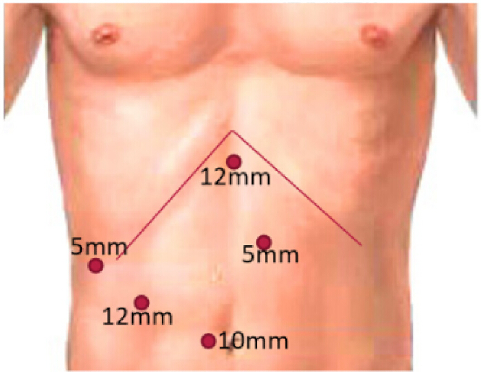
Figure 1: Schematic diagram of trocar configuration Please click here to view a larger version of this figure.
4. Critical procedural steps
- Divide the round and falciform ligaments following abdominal access using a monopolar energy device.
- Dissect the second hilum to expose the caval fossa, achieving full visualization of the right hepatic vein (RHV). Employ a 14-French catheter at the first hepatic hilum to pre-position the Pringle maneuver tourniquet.
- Divide the right triangular ligament and fully mobilize the right liver from its dorsal inferior aspect to expose the inferior vena cava (IVC) by carefully separating the areolar tissue.
- Dissect the dorsal superior border of the right liver toward the RHV. Divide the Makuuchi ligament, permitting circumferential exposure of the RHV, which is then encircled with a silk ligature (Figure 2).
NOTE: Circumferential dissection of major hepatic veins should be performed with extreme care to avoid catastrophic hemorrhage. - With a temporary Pringle occlusion, bluntly dissect the Glissonean pedicle to the dorsal S7 subsegment (G7-1) along the avascular plane of Laennec's capsule and encircle it with silk.
NOTE: During inflow occlusion, monitor hepatic ischemia time closely to prevent postoperative liver dysfunction. - After releasing the Pringle clamp, apply a non-crushing vascular clamp to G7-1. Observe and mark clear ischemic demarcation of the dorsal S7 territory with electrocautery (Figure 3A,B).
- Identify Cantlie's line (midplane of the liver) as the right/left hemiliver boundary by tracing the course of the middle hepatic vein and marking it, concurrently delineating the cranial/caudal right liver division (Figure 4A,B).
- Using non-traumatic forceps, adjust and mark the ventral resection line for S7+S8d (vertical limb of the reverse-L plane): Mark a point 2.5 cm lateral to the middle hepatic vein (MHV) guided by intraoperative visual estimation and preoperative imaging fusion, ensuring > 2 cm resection margins while preventing exposure of the MHV (Figure 5).
- Expose and transect the RHV root using a vascular stapler after confirming safe circumferential dissection, revealing ischemic discoloration in S7 and congested areas in partial S6 and S5.
NOTE: Prior to stapler application, ensure complete and safe isolation of the vessel. - Extrahepatically dissect the right hepatic pedicle (RHP) from portal branch IV to VI along Laennec's capsule. Implement silk-loop-guided right hemihepatic inflow occlusion to demonstrate a sharp Cantlie's line (Figure 6).
- Proceed with parenchymal transection along both the modified ventral resection line (S7 ischemic boundary) and the modified dorsal resection line (horizontal limb of the reverse-L plane) using a combination of ultrasonic aspirator and bipolar sealer.
- With both planes established, perform progressive parenchymal dissection, encountering and transecting the middle right hepatic vein (MRHV), the superior right hepatic vein (SRHV), the secondary G7 branch (G7-2) and the ventral S8 vein (V8v) between clips or with staplers, while maintaining meticulous preservation of the inferior right hepatic vein (IRHV) throughout by direct visualization and avoiding traction. (Figure 7A-D).
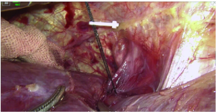
Figure 2: Exposure of the right hepatic vein (RHV) with silk-loop guidance in preparation for transection. Please click here to view a larger version of this figure.
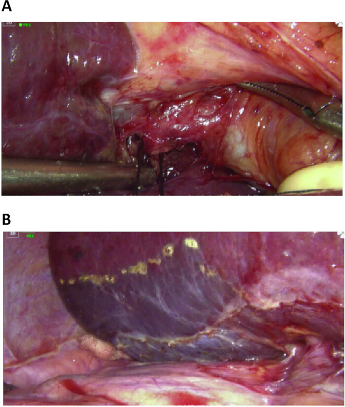
Figure 3: Blunt dissection along Laennec's capsule and ischemic demarcation of segment S7. (A) Blunt dissection along Laennec's capsule to isolate the G7-1 subsegmental pedicle with silk-loop encirclement. (B) Ischemic demarcation of segment S7 following bulldog clamp application to the G7-1 subsegmental pedicle Please click here to view a larger version of this figure.
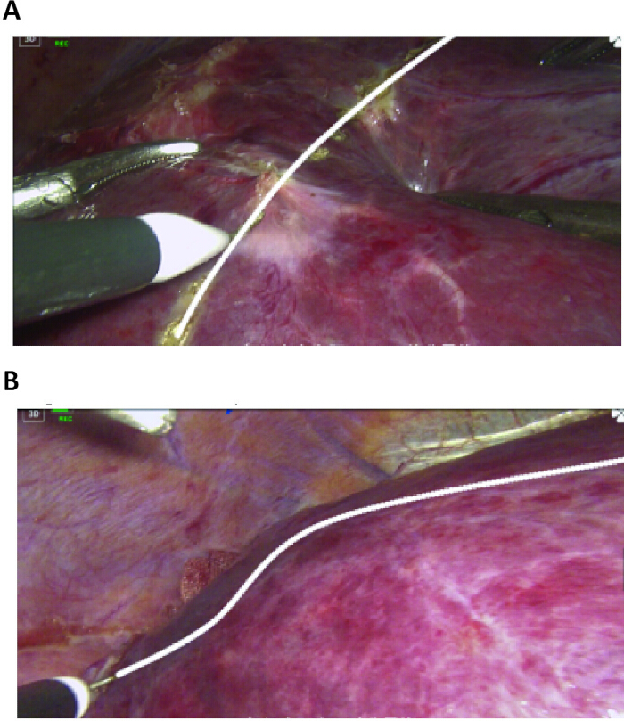
Figure 4: Identification of Cantlie's line. (A) Cantlie's line demarcating the right and left hemilivers. (B) The boundary between the cranial and caudal sectors of the right liver. Please click here to view a larger version of this figure.
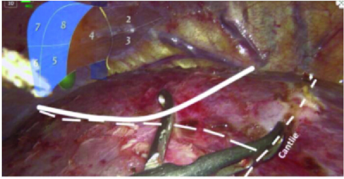
Figure 5: Modified ventral resection line for segment S8d. Please click here to view a larger version of this figure.
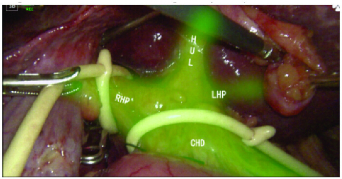
Figure 6: Occlusion of the right hepatic pedicle. Please click here to view a larger version of this figure.
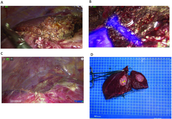
Figure 7: Parenchymal transection. (A) Complete reverse-L-shaped transection plane. (B) Transection surface demonstrating the dominant inferior right hepatic vein (IRHV). (C) Post-resection demonstration of the reverse-L-shaped transection surface. (D) Resected specimen demonstrating >2 cm tumor-free margins. Please click here to view a larger version of this figure.
5. Postoperative monitoring and management
- On postoperative day (POD) 1, initiate continuous monitoring with supplemental low-flow oxygen therapy.
- Initiate an Enhanced Recovery After Surgery (ERAS) protocol on POD 1, permitting liquid diet initiation and bedside ambulation.
- Administer hepatoprotective agents, albumin, antiinflammatory drugs, analgesics, and low-dose spironolactone for diuretic therapy.
- Perform laboratory surveillance encompassing: Liver function (Alanine aminotransferase [ALT], Aspartate aminotransferase [AST], Total bilirubin [TBIL], Direct bilirubin [DBIL]) and coagulation profile (Prothrombin time [PT], Activated partial thromboplastin time [APTT], Thrombin time [TT], Fibrinogen [FIB]).
Access restricted. Please log in or start a trial to view this content.
Results
he procedure was completed within 270 min with minimal intraoperative blood loss (approximately 100 mL), obviating transfusion requirements. Right hemihepatic Pringle maneuver was applied three times (total duration: approximately 40 min), and the first hilum Pringle maneuver was performed three times (total duration: approximately 50 min). No short-term complications occurred. The patient maintained hemodynamic stability and achieved an uneventful recovery, discharging on postoperative day (POD) 9.
Access restricted. Please log in or start a trial to view this content.
Discussion
Comprehensive understanding of the diverse venous drainage patterns in the right posterior sector is critical9. Makuuchi et al. first systematically established in 1987 that preserving the inferior right hepatic vein (IRHV) is pivotal during right hepatectomy, particularly when transection of the right hepatic vein (RHV) is required, as the IRHV becomes the sole venous outflow conduit for segment S610. Subsequent studies have validated the significance of IRHV preservation<...
Access restricted. Please log in or start a trial to view this content.
Disclosures
The authors have nothing to disclose.
Acknowledgements
This study was financially supported by the Sichuan Medical Science and Technology Innovation Research Association (Project Code: YCH-KY-YCZD2024-075) and the Health Commission of Sichuan Province Medical Science and Technology Program (Project No. 24WSXT089).
Access restricted. Please log in or start a trial to view this content.
Materials
| Name | Company | Catalog Number | Comments |
|---|---|---|---|
| Electrocautery | Hangzhou Kangji Medical Instrument Co., Ltd | KJ-SJ0205 | Sterile,dry heat sterilized, reusable |
| Gold finger | Hangzhou Kangji Medical Instrument Co., Ltd | https://www.kangjimed.com/search/gold%20finger.html | 101.237Φ10 ´ 350 mm (Sterile,dry heat sterilized, reusable) |
| Harmonic Scalpel | Johnson & Johnson MedTech | https://www.jnjmedtech.com/en-US/product/harmonic-1100-shears | Sterile, ethylene oxide sterilized, disposable |
| Non-absorbable suture | Johnson & Johnson MEDICAL (CHINA) Ltd | 2-0/W2512 | Sterile, ethylene oxide sterilized, disposable |
| Non-traumatic forceps | Hangzhou Kangji Medical Instrument Co., Ltd | https://www.kangjimed.com/search/Non-traumatic%20forceps.html | Φ10×260 (Sterile,dry heat sterilized, reusable) |
| Single-Use Linear Cutter | Fengh Medical Co., Ltd | TCMM45Y Standard Beak Type Linear Cutter | Sterile, ethylene oxide sterilized, disposable |
| Soft rubber ureteric catheter | Yangzhou Jinhuan Medical Appliance factory | http://www.yzjinhuan.com/portal.php | Type A 5.3 mm (16 Fr) (Sterile, ethylene oxide sterilized, disposable) |
| Trocar | Zhejiang Geyi Medical Instrument Co.,Ltd | https://www.geyi-medical.com/search/trocar.html | GYTR-I Φ5/Φ10/Φ12 (Sterile, ethylene oxide sterilized, disposable) |
References
- Fujiyama, Y., et al. Latest findings on minimally invasive anatomical liver resection. Cancers (Basel). 15 (8), 2218(2023).
- Takamoto, T., Makuuchi, M. Precision surgery for primary liver cancer. Cancer Biol Med. 16 (3), 475-485 (2019).
- Wakabayashi, G., et al. The Tokyo 2020 terminology of liver anatomy and resections: Updates of the Brisbane 2000 system. J Hepatobiliary Pancreat Sci. 29 (1), 6-15 (2022).
- Sugioka, A., Kato, Y., Tanahashi, Y. Systematic extrahepatic glissonean pedicle isolation for anatomical liver resection based on Laennec's capsule: Proposal of a novel comprehensive surgical anatomy of the liver. J Hepatobiliary Pancreat Sci. 24 (1), 17-23 (2017).
- Cho, J. Y., et al. Practical guidelines for performing laparoscopic liver resection based on the second international laparoscopic liver consensus conference. Surg Oncol. 27 (1), A5-A9 (2018).
- Lopez-Lopez, V., et al. Laparoscopic liver resection of segments 7 and 8: From the initial restrictions to the current indications. J Minim Invasive Surg. 23 (1), 5-16 (2020).
- Lee, J. W., Choi, S. H., Kim, S., Kwon, S. W. Laparoscopic liver resection for segment VII lesion using a combination of rubber band retraction method and flexible laparoscope. Surg Endosc. 34 (2), 954-960 (2020).
- Hu, M., et al. Efficacy and safety of indocyanine green fluorescence navigation versus conventional laparoscopic hepatectomy for hepatocellular carcinoma: A systematic review and meta-analysis. Surg Endosc. 39 (3), 1681-1695 (2025).
- Shilal, P., Tuli, A. Anatomical variations in the pattern of the right hepatic veins draining the posterior segment of the right lobe of the liver. J Clin Diagn Res. 9 (3), AC08-AC12 (2015).
- Makuuchi, M., Hasegawa, H., Yamazaki, S., Takayasu, K. Four new hepatectomy procedures for resection of the right hepatic vein and preservation of the inferior right hepatic vein. Surg Gynecol Obstet. 164 (1), 68-72 (1987).
- Machado, M. A., Bacchella, T., Makdissi, F. F., Surjan, R. T., Machado, M. C. Extended left trisectionectomy severing all hepatic veins preserving segment 6 and inferior right hepatic vein. Eur J Surg Oncol. 34 (2), 247-251 (2008).
- Xing, X., Li, H., Liu, W. G. Clinical studies on inferior right hepatic veins. Hepatobiliary Pancreat Dis Int. 6 (6), 579-584 (2007).
- Nakayama, H., et al. Verification of inferior right hepatic vein-conserving segments 7 to 8 resection of the liver. Int Surg. 100 (4), 683-688 (2015).
- Terayama, M., et al. Preserving inferior right hepatic vein enabled bisegmentectomy 7 and 8 without venous congestion: A case report. Surg Case Rep. 7 (1), 101(2021).
- Kamiyama, T., Kakisaka, T., Orimo, T. Current role of intraoperative ultrasonography in hepatectomy. Surg Today. 51 (12), 1887-1896 (2021).
- Bezzi, M., et al. Laparoscopic and intraoperative ultrasound. Eur J Radiol. 27 Suppl 2, S207-S214 (1998).
- Wang, J., Xu, Y., Zhang, Y., Tian, H. Safety and effectiveness of fluorescence laparoscopy in precise hepatectomy: A meta-analysis. Photodiagnosis Photodyn Ther. 42, 103599(2023).
Access restricted. Please log in or start a trial to view this content.
Reprints and Permissions
