This study presents a robust method to isolate axonal mRNAs using porous membrane inserts, enabling total neuron vs. neurite separation and RNA purification. Combined with RTddPCR, the approach allows absolute quantification of low-copy transcripts, facilitating studies of mRNA transport and local translation with high sensitivity, reproducibility, and broad experimental applicability.
Method Article
Isolation and Quantification of Axonal mRNAs Using Porous Membrane Inserts and RTddPCR
* These authors contributed equally
In This Article
Summary
Abstract
The spatial dynamics of mRNA localization and translation within neurons are essential for various mechanisms of neuronal function, including neuronal connectivity, synaptic plasticity, and response to injury. Due to the extreme polarity of neurons, many of these functions rely on the ability of axons to locally translate specific transcripts. However, quantifying these subcellular RNA populations remains technically challenging. Here, we describe a reproducible approach for obtaining separate somatic and axonally enriched compartments from cultured rodent neurons and for quantifying compartment-specific mRNA expression. Primary rodent embryonic or adult neurons were cultured on inserts with a porous membrane of size 1-3 µm. These membranes are only permissive to axons, allowing physical separation of the somatic and axonal compartments. RNA was then isolated from the whole neuron and axon-enriched fractions separately, which were further used for reverse transcriptase droplet digital PCR (RTddPCR) with gene-specific primers. This system offers an absolute quantitative comparison between subcellular compartments, enabling high-sensitivity detection of localized transcripts. This approach measures steady-state RNA abundance and facilitates examination of axonal RNA changes over time in response to neurotrophic factors, stress, or injury models. The combination of physical compartmentalization and RTddPCR analysis reduces cross-contamination and gives exact copy numbers of rare transcripts, offering high sensitivity, reproducibility, and detection of low-copy-number mRNAs that control axon growth and regeneration. This method also works with downstream tests, such as measuring protein synthesis, studying RNA stability, and doing perturbation experiments using siRNA or drugs that block certain proteins. Importantly, this technique can be adapted for different neuronal subtypes, developmental stages, or injury models. In general, this approach is a flexible, sensitive, and reproducible way to study the molecular basis of axonal mRNA localization and how it affects neuronal function and disease mechanisms.
Introduction
Neurons are one of the most structurally complex and polarized cells in biology. They have long processes that can sometimes reach distances exceeding a meter. This polarity complicates cellular communication and protein homeostasis in distinct manners. A refined approach to address these requirements is to transport mRNAs to remote compartments such as axons and dendrites, facilitating their translation in a regulated manner in response to localized signals1,2,3. Local translation enables axons to synthesize proteins in a spatiotemporal manner. This process is crucial for axonal development, synaptic plasticity, regeneration after injury, and responses to developmental and extracellular stimuli4,5,6,7,8,9,10,11,12,13,14,15.
The ability to analyze the functions of localized mRNAs in axons is crucial for understanding their roles in both normal neuronal function and in the context of pathophysiology. Dysregulation of axonal mRNA translation has been linked to various neurodegenerative diseases16, such as amyotrophic lateral sclerosis (ALS)17,18,19, spinal muscular atrophy (SMA)20,21, and Alzheimer's disease (AD)22. Axonal regeneration following damage is heavily dependent on the rapid, precise translation of cytoskeletal proteins, signaling molecules, and receptors3,23,24,25,26,27,28. Despite these novel concepts, the discipline continues to face challenges in effectively quantifying and capturing axon-specific mRNA populations.
One of the challenges of axonal transcriptomics is getting the soma and axons to separate cleanly. As most of the mRNAs are enriched in the soma, even small amounts of contamination from the cell body can affect the results when assessing axonal content of a particular mRNA. Conventional techniques, including microfluidic chambers29,30,31,32 or Campenot chambers33, provide directional axonal growth and clear separation between the two compartments, but axonal yield can be too low for biochemical studies such as bulk RNA sequencing (RNA-seq), RNA co-immunoprecipitation followed by RNA sequencing, or quantitative polymerase chain reaction (qPCR). Bulk RNA-seq and qPCR, however beneficial, frequently lack the requisite sensitivity to accurately identify low-copy transcripts in axons, resulting in the incorrect estimation of physiologically significant species.
To address these challenges, we and others have used permeable membrane inserts for the physical separation of axons and somata34,35,36,37,38,39,40,41. These inserts let neurons develop on a microporous surface, and axons can extend into the bottom compartment through them, but not the soma. This basic but effective architecture makes it possible to culture a huge number of neurons with a clear physical separation of soma and axons. The membrane-based method is important because it avoids the technical problems that come with microfluidic devices and gives high quantities of axonal material that can be used for molecular and biochemical studies. The insert system is also easy to use for things like mechanical injury, which makes it useful in many different experimental settings related to neural repair. The method described here further optimizes neuronal yield and purity by refining culture conditions, adjusting membrane pore characteristics, and incorporating Reverse transcriptase droplet digital PCR (RTddPCR)-based validation for low-yield axonal RNA samples.
It is also important to make sure that axonal fractions are pure. We only extract axons from the lower chamber after carefully removing any leftover somatic material from the upper surface. Primer validation shows strong enrichment of axonal markers and no or negligible somatic markers. Molecular confirmation reinforces this distinction: axonal fractions are enriched with recognized axonal mRNAs such as Gap43, and lower expression of Actγ42. This shows that the insert system makes axonal samples that have negligible soma contents in them, which are good for further examination.
After isolating enriched axonal fractions, the subsequent hurdle is to measure mRNAs that exist in extremely low copy numbers. The little starting material from axonal fractions and the presence of low copy number mRNAs in the axons push the sensitivity limits of conventional reverse transcriptase quantitative PCR (RT-qPCR), which uses standard curves for quantification. Moreover, RT-qPCR also does not provide the absolute copy numbers of a specific transcript. RTddPCR, on the other hand, separates cDNA samples into thousands of droplets, which helps to get an exact transcript count using Poisson statistics. This makes it possible to reliably find transcripts even if only a few copies of the transcripts might be present in a single nanogram of total RNA5,6,7,43.
In this manuscript, we provide a streamlined approach, which includes methods to culture different adult or embryonic rodent neurons on inserts, isolation of RNA from whole neuron vs. axonal enriched compartments, check for axonal purity, and RTddPCR-based absolute quantification of mRNA copies. These methods can be utilized to determine the levels of specific mRNAs, which localize to axons under basal conditions, and how they change upon axotomy or in response to growth factor stimulation or neurotoxic signals. By combining this method with protein co-immunoprecipitations, one can also assess the dynamics of ribonuclear protein (RNP) complexes in axons. For example, we have extensively used this method to study the mRNAs, which are present in the axonal Ras GTPase-activating protein-binding protein 1 (G3BP1) granules. G3BP1 is a core component of stress granules44, and our previous studies have shown that it inhibits axonal protein synthesis and, in turn, blocks both PNS and CNS axon regeneration5. Moreover, this method can be utilized to investigate the role of axonal translation dysregulation in various pathophysiological conditions, including ALS, SMA, and AD.
The goal of this study is to create and test a method for measuring axonal mRNAs that is sensitive, reproducible, and scalable. By combining compartmentalized insert-based cultures with RTddPCR-based quantification, we provide the field a solution to the enduring problems of axonal transcriptomics. This methodology will not only enable essential discoveries regarding local translation but also establish a basis for translational investigations focused on therapeutic interventions in neural repair and neurodegeneration.
Protocol
1. Preparing the inserts for cell seeding
- Place the inserts with the appropriate pore size in a 6-well plate.
NOTE: Use a 1 μm pore size for separating axons from the soma and a 3 μm pore size for separating all neurites (axons and dendrites) from the soma. - Add 100 μg/mL poly-L-lysine (PLL) to the 6-well plate in a way that it touches the bottom of the inserts, and to the top of the inserts (2 mL/well and 1 mL/insert).
- Incubate overnight at 37 °C.
- Wash the wells (bottom of the inserts) and the top of the inserts with sterile ultrapure water - 4 washes × 5 min.
- For dorsal root ganglion (DRG) neurons only, add 5 µg/mL laminin (2 mL/well and 1 mL/insert) and incubate for at least 1 h at 37 °C. Make sure both the top and the bottom of the insert are evenly coated.
- Wash with sterile phosphate-buffered saline (PBS) (pH 7.4) containing 100 U/mL Penicillin/Streptomycin - 2 washes × 5 min.
- Proceed with the dissection and seed around 1 × 10⁶ cells/insert for embryonic cortical5, hippocampal45, and basal forebrain cholinergic neurons31,32. For adult rat DRGs, plate 6-8 DRGs/insert5,7 (Figure 1).
2. Collection of neuronal subcellular fractions from 6-well inserts
- Aliquot 250 µL of TRIzol each in a 1.5 mL microcentrifuge tube per insert and one well/insert of a 6-well plate. Keep it aside.
- In another 6-well plate, add 2 mL/well of sterile PBS. The number of wells/plates should be proportional to the number of inserts.
- Aspirate culture media from the top and bottom of the insert and transfer the insert to a 6-well plate containing PBS.
- Using forceps, gently place the insert and add 2 mL of PBS on top of the insert. Aspirate media from the top and bottom of the insert and repeat. In total, wash twice with PBS and leave it in fresh PBS (1 mL and 2 mL at the top and bottom of the insert, respectively).
- Isolation of the whole neuron fraction
- Using a sterile cell scraper, scrape the whole neuron fraction (top of the insert). Apply just enough pressure so the cells start to come off, but the membrane does not break (Figure 1).
- Collect the whole neuron from the insert and transfer it into a 1.5 mL microcentrifuge tube.
- Centrifuge the tube at 10000-15000 g for 2 min.
- Discard the supernatant and resuspend the lysate in 250 µL of TRIzol.
- Label this tube as the whole neuron fraction.
- Continue with the neurite fraction collection.
- Isolation of the neurite fraction
- Take a sterile cotton swab, and using one end, move in a zig-zag pattern from top to bottom very slowly on the whole neuron side. Rotate the insert 90 degrees and repeat the process with the other end of the swab (if using a double-sided swab, or use a new swab for a single-sided one). Discard this swab (Figure 1).
- Use a new swab and move in concentric circles, starting from the middle of the insert and going outward. Make sure to clean the walls as well (circumference).
- Invert the membrane insert (neurite side facing up) and cut the membrane using a new sterile scalpel blade while holding it with forceps. Make sure to leave ~2-3 mm in the periphery.
- Place the cut membrane in the 6-well plate containing TRIzol with the neurite side facing down. Make sure it is submerged.
- Collect the TRIzol containing the neurite lysate from the 6-well plate and transfer to a 1.5 mL microcentrifuge tube.
- For both whole neuron and neurite lysates, proceed with RNA isolation or save the lysates at -80 °C for processing them later.
3. RNA isolation and quantification
- RNA isolation
NOTE: For this part, always work in an RNase-free environment with dedicated, sterile plasticware and gloves.- Add 0.2 mL of chloroform per 1 mL of TRIzol, shake vigorously for 15 s, and incubate for 2-3 min at room temperature. Centrifuge at 12,000 × g for 15 min at 4 °C to separate into layers: organic, interphase, and aqueous (containing RNA).
- Transfer the upper aqueous phase to a new tube, avoiding the interphase. To precipitate the RNA, add 1 volume of isopropanol and mix gently. Incubate for 10 min at room temperature (or longer at -20 °C for higher yield) to precipitate RNA. Centrifuge at 12,000 × g for 10 min at 4 °C to pellet RNA.
- Discard the supernatant and wash the pellet with 1 mL of 75% ethanol. Briefly vortex and centrifuge at 7,500 × g for 5 min at 4 °C.
- Dissolving RNA: Carefully remove the ethanol wash and air-dry the pellet for 5-10 min, avoiding complete dryness. Resuspend the pellet in a small volume of RNase-free water or 1x Tris-EDTA (TE) buffer, incubating at 55-60 °C for complete dissolution. Store purified RNA at -80 °C.
- RNA quantification using the RiboGreen assay
NOTE: The RiboGreen assay (below) uses a fluorescent dye specific for nucleic acids, offering higher sensitivity, specificity, and the ability to detect intact RNA than UV absorbance methods. Consider DNase treatment if DNA contamination is an issue.- Prepare 1x TE buffer, dilute the provided 20x TE buffer 20-fold with RNase-free water. Prepare RiboGreen working solutions and protect the light-sensitive dye with foil. Dilute the concentrated dye 1:200 in 1x TE for the high-range assay (10 ng/mL-1 µg/mL), or 1:2000 in 1x TE for the low-range assay (2.5 ng/mL-50 ng/mL). Prepare RNA standards by serial dilution of the kit's RNA standard in 1x TE to cover the expected sample range. Run standards in triplicate.
- Prepare samples by diluting RNA samples in 1x TE to fit the assay's dynamic range. Run samples in triplicate.
- Add equal volumes of RiboGreen working solution and RNA standard or sample (e.g., 100 µL each). Incubate for 2-5 min at room temperature, protected from light. Measure fluorescence using the appropriate settings (excitation ~500 nm, emission ~525 nm). Measure and subtract a reagent blank reading (1x TE + RiboGreen dye).
- Analyze and quantify: Plot a standard curve using the fluorescence values of the standards versus their concentrations. Use this curve to determine the RNA concentration in the samples (Figure 2).
4. cDNA synthesis
- Thaw all components on ice. Gently mix each tube by flicking and spin down briefly before use. In a PCR tube on ice, combine the following components for each reaction:
RT master mix (5x): 4 µL
RNA Sample: variable (up to 1 µg total RNA)
Nuclease-free Water: to 20 µL total volume
NOTE: For a no-template control (NTC), replace the RNA sample with nuclease-free water. - Gently mix and spin down the tubes to collect the contents at the bottom and run the thermocycler program.
Primer annealing: 25 °C for 2 min
cDNA synthesis: 55 °C for 10 min
Heat inactivation: 95 °C for 1 min
5. Primer designing and validation
NOTE: RTddPCR primer design generally follows the same principles as quantitative qPCR, but requires additional attention to ensure clear separation of positive and negative droplets. Use primer design tools from NEB, IDT, or ThermoFisher to facilitate this process. A successful RTddPCR assay is defined by high specificity and robust amplification, which generates a distinct separation between amplified (positive) and unamplified (negative) droplets. The following NCBI RefSeq accession IDs were used for designing PCR primers:
Actg: NM_001127449.1
Forward 1: CAGTCTAACAGGGTGGGAAAG
Reverse 1: CCAACTCAAGGCAAGTAACAAC
Amplicon Length: 98
Forward 2: GTAGTGATCTGTGCAGGGTATT
Reverse 2: GACTTTCCCACCCTGTTAGAC
Amplicon Length: 118
Gap43: NM_017195.3
Forward 1: GGAAATTCTTGCACTGTACCC
Reverse 1: GACTCCTCAGAACGGAACAT
Amplicon Length: 122
Forward 2: CAGTCATCTTGGGAAATTCTTGC
Reverse 2: CATTGCACACACACACTTGG
Amplicon Length: 116
- Design a primer with the recommended primer length of 18-30 base pairs and a GC content of 40-60%. Ensure the melting temperature (Tm) falls between 55-65 °C, and the forward and reverse primers are within 2-5 °C of each other. Incorporate a GC clamp, one or two G or C bases within the last five bases at the 3′ end, to enhance binding specificity, but avoid long runs of Gs or Cs.
- Validate primer specificity using alignment tools such as National Center for Biotechnology Information (NCBI) Basic Local Alignment Search Tool (BLAST) to ensure that primers bind only to the intended target sequence. Design amplicons between a size of 50-200 base pairs, with ~100 base pairs as optimal, as shorter products amplify more efficiently while remaining distinguishable from primer-dimers.
NOTE: In this study, the short PCR products are typically amplified with higher efficiency than longer ones in RT-ddPCR. - Minimize primer dimers by designing primers with 50-60% GC content, avoiding secondary structures, and 3' complementarity. During primer validation, use agarose gel electrophoresis, and always use a no-template control to assess primer dimers.
- Avoid designing primers in regions of the template with a high likelihood of forming secondary structures, as this can interfere with amplification efficiency.
6. RTddPCR
NOTE: Use the QX200 ddPCR System (Bio-Rad) to perform RTddPCR according to the manufacturer's instructions and as described previously by Das et al. (2022)43, with a few minor adjustments to improve assay reproducibility.
- Prepare RTddPCR reactions with ddPCR ready-to-use universal mix and target-specific primers using appropriate cDNA.
- Generate the droplets using the droplet generator.
- Read the endpoint fluorescence with the droplet reader, and analyze the data with the built-in software.
- Employ the following quality control measures to ensure precise quantification and reproducible droplet formation:
- Run primer sets consistently in the same row or column to minimize plate effects.
- Prepare master mixes to reduce pipetting of small volumes.
- Pipette at a low flow rate to prevent bubbles and use multichannel pipettes for uniform sample dispensing.
- Handle plates gently after droplet generation and seal immediately to prevent evaporation.
- Prepare all reactions on ice and avoid repeated freeze-thaw cycles for reagents.
- Include no-template controls for each primer set to monitor contamination.
- Exclude wells containing fewer than 10,000 accepted droplets from analysis.
- Perform technical replicates for all samples to ensure reproducibility.
- Manually inspect droplet fluorescence amplitude plots to verify the accuracy of automatic thresholding.
Results
Primary embryonic rodent cortical neurons, hippocampal neurons, and BFCNs plated on PLL-coated 1 µm inserts adhere robustly and extend neurites across the membrane by 5-7 days in vitro (DIV). By 11 DIV, cultures display dense networks. For adult rat DRGs, adding a coat of laminin to PLL-coated 3 µm inserts helps achieve comparable axonal extension by 5 DIV. These observations confirm the ability of inserts to support long-term neuronal growth and provide sufficient axonal material for downstream RNA extraction. In case of need to scale, we combine multiple inserts to provide us with enough material.
TRIzol extraction from individual inserts yields sufficient RNA for reverse transcription and subsequent ddPCR. RiboGreen quantification confirms consistent RNA yields from whole neuron fractions (212.85 ng/insert) and lower yields from neurite fractions (42.75 ng/insert) (Figure 2). We generally get ~5-7 fold RNA from the whole neuron fraction vs. the neurite enriched fraction.
All primers are validated before RTddPCR experiments. For each target transcript, at least two primer sets are ordered and tested using conventional PCR on cDNA generated from the same neuronal samples. The primer pair producing the strongest and most specific band is selected for RTddPCR (Figure 3A). For instance, we chose primer set 1 for Gap43. In cases of ambiguous results, such as those obtained for Actγ, we perform a gradient PCR to optimize annealing temperatures (Figure 3B). This ensures that only well-validated primers are being used for absolute quantification, reducing the likelihood of artifacts in droplet separation.
The scraping and swabbing procedure reliably separates somatic and neuritic compartments. RNA isolated from the whole neuron fractions shows enrichment of transcripts, such as Actγ46,47,48,49 (Figure 4). In contrast, axon/neurite fractions yield markedly lower total RNA, consistent with their restricted volume, but show enrichment of transcripts known to localize to distal processes, such as Gap43 (Figure 4). Minimal cross-detection of soma-restricted transcripts indicates high compartmental purity when inserts are handled carefully and plated at optimal density.
Positive results are characterized by: (1) intact neuronal morphology with clear axonal extension, (2) clean separation of compartments without rupture of the membrane, (3) validated primers that produce distinct separation between positive and negative droplets, and (4) strong RTddPCR signals for axonal transcripts. Suboptimal experiments result in low RNA yield, cross-contamination evidenced by soma markers in neurite fractions, or RTddPCR artifacts such as increased droplet "rain" due to primer dimerization. These issues are most often linked to insert membrane damage, excessive scraping pressure, or inadequate primer annealing temperature optimization.
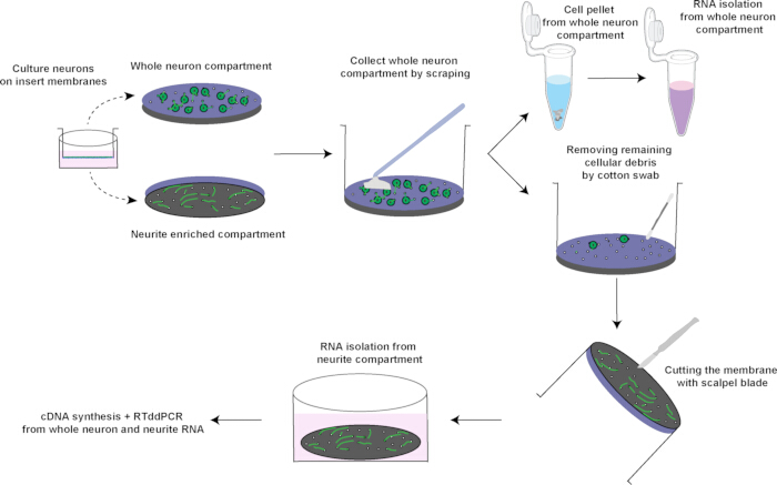
Figure 1: Overview of compartment-specific RNA isolation using membrane inserts. Rodent neurons were cultured on semipermeable inserts with a pore size of 1-3 µm, which allows neurite extension while restricting soma. For whole neuron preparation, cells from the upper compartment were scraped off using a sterile cell scraper, pelleted, and then lysed in TRIzol for RNA isolation, cDNA synthesis, and RTddPCR. Residual cellular debris was removed from the insert membrane with a sterile cotton swab. For neurite preparation, the insert membrane now containing only neurites was inverted and cut with a scalpel blade, and immersed directly in TRIzol, followed by RNA isolation, cDNA synthesis, and RTddPCR. Please click here to view a larger version of this figure.

Figure 2: Standard RNA concentration curve using RiboGreen assay. (A) Fluorescence intensity was measured using the RiboGreen RNA quantification assay across a dilution series of RNA standards. A linear regression analysis revealed a strong correlation between RNA concentration and fluorescence signal (R² = 0.9956). The R² value close to one demonstrates excellent linearity and reliability of the RiboGreen assay for accurate RNA quantification. (B) Using the equation (y=19.738x + 14.905) for the slope obtained in A, total RNA was calculated for each fraction. Please click here to view a larger version of this figure.
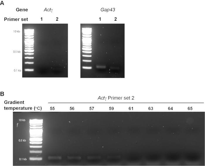
Figure 3: Primer validation by PCR and agarose gel electrophoresis. (A) PCR amplification products using cDNA for Actγ and Gap43 were resolved on a 2% agarose gel to assess primer specificity. For each gene, two independent primer sets (set 1 and set 2) were tested. DNA size markers (ladders) are shown in adjacent lanes with molecular weights indicated (10 kb, 0.5 kb, 0.1 kb). Distinct single bands of the expected size confirm successful amplification and specificity of the selected primer sets. (B) PCR amplification was performed using Actγ primer set 2 across a temperature gradient (55-65 °C) to determine the optimal annealing temperature. Amplified products were resolved on a 2% agarose gel alongside a DNA ladder (lane 2, sizes indicated). Consistent single bands of the expected size were observed across the tested range, with the strongest amplification detected at 55 °C, suggesting it to be the optimal temperature for this primer set. Please click here to view a larger version of this figure.

Figure 4: RTddPCR analysis of selected transcripts and quantification of mRNA copies. (A,B) Representative one-dimensional amplitude plots showing droplet separation for (A) Actγ, and (B) Gap43. Each dot represents a single droplet, with blue droplets indicating positive amplification and gray droplets indicating negative amplification. The Y-axis represents amplitude, and the X-axis indicates the well position in the ddPCR plate. Clear separation between positive (blue) and negative (gray) droplets confirms robust detection of the target transcripts with the indicated primer sets. (C) Table summarizing the number of copies/µL and copies per ng of total RNA for Actγ and Gap43 transcripts in whole neuron and neurite fractions. Copy number estimates were obtained from absolute quantification using ddPCR droplet counts (shown in panels A,B). Please click here to view a larger version of this figure.
Discussion
Several steps are crucial to reproducibility. The insert coating must be uniform to promote neuronal adhesion; incomplete coating leads to poor neurite/axonal growth. Cell plating density is equally important, since excessive density increases dendritic crossing, whereas sparse plating yields insufficient axonal RNA. The scraping-swabbing sequence must be performed with precision: too little pressure leaves somatic contamination, while too much pressure risks damaging the insert.
Primer validation represents another critical step. Running two independent primer sets for each transcript allows selection of the most reliable pair, while gradient PCR can further refine annealing temperatures. This practice reduces primer-dimer formation, increases droplet clarity, and ensures reproducible quantification across biological replicates. While this approach achieves high compartmental purity, trace contamination from somatic material cannot be fully excluded. Axonal RNA yields are inherently low, limiting the scope of analyses that require high input, such as full transcriptome sequencing. A critical limitation of this method is its inability to address transcript localization within distal vs proximal axons. Although glial or endothelial extensions through membrane pores have been reported under certain conditions, our culture system minimizes this possibility. In adult DRG cultures, the addition of cytosine β-D-arabinofuranoside (Ara-C)5,7 and the use of serum-free media for other neuronal cultures (cortical, hippocampal, and BFCNs)5,45 effectively restricts glial and endothelial cell proliferation. Additionally, the rigid polycarbonate or polyethylene terephthalate (PC/PET) membranes used here are non-elastic and non-permissive to active cell migration. Moreover, we use 3 µm pore size inserts for DRG cultures to allow the passage of large-diameter axons. In contrast, for cortical or BFCN cultures, we use 1 µm pore size inserts, which further restricts the migration of glia. These properties distinguish our setup from PDMS-based microdevices that support endothelial migration through flexible pores. Consistent with prior reports34,35,36,40, the molecular validation in this study confirms strong enrichment of axonal transcripts (e.g., Gap43) and negligible detection of somatic markers (e.g., Actγ), supporting the neuronal specificity of the material traversing the membrane. As the physical constraint does not completely abolish glia migration, the researchers should also use Gfap as a negative marker.
Compared to microfluidic devices, the insert system is simpler, more scalable, and compatible with routine culture workflows. It avoids specialized equipment while still enabling reproducible axon-soma separation. By coupling inserts with RTddPCR, this method provides both accessibility and high sensitivity, enabling absolute quantification of transcripts that are often below qPCR detection thresholds. This protocol is well-suited for probing changes in axonal transcript localization, assessing local translation in response to developmental, neurotoxic, or injury stimuli, and also studying RNA transport defects implicated in neurodegenerative diseases or neurodevelopmental disorders. Future refinements may include multiplexed RTddPCR for simultaneous quantification of multiple mRNAs, or integration with low-input RNA sequencing approaches to expand transcriptome coverage. In addition, adapting this system for human iPSC-derived neurons may extend its applications to disease modeling and regenerative studies.
Disclosures
PKS holds US patents on the use of the G3BP1 cell-permeable peptide for axon regeneration and neurodegeneration. The other authors declare no conflicts of interest.
Acknowledgements
This work was supported by grants from the Merkin Peripheral Neuropathy and Nerve Regeneration Center (to P.K.S.).
Materials
| Name | Company | Catalog Number | Comments |
|---|---|---|---|
| Cell culture Inserts for 6-well plate, 1.0 μm Pore Size, Sterile | Corning | 353102 | Consumables |
| Cell culture Inserts for 6-well plate, 3.0 μm Pore Size, Sterile | CELLTREAT | 230603 | Consumables |
| Cell Lifter with Narrow Blade | VWR | 76036-006 | Consumables |
| CELLSTAR 6-well Tissue culture plates | VWR | 82050-842 | Consumables |
| ddPCR 96-Well Plates | Bio-rad | 12001925 | Consumables |
| ddPCR Droplet Reader Oil | Bio-rad | 1863004 | Reagents |
| DG8 Cartridges for QX200/QX100 Droplet Generator | Bio-rad | 1864008 | Consumables |
| Laminin | Gibco/Fisher Scientific | 23017-015 | Reagents |
| LunaScript RT SuperMix Kit | NEB | E3010L | Reagents |
| Microplates for Fluorescence-based, 96-well, black | Thermo Fisher | M33089 | Consumables |
| PCR Plate Heat Seal, foil, pierceable | Bio-rad | 1814040 | Consumables |
| Poly-Lysine | Sigma | P1274 | Reagents |
| PTC Tempo 96 Thermal Cycler | Bio-rad | 12015382 | Equipment |
| QuantaSoft software | Bio-Rad | PCR data analysis software | |
| Quant-it RiboGreen Reagent and RNA Assay Kit | Thermo Fisher | R11490 | Reagents |
| QX200 ddPCR EvaGreen Supermix | Bio-rad | 1864034 | Reagents |
| QX200 Droplet Generation Oil for EvaGreen | Bio-rad | 1864005 | Reagents |
| QX200 Droplet Generator | Bio-rad | 17005227 | Equipment |
| QX200 Droplet Reader | Bio-rad | 17005228 | Equipment |
| TRIzol Reagent | Invitrogen | 15-596-026 | Reagents |
References
- Agrawal, M., Welshhans, K. Local translation across neural development: A focus on radial glial cells, axons, and synaptogenesis. Front Mol Neurosci. 14, 717170(2021).
- Dalla Costa, I., et al. The functional organization of axonal mRNA transport and translation. Nat. Rev. Neurosci. 22 (2), 77-91 (2021).
- Sahoo, P. K., Smith, D. S., Perrone-Bizzozero, N., Twiss, J. L. Axonal mRNA transport and translation at a glance. J. Cell Sci. 131 (8), (2018).
- Alber, S., et al. PTBP1 regulates injury responses and sensory pathways in adult peripheral neurons. Sci Adv. 9 (30), eadi0286(2023).
- Sahoo, P. K., et al. Disruption of G3BP1 granules promotes mammalian CNS and PNS axon regeneration. Proc Natl Acad Sci. 122 (9), e2411811122(2025).
- Sahoo, P. K., et al. A Ca2+-dependent switch activates axonal casein kinase 2α; translation and drives G3BP1 granule disassembly for axon regeneration. Curr Biol. 30 (24), 4882-4895.e6 (2020).
- Sahoo, P. K., et al. Axonal G3BP1 stress granule protein limits axonal mRNA translation and nerve regeneration. Nat Commun. 9 (1), 3358(2018).
- Terenzio, M., et al. Locally translated mTOR controls axonal local translation in nerve injury. Science. 359 (6382), 1416-1421 (2018).
- Van Erp, S., et al. Age-related loss of axonal regeneration is reflected by the level of local translation. Exp Neurol. 339, 113594(2021).
- Zahavi, E. E., et al. Repeat-element RNAs integrate a neuronal growth circuit. Cell. 188 (16), 4350-4365.e22 (2025).
- Rajgor, D., Welle, T. M., Smith, K. R. The coordination of local translation, membranous organelle trafficking, and synaptic plasticity in neurons. Front Cell Dev Biol. 9, 711446(2021).
- Oliveira, M. M., Klann, E. A deep dive into local mRNA translation in neurons. Proc Natl Acad Sci. 118 (45), e2117116118(2021).
- Sun, C., et al. The prevalence and specificity of local protein synthesis during neuronal synaptic plasticity. Sci Adv. 7 (38), eabj0790(2021).
- Castillo, P. E., Jung, H., Klann, E., Riccio, A. Presynaptic protein synthesis in brain function and disease. J Neurosci. 43 (45), 7483-7488 (2023).
- Younts, T. J., et al. Presynaptic protein synthesis is required for long-term plasticity of GABA release. Neuron. 92 (2), 479-492 (2016).
- Nagano, S., Araki, T. Axonal transport and local translation of mRNA in neurodegenerative diseases. Front Mol Neurosci. 14, 697973(2021).
- Altman, T., et al. Axonal TDP-43 condensates drive neuromuscular junction disruption through inhibition of local synthesis of nuclear encoded mitochondrial proteins. Nat Commun. 12 (1), 6914(2021).
- Bar Avi, O., Perlson, E. Navigating the pathways: Tar-DNA-binding-protein-43 aggregation, axonal transport, and local synthesis in amyotrophic lateral sclerosis pathology. Neural Regen. Res. 20 (10), 2921-2922 (2025).
- Ionescu, A., Altman, T., Perlson, E. Looking for answers far away from the soma-the (un)known axonal functions of TDP-43, and their contribution to early NMJ disruption in ALS. Mol. Neurodegener. 18 (1), 35(2023).
- Fallini, C., Bassell, G. J., Rossoll, W. Spinal muscular atrophy: The role of SMN in axonal mRNA regulation. Brain Res. 1462, 81-92 (2012).
- Rehorst, W. A., et al. Muscle regulates mTOR dependent axonal local translation in motor neurons via CTRP3 secretion: Implications for a neuromuscular disorder, spinal muscular atrophy. Acta Neuropathol Commun. 7 (1), 154(2019).
- Twiss, J. L., Buchanan, C. N. Regulation of subcellular protein synthesis for restoring neural connectivity. Int J Mol Sci. 26 (15), 7283(2025).
- Gomes, C., et al. Axonal localization of neuritin/cpg15 mRNA is limited by competition for HuD binding. J Cell Sci. 130 (21), 3650-3662 (2017).
- Lee, S. J., et al. hnRNPs interacting with mRNA localization motifs define axonal RNA regulons. Mol Cell Proteomics. 17 (11), 2091-2106 (2018).
- Lee, S. J., et al. Selective axonal translation of the mRNA isoform encoding prenylated cdc42 supports axon growth. J Cell Sci. 134 (7), jcs251967(2021).
- Merianda, T. T., et al. Axonal amphoterin mRNA is regulated by translational control and enhances axon outgrowth. J Neurosci. 35 (14), 5693-5706 (2015).
- Patel, P., et al. Intra-axonal translation of Khsrp mRNA slows axon regeneration by destabilizing localized mRNAs. Nucleic Acids Res. 50 (10), 5772-5792 (2022).
- Smith, T. P., Sahoo, P. K., Kar, A. N., Twiss, J. L. Intra-axonal mechanisms driving axon regeneration. Brain Res. 1740, 146864(2020).
- Huang, N., Sheng, Z. H. Microfluidic devices as model platforms of CNS injury-ischemia to study axonal regeneration by regulating mitochondrial transport and bioenergetic metabolism. Cell Regen. 11 (1), 33(2022).
- Ionescu, A., Perlson, E. Microfluidic neuromuscular co-culture system for tracking cell-to-cell transfer and axonal transport of labeled proteins. Methods Mol Biol. 2431, 145-161 (2022).
- Dasgupta, S., et al. Cortical brain injury causes retrograde degeneration of afferent basal forebrain cholinergic neurons via the p75NTR. eNeuro. 10 (8), (2023).
- Dasgupta, S., Pandya, M. A., Friedman, W. J. Microfluidic cultures of basal forebrain cholinergic neurons for assessing retrograde cell death by live imaging. Bio-protoc. 15 (1), e5149(2025).
- Bi, J., Tsai, N. -P., Lin, Y. -P., Loh, H. H., Wei, L. -N. Axonal mRNA transport and localized translational regulation of κ-opioid receptor in primary neurons of dorsal root ganglia. Proc Natl Acad Sci. 103 (52), 19919-19924 (2006).
- Hirai, T., et al. Aberrant plasticity of peripheral sensory axons in a painful neuropathy. Sci Rep. 7 (1), 3407(2017).
- Wu, K. Y., et al. Local translation of RhoA regulates growth cone collapse. Nature. 436 (7053), 1020-1024 (2005).
- Cox, L. J., Hengst, U., Gurskaya, N. G., Lukyanov, K. A., Jaffrey, S. R. Intra-axonal translation and retrograde trafficking of CREB promotes neuronal survival. Nat Cell Biol. 10 (2), 149-159 (2008).
- Chen, H. C. Boyden chamber assay. Methods Mol Biol. 294, 15-22 (2005).
- Brown, K. C., et al. An experimental protocol for the Boyden chamber invasion assay with absorbance readout. Bio Protoc. 14 (15), e5040(2024).
- Willis, D. E., Twiss, J. L. Profiling axonal mRNA transport. Methods Mol Biol. 714, 335-352 (2011).
- Zheng, J. -Q., et al. A functional role for intra-axonal protein synthesis during axonal regeneration from adult sensory neurons. J. Neurosci. 21 (23), 9291-9303 (2001).
- Shigeoka, T., et al. On-site ribosome remodeling by locally synthesized ribosomal proteins in axons. Cell Rep. 29 (11), 3605-3619.e10 (2019).
- Sun, Y., Joyce, P. A. Application of droplet digital PCR to determine copy number of endogenous genes and transgenes in sugarcane. Plant Cell Rep. 36 (11), 1775-1783 (2017).
- Das, A., Das, D., Panda, A. C. Quantification of circular RNAs using digital droplet PCR. J Vis Exp. (187), e64464(2022).
- Desai, M., Gulati, K., Agrawal, M., Ghumra, S., Sahoo, P. K. Stress granules: Guardians of cellular health and triggers of disease. Neural Regener Res. 21 (2), 588-597 (2026).
- Agrawal, M., et al. Restoring DSCAM expression rescues neuronal morphology and axon guidance deficits in Down syndrome. bioRxiv. , (2025).
- Bassell, G. J., et al. Sorting of β-actin mRNA and protein to neurites and growth cones in culture. J Neurosci. 18 (1), 251-265 (1998).
- Moradi, M., et al. Differential roles of α-, β-, and γ-actin in axon growth and collateral branch formation in motoneurons. J Cell Biol. 216 (3), 793-814 (2017).
- Willis, D. E., et al. Extracellular stimuli specifically regulate localized levels of individual neuronal mRNAs. J Cell Biol. 178 (6), 965-980 (2007).
- Zhang, H. L., et al. Neurotrophin-induced transport of a β-actin mRNP complex increases β-actin levels and stimulates growth cone motility. Neuron. 31 (2), 261-275 (2001).
Reprints and Permissions
