Here, we present a case of robotic-assisted splenectomy for pediatric hereditary spherocytosis.
A subscription to JoVE is required to view this content. Sign in or start your free trial.
Case Report
Robot-Assisted Laparoscopic Splenectomy In Children: A Case Report with Literature Review
* These authors contributed equally
In This Article
Summary
Abstract
In March 2025, a pediatric patient diagnosed with hereditary spherocytosis (HS) was admitted to the Department of Gastrointestinal Surgery at Xiangyang Central Hospital, affiliated with Hubei University of Arts and Sciences. A total robotic splenectomy was elected as the definitive surgical intervention. The procedure was successfully executed, with a total operative time of 145 min. This duration was strategically allocated, comprising 35 min for the initial system docking, followed by 110 min of efficient console time for splenic dissection and removal. A key highlight of the operation was its exceptional hemostatic control, with an estimated blood loss of merely 2 mL, underscoring the precision afforded by the robotic platform. Postoperative ultrasonographic evaluation of the abdomen, liver, and spleen revealed no significant abnormalities. This case suggests that robotic-assisted surgery using the Da-Vinci platform is a feasible approach for a pediatric patient with hereditary spherocytosis requiring spleen resection, providing valuable technical insights for similar procedures in children.
Introduction
Hereditary spherocytosis is an inherited disorder affecting erythrocyte membrane proteins (including proteins ankyrin, band 3, β spectrin, α spectrin, or protein 4.2), ultimately resulting in a lack of membrane surface area1. Due to the inherent defects of the membrane of spheroid erythrocytes, they are easily retained and destroyed in the spleen. The clinical manifestations are anemia, jaundice, and splenomegaly, and hemolytic crisis may occur in acute attacks. Jaundice and anemia usually disappear in a short period after splenectomy, and anemia can be completely and permanently corrected2. For patients requiring surgical intervention, laparoscopic splenectomy (LS) was the clinical choice before the application of robot-assisted technology and has been widely used in clinical practice. But it remains a complex and difficult procedure, which places high demands on experienced users with good laparoscopic skills. Compared with traditional laparoscopy, robot-assisted splenectomy has the following advantages, including increased flexibility, 3D vision, motion level, and improved ergonomics3. However, the application of robotic surgical systems in pediatric surgery is still in its infancy, and there are few studies on robot-assisted splenectomy in children in China. This study aims to investigate the feasibility of robot-assisted splenectomy in children.
Case Presentation:
A 7-year-old girl, who had been diagnosed with hereditary spherocytosis for 3 years, was admitted to our hospital. Her height and weight were 125 cm and 25.3 kg, respectively. She had a history of fava bean allergy.
Diagnosis, Assessment, and Plan
Physical examination revealed slightly yellowish skin and sclera, flat abdomen, no tenderness, rebound pain, no palpable abdominal mass, and the spleen was 2 cm below the subcostal line of the left clavicle and 4–5 cm below the subcostal line of the left anterior axillary line. Shifting dullness was negative, and bowel sounds were acceptable. Blood routine and biochemical results showed: red blood cell count: 3.05 × 10¹²/L, hemoglobin determination: 96 g/L, total bilirubin: 118.1 µmol/L, direct bilirubin: 10.9 µmol/L, indirect bilirubin: 107.2 µmol/L. Color Doppler ultrasound of the liver, gallbladder, and spleen showed splenomegaly, dense light spots in the liver parenchyma, uneven gallbladder wall, and cholestasis. The initial diagnosis was hereditary spherocytosis, moderate splenomegaly, mild anemia, and jaundice. Therefore, we decided that the patient would undergo laparoscopic splenectomy.
Access restricted. Please log in or start a trial to view this content.
Protocol
This protocol was approved by the Clinical Research Ethics Committee of the Xiangyang Central Hospital, Affiliated Hospital of Hubei University of Arts and Sciences. Written informed consent was obtained from the patient.
1. Preoperative preparations
- Have the patient fast for a period of 6 h prior to the procedure, and have the patient abstain from drinking for a further 2 h. In addition, administer prophylactic antibiotics.
2. Patient positioning and trocar placement
- Following the success of the anesthesia, position the patient in the lateral decubitus position.
- Prepare the skin and sterile drape the operative field prior to incision. Make a 0.8 cm incision in the umbilicus to create an optical access port.
- Insufflate the abdomen to establish pneumoperitoneum, with a pressure of 10 mmHg. Introduce a 0.8 cm Trocar and examine the peritoneal cavity, revealing significant hepatomegaly.
- Locate the operative sites: the primary operative site in the right upper abdomen, between the lateral and midline lines, and the secondary site in the left lower abdomen, at the level of the umbilicus (diameter 8 mm).
- Make a 1.2 cm incision to create an assistant port, into which a trocar of the appropriate size was inserted (see Figure 1 ). Perform the entire surgical procedure using the da Vinci Xi system.
NOTE: This system consists of four interconnected mechanical arms: the central arm (R3) is used for 30° hard tissue endoscopy; the second arm (R2) is used for a double-ended clamp; and the fourth arm (R4) is used for an electrosurgical hook. The first mechanical arm is kept in a non-activated state. - After the 12 mm auxiliary tube has been inserted through the incision, perform the procedure.
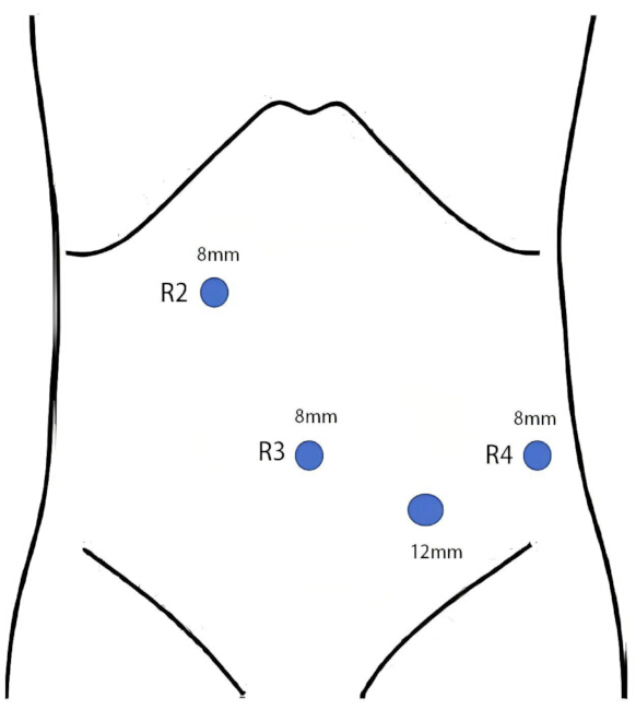
Figure 1: Schematic diagram of trocar placement for pediatric robot-assisted splenectomy. Please click here to view a larger version of this figure.
3. Laparoscopic splenectomy
- Use an ultrasonic scalpel to separate the spleen and stomach ligament. Then, separate the short gastric vessels and the superior splenic vessels branch by branch to expose the splenic hilum.
- Separate the adhesion between the tail of the pancreas and the lower spleen carefully with an electric hook. Separate the splenic artery at the upper edge of the pancreas to expose and ligate it with a silk thread, and then ligate it by Hem-o-lok.
- Use an ultrasonic scalpel to separate the splenocolonic ligament, splenophrenic ligament, and splenorenal ligament, paying attention to avoid injury to the splenic hilum and pancreatic tail. Dissect the splenic vein after Hem-o-lok clamping.
- After the spleen is completely dissociated, withdraw the instruments and robotic arms.
4. Spleen extraction
- Mobilize the spleen into an endoscopic retrieval bag and morcellate under direct visualization.
- Extract specimens through the umbilical port site following extension to 20 mm.
- Perform a meticulous inspection to confirm hemostasis at the splenic bed, pancreatic tail, and gastric greater curvature.
- Place a 19-Fr closed-suction drain in the splenic fossa via the left lateral 12-mm trocar under laparoscopic guidance. Submit all specimens for histopathological analysis.
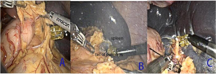
Figure 2: Intraoperative view during robotic pediatric splenectomy using the da Vinci Xi system. (A) Transection of the short gastric vessels. (B) Dissection of the pancreatic tail from the inferior pole of the spleen. (C) Ligation of the splenic pedicle vessels. Please click here to view a larger version of this figure.
Access restricted. Please log in or start a trial to view this content.
Results
Total operative time was 145 min (docking: 35 min; console: 110 min) with estimated blood loss of 2 mL. The patient maintained hemodynamic stability throughout the perioperative period. On postoperative day 1, pyrexia (38.6 °C max) resolved with intravenous cefoperazone and acetaminophen. Flatus was observed on postoperative day 2. A liquid diet was initiated on postoperative day 3. The abdominal drainage tube was removed on postoperative day 4. A blood test rechecked on postoperative day 3 revealed an increase in platel...
Access restricted. Please log in or start a trial to view this content.
Discussion
Hereditary spherocytosis (HS) is a prevalent autosomal dominant hemolytic disorder characterized by spherical erythrocytes in peripheral blood. These microspherocytes undergo splenic sequestration and extravascular hemolysis, resulting in chronic anemia and unconjugated hyperbilirubinemia. Splenectomy remains the definitive management for symptomatic HS, with resolution of hemolytic parameters postoperatively1. According to established clinical guidelines, splenectomy is not indicated for patients...
Access restricted. Please log in or start a trial to view this content.
Disclosures
The authors declare that they have no known competing financial interests or personal relationships that could have appeared to influence the work reported in this paper.
Acknowledgements
We express profound gratitude to all individuals who have contributed to the success of this research study and acknowledge the exceptional dedication and professionalism exhibited by all those involved. This study was funded by the Baseline Health Project of Hubei Province (Grant numbers: JCWJKJCX2025XY1).
Access restricted. Please log in or start a trial to view this content.
Materials
| Name | Company | Catalog Number | Comments |
|---|---|---|---|
| Absorbable ligature clip | Hangzhou KANGJI Medical Instrument co., LTD | KJ-JZJ02ML | |
| Fenestrated bipolar forceps | Intuitive Surgical | 471205 | |
| Leonardo Da Vinci surgical robot | Intuitive Surgical | Xi system | |
| Permanent Cautery Hook | Intuitive Surgical | 3519 | |
| Ultrasound knife | INNOLCON, Medical Science and Technology (suzhou) co., LTD | N/A |
References
- Perrotta, S., Gallagher, P. G., Mohandas, N. Hereditary spherocytosis. Lancet. 372 (9647), 1411-1426 (2008).
- Iolascon, A., et al. Recommendations regarding splenectomy in hereditary hemolytic anemias. Haematologica. 102 (8), 1304-1313 (2017).
- Zhang, Y., et al. Robotic-assisted and laparoscopic splenectomy in children: a single center comparative study. J Laparoendosc Adv Surg Tech A. 34 (6), 541-545 (2024).
- Gallagher, P. G. Abnormalities of the erythrocyte membrane. Pediatr Clin North Am. 60 (6), 1349-1362 (2013).
- Li, M., Li, S. L. Guidelines for pediatric laparoscopic splenectomy (2020 edition). J Clin Pediatr Surg. 20 (1), 6-13 (2021).
- Talamini, M. A., Chapman, S., Horgan, S., Melvin, W. S. A prospective analysis of 211 robotic-assisted surgical procedures. Surg Endosc. 17 (10), 1521-1524 (2003).
- Ojima, T., et al. Short-term outcomes of robotic gastrectomy vs laparoscopic gastrectomy for patients with gastric cancer: a randomized clinical trial. J Clin Oncol. 41 (4 Suppl), 344-344 (2023).
- Sheetz, K. H., Claflin, J., Dimick, J. B. Trends in the adoption of robotic surgery for common surgical procedures. JAMA Netw Open. 3 (1), e1918911(2020).
- Zhang, Y. B., et al. Robot-assisted laparoscopic splenectomy in children: a report of 3 cases with review of the literature. J Clin Pediatr Surg. 20 (8), 718-723 (2021).
- Shelby, R., et al. A comparison of robotic-assisted splenectomy and laparoscopic splenectomy for children with hematologic disorders. J Pediatr Surg. 56 (5), 1047-1050 (2021).
- Isshiki, K., et al. Long-term efficacy and safety profile of splenectomy for pediatric chronic immune thrombocytopenia. Int J Hematol. 117 (5), 774-780 (2023).
- Ghidini, F., et al. Robot-assisted versus laparoscopic approach for splenectomy in children: systematic review and meta-analysis. J Laparoendosc Adv Surg Tech A. 32 (11), 1203-1210 (2022).
- Pincez, T., et al. Long-term follow-up of subtotal splenectomy for hereditary spherocytosis: a single-center study. Blood. 127 (12), 1616-1618 (2016).
- Rosman, C. W. K., Broens, P. M. A., Trzpis, M., Tamminga, R. Y. J. A long-term follow-up study of subtotal splenectomy in children with hereditary spherocytosis. Pediatr Blood Cancer. 64 (10), e26592(2017).
- Wang, X. L., Zhang, G. W., Liu, H., Yang, Y. R. Hem-o-lok clip migration to the duodenum after laparoscopic cholecystectomy: a case report. J Hepatopancreatobiliary Surg. 36 (11), 695-696 (2024).
- Mbaka, M. I., Robl, E., Camps, J. I. Laparoscopic versus robotic-assisted splenectomy in the pediatric population: our institutional experience. Am Surg. 83 (9), 358-359 (2017).
- Delgado-Miguel, C., Camps, J. I. Robotic-assisted versus laparoscopic splenectomy in children: a cost-effectiveness study. J Robot Surg. 18 (1), 51(2024).
- Mehdorn, A. S., et al. Pancreatic fistula and biochemical leak after splenectomy: incidence and risk factors–a retrospective single-center analysis. Langenbecks Arch Surg. 407 (6), 2517-2525 (2022).
- Bassi, C., et al. The 2016 update of the International Study Group (ISGPS) definition and grading of postoperative pancreatic fistula: 11 years after. Surgery. 161 (3), 584-591 (2017).
- Liu, Y., et al. Hereditary spherocytosis before and after splenectomy and risk of hospitalization for infection. Pediatr Res. 93 (5), 1336-1341 (2023).
- Soyer, T., Ciftci, A. O., Tanyel, F. C., Senocak, M. E. Portal vein thrombosis after splenectomy in pediatric hematologic disease: risk factors, clinical features, and outcome. J Pediatr Surg. 41 (11), 1899-1902 (2006).
- Crary, S. E., Buchanan, G. R. Vascular complications after splenectomy for hematologic disorders. Blood. 114 (14), 2861-2868 (2009).
- Robinette, C. D., Fraumeni, J. F. Jr Splenectomy and subsequent mortality in veterans of the 1939-45 war. Lancet. 2 (8029), 127-129 (1977).
Access restricted. Please log in or start a trial to view this content.
Reprints and Permissions
