This study evaluates a structured protocol for ultrasound-guided acupotomy combined with extracorporeal shock wave therapy for nonspecific low back pain, focusing on pain relief and improvement in lumbar function.
Research Article
A Comparative Clinical Evaluation of Ultrasound-guided Acupotomy Combined with Extracorporeal Shock Wave Therapy for Nonspecific Low Back Pain
In This Article
Summary
Abstract
Nonspecific low back pain (NLBP) is a prevalent condition associated with pain and functional limitation. This study evaluated the therapeutic efficacy and procedural feasibility of ultrasound-guided acupotomy combined with extracorporeal shock wave therapy (ESWT) compared with monotherapy approaches for NLBP. A total of 90 patients with NLBP were randomly allocated to three groups (n = 30 each). In the acupotomy group, trigger points within the lumbar erector spinae muscles were identified by palpation and treated using acupotomy. In the shock wave group, ESWT was applied to painful areas using an energy density of 0.10–0.20 mJ/mm2 at a frequency of 10 Hz. In the combined group, ESWT and acupotomy were performed sequentially under ultrasound guidance. Clinical outcomes were evaluated using the visual analog scale (VAS), Oswestry Disability Index (ODI), and Japanese Orthopaedic Association (JOA) scores before treatment and at a 2-month follow-up. Therapeutic efficacy was additionally assessed in accordance with the Traditional Chinese Medicine Syndrome Diagnosis and Efficacy Standard. Baseline scores were comparable among groups (p > 0.05). After treatment, the combined group demonstrated significantly lower VAS and ODI scores, higher JOA scores, and a higher overall effective rate (93.33%) compared with the monotherapy groups (p < 0.05). This study presents a structured protocol for combining ultrasound-guided acupotomy with ESWT for NLBP and demonstrates improved short-term clinical outcomes compared with monotherapy.
Introduction
Nonspecific low back pain (NLBP) is a general term used to describe a type of low back pain in which no clear pathological or anatomical changes can be identified upon diagnosis, and for which there is no definitive clinical cause or objective test findings. If symptoms have persisted for three months or longer, the condition is diagnosed as chronic nonspecific low back pain1. Although most patients with NLBP present with acute and self-limited symptoms, NLBP is often persistent and difficult to treat because of its multifactorial causes2. Approximately 7.3% of the global population is affected by NLBP, and nearly 500 million people take time off work each year due to NLBP, creating a substantial socioeconomic burden3. The incidence of NLBP is closely associated with age, with prevalence increasing significantly after 30 years of age and peaking between 41 and 55 years4. In China, the prevalence of NLBP has increased annually and has shown a trend toward younger patient populations.
Current rehabilitation strategies for NLBP include pharmacological, psychological, and physical therapies5. Extracorporeal shock wave therapy (ESWT) is an established non-invasive treatment modality for chronic musculoskeletal pain conditions, including NLBP6. Clinical studies have shown that ESWT can increase blood flow in the lumbopelvic region, reduce levels of inflammatory cytokines (such as TNF-α and IL-1β), and improve pain and functional scores7,8,9. ESWT delivers high-energy acoustic waves to targeted tissues and has been associated with pain relief and functional improvement in patients with chronic low back pain6,7,8,9.
Acupotomy is a minimally invasive intervention derived from traditional acupuncture techniques combined with modern soft tissue release methods10. Acupotomy has been used in the treatment of various chronic pain conditions, including NLBP, neck pain, shoulder pain, chronic headache or migraine, and osteoarthritis11,12. Conventional acupotomy is commonly performed using palpation-guided localization based on areas of tenderness or muscle tightness. However, blind localization may increase procedural variability and the risk of injury to surrounding structures13. Ultrasound can distinguish the fine structures and echogenic changes in subcutaneous tissues, precisely locate muscles, and provide real-time, dynamic observation of muscle changes during movement. Existing clinical evidence indicates14 that ultrasound‑guided acupotomy is superior to traditional manual palpation in improving pain symptoms and enhancing lumbar spine function. Ultrasound guidance enables precise identification of deep myofascial trigger points and real‑time avoidance of blood vessels and nerves, thereby enhancing treatment safety.
The combination of ultrasound-guided acupotomy and ESWT may provide complementary therapeutic effects for patients with NLBP11,13. Although both interventions have been applied individually in the management of NLBP, standardized procedural descriptions for their combined use under ultrasound guidance remain limited6,12. Therefore, this study enrolled patients with NLBP admitted to our hospital between January 2025 and November 2025 to evaluate the therapeutic efficacy of ultrasound-guided acupotomy combined with ESWT. In addition, this study presents a structured protocol describing treatment sequencing, ultrasound-guided needle insertion, and procedural monitoring during combined therapy.
Protocol
This study was conducted in accordance with the Declaration of Helsinki and was approved by the Ethics Committee of the Second Affiliated Hospital of Qiqihar Medical University (No. [2024]92). Written informed consent was obtained from all participants. The research tools used in this protocol are listed in the Table of Materials.
1. Study participants
A total of 90 patients with NLBP admitted to the Second Affiliated Hospital of Qiqihar Medical University between January 2025 and November 2025 were enrolled in this randomized controlled trial and allocated to three groups (n = 30 each): the acupotomy group, the shock wave group, and the combined group (ultrasound-guided acupotomy plus ESWT).
Eligible participants were adults aged 18–60 years with a diagnosis of NLBP based on the 2016 Chinese Expert Consensus on the Diagnosis and Treatment of Acute/Chronic Nonspecific Low Back Pain16 and the 2017 American College of Physicians guideline for low back pain17, a pain duration of at least 3 months, and normal cognitive and communication abilities sufficient to cooperate with treatment and outcome evaluation.
Patients were excluded if they had specific spinal diseases (including infection, tumor, fracture, lumbar disc herniation, ankylosing spondylitis, or spina bifida occulta), severe osteoporosis, severe systemic disease, a history of psychiatric illness, a bleeding tendency or current anticoagulant use, pregnancy or lactation, or a body mass index (BMI) ≥28 kg/m2.
Participants were randomly allocated using a computer-generated randomization sequence. Allocation concealment was performed using sealed opaque envelopes prepared by an independent researcher who was not involved in recruitment or treatment. Due to the nature of the interventions, participant blinding was not feasible, particularly in the acupotomy and combined groups.
2. Treatment methods
All participants received functional and activities-of-daily-living training during the study period. Clinical assessments were performed within 24 h before treatment initiation and after 2 months of treatment using the visual analog scale (VAS), Japanese Orthopaedic Association (JOA) score, and Oswestry Disability Index (ODI). The study workflow and treatment sequence are shown in Supplementary Figure 1.
- Acupotomy group
Have the patient assume a comfortable position, preferably prone, and palpate the erector spinae muscles in the lower back to locate the hardest point, which will serve as the treatment site for the needle knife procedure. After standard skin disinfection and sterile draping, a sterile disposable acupotomy needle was inserted with the blade aligned parallel to the longitudinal axis of the muscle fibers.
The needle was advanced to the target depth, and the release technique was performed using 2–3 longitudinal cutting motions combined with transverse peeling movements to release adhesions and scar tissue. A sensation of tissue loosening was considered indicative of adequate release. After needle withdrawal, local pressure was applied with sterile gauze for approximately 10 min to achieve hemostasis, followed by application of a sterile dressing. Participants were instructed to avoid strenuous activity for 24 h and to keep the puncture site dry for 48 h. Acupotomy was performed once weekly.
- Shock wave group
Participants were positioned prone with the lumbar region exposed. ESWT was administered using a 15 mm probe with an initial energy flux density of 0.10 mJ/mm2 and a frequency of 10 Hz. Treatment intensity was adjusted according to patient tolerance.
Avoiding direct application over the spine, the probe was moved slowly across the painful lumbar region, delivering 1,500–2,000 pulses to each treatment area. Two to three painful regions were treated during each session. Final treatment parameters were recorded for each participant. ESWT was administered twice weekly with an interval of at least 48 h between sessions. - Combined group
In the combined group, ESWT and acupotomy were performed sequentially under ultrasound guidance. ESWT was administered twice weekly, and acupotomy was performed once weekly. During the first weekly treatment session, acupotomy was performed 5–10 min after completion of ESWT on the same day. During the second weekly session, only ESWT was administered.
Ultrasound coupling gel was applied, and a high-frequency linear transducer was used to visualize the lumbar anatomy and treatment region. ESWT was performed under ultrasound guidance. Following ESWT, the treatment area was reassessed. Acupotomy proceeded only if there was no visible redness, bruising, or hematoma and if the participant reported acceptable pain tolerance with a VAS score ≤4. If these criteria were not met, treatment was postponed and documented accordingly.
For ultrasound-guided acupotomy, the participant remained in the prone position. Following repeat skin disinfection and sterile draping, the ultrasound transducer was covered with a sterile sleeve. The target treatment area within the erector spinae muscles was re-identified under ultrasound guidance while avoiding adjacent blood vessels and nerves. Using an in-plane approach, the needle was inserted at approximately 45° under continuous ultrasound guidance to maintain visualization of the needle shaft and tip.
The needle was advanced to the target tissue region, and the release technique was performed under real-time ultrasound observation. A successful release was identified by the visible separation of fascial or muscular layers, appearing as hypoechoic clefts, and by reduced echogenicity within dense soft tissue structures. Following completion of the procedure, the needle was withdrawn, hemostasis was achieved, and a sterile dressing was applied. Post-procedure care instructions were consistent with those used in the acupotomy group.
3. Observation indicators
Baseline demographic and clinical characteristics, including age, sex, disease duration, education level, and BMI, were recorded. Clinical outcomes were evaluated using the following scales:
VAS: 0–10 points, where 0 indicates no pain, and 10 indicates maximal pain;
ODI: a 10-item questionnaire assessing lumbar dysfunction, with higher scores indicating greater disability;
JOA: a comprehensive assessment of symptoms, signs, and daily functional limitation in patients with NLBP, with lower scores indicating greater dysfunction.
All scales were assessed before treatment and after 2 months of treatment.
Therapeutic efficacy was evaluated according to the State Administration of Traditional Chinese Medicine Syndrome Diagnosis and Efficacy Standard17. Outcomes were categorized as invalid, improvement, significant effect, or cure. The effective rate was defined as the sum of improvement, significant effect, and cure. Adverse events were monitored throughout the treatment and follow-up periods. Recorded adverse events included local bleeding, hematoma, infection, skin irritation, nerve palsy, and muscle spasm. Participants were evaluated during each treatment session and instructed to report delayed symptoms during follow-up assessments.
4. Statistical methods
Sample size estimation was performed using software based on an expected medium effect size (Cohen’s f = 0.25), a statistical power of 0.80, and a two-sided significance level (α) of 0.05. The minimum required sample size for three-group comparisons was calculated as 42 participants (14 per group). Considering an anticipated dropout rate of approximately 10%, at least 46–48 participants were required. A total of 90 participants (30 per group) were ultimately enrolled.
All statistical analyses were performed using SPSS software. Normality of continuous variables was assessed using the Shapiro–Wilk test. Normally distributed data are presented as mean ± standard deviation (SD) and were analyzed using one-way analysis of variance (ANOVA), followed by Tukey’s honestly significant difference (HSD) post hoc test. Non-normally distributed data are presented as median (interquartile range) and were analyzed using the Kruskal–Wallis test followed by Dunn’s test with Bonferroni correction. Within-group comparisons before and after treatment were analyzed using the paired t-test or the Wilcoxon signed-rank test as appropriate. Categorical variables are presented as counts and percentages and were analyzed using the chi-squared (χ2) test or Fisher’s exact test. A two-sided p-value < 0.05 was considered statistically significant.
Results
A total of 90 patients were enrolled in this study and randomly allocated to three groups, with 30 participants in each group. The flowchart of patient enrolment and allocation is shown in Figure 1.
In the acupotomy group, there were 18 men and 12 women, with a mean age of 47.73 ± 8.68 years. In the shock wave group, there were 16 men and 14 women, with a mean age of 47.70 ± 10.51 years. In the combined group, there were 15 men and 15 women, with a mean age of 47.17 ± 7.30 years. No statistically significant differences were observed among the three groups with respect to sex, age, disease duration, education level, BMI, or other baseline characteristics (p > 0.05; Table 1).
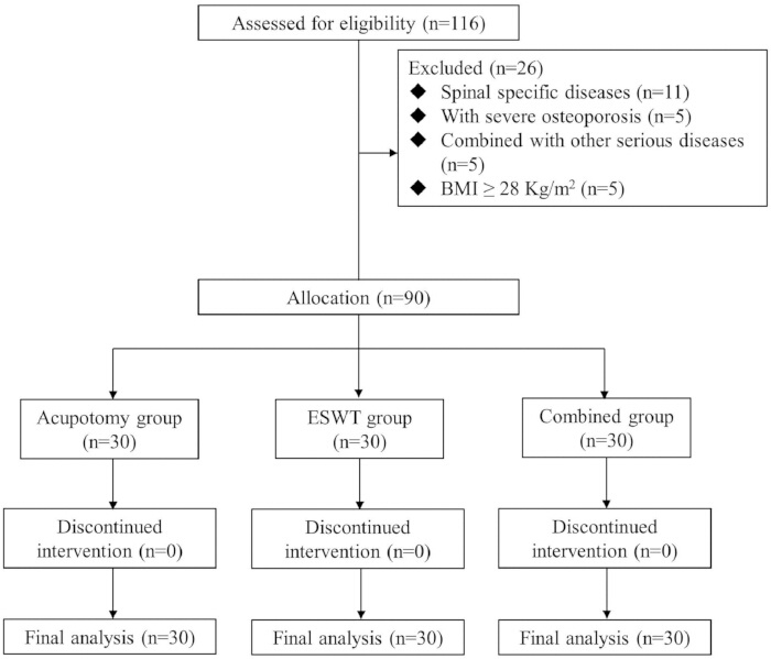
Figure 1: Flowchart of patient enrolment and allocation. A total of 90 patients with nonspecific low back pain were enrolled and randomly allocated to three groups (n = 30 per group): acupotomy alone, extracorporeal shock wave therapy (ESWT) alone, and ultrasound-guided acupotomy combined with ESWT. Patients meeting the exclusion criteria, including spinal-specific diseases, severe osteoporosis, and bleeding tendency, were excluded. All participants completed the 2-month treatment and follow-up period. Please click here to view a larger version of this figure.
| Characteristic | Acupotomy Group (n = 30) | Shock Wave Group (n = 30) | Combined Group (n = 30) | F/χ² | p Value |
| Male sex, n (%) | 18 (60.00) | 16 (53.33) | 15 (50.00) | 0.627 | 0.731 |
| Age (years) | 47.73 ± 8.68 | 47.70 ± 10.51 | 47.17 ± 7.30 | 0.038 | 0.963 |
| Disease duration (months) | 16.07 ± 7.10 | 16.50 ± 10.27 | 17.03 ± 7.78 | 0.097 | 0.907 |
| Education level, n (%) | 1.518 | 0.468 | |||
| High school or below | 23 (76.67) | 20 (66.67) | 24 (80.00) | ||
| Above high school | 7 (23.33) | 10 (33.33) | 6 (20.00) | ||
| BMI (kg/m²) | 23.02 ± 3.43 | 23.61 ± 4.36 | 22.73 ± 3.04 | 0.458 | 0.634 |
| Note: Continuous variables are presented as mean ± standard deviation (SD). Categorical variables are presented as number (%). Between-group comparisons were performed using one-way analysis of variance (ANOVA) for continuous variables and the chi-squared (χ²) test for categorical variables. BMI, body mass index. | |||||
Table 1: Baseline demographic and clinical characteristics of the three treatment groups. Data include sex, age, disease duration, education level, and body mass index (BMI). Continuous variables are presented as mean ± standard deviation (SD), and categorical variables are presented as counts and percentages. Between-group comparisons were performed using one-way analysis of variance (ANOVA) for continuous variables and the chi-squared (χ2) test for categorical variables. No statistically significant baseline differences were observed among the groups (all p > 0.05).
Primary Outcomes
VAS Scores
Before treatment, the VAS scores in the acupotomy, shock wave, and combined groups were 5.77 ± 0.68, 5.93 ± 0.64, and 5.90 ± 0.80, respectively, with no significant differences among the groups (p = 0.632).
After 2 months of treatment, the VAS scores were 3.97 ± 0.72, 3.70 ± 0.79, and 2.07 ± 0.70, respectively, and the difference among the groups was statistically significant (p < 0.001). Post-hoc Tukey analysis showed that the combined group had significantly lower VAS scores than the acupotomy group (mean difference: −1.90; 95% confidence interval [CI]: −2.45 to −1.35; p < 0.001) and the shock wave group (mean difference: −1.63; 95% CI: −2.18 to −1.08; p < 0.001). No significant difference was observed between the acupotomy and shock wave groups (mean difference: 0.27; 95% CI: −0.28–0.82; p = 0.487).
ODI Scores
Before treatment, the ODI scores in the acupotomy, shock wave, and combined groups were 33.33 ± 9.30, 33.40 ± 8.74, and 34.13 ± 7.50, respectively, with no significant differences among the groups (p = 0.873).
After 2 months of treatment, the ODI scores were 19.77 ± 6.93, 17.93 ± 8.70, and 15.43 ± 7.11, respectively, with a statistically significant difference among the groups (p = 0.031). Post-hoc Tukey analysis showed that the combined group had significantly lower ODI scores than the acupotomy group (mean difference: −4.34; 95% CI: −7.82 to −0.86; p = 0.009). No statistically significant differences were observed between the combined group and shock wave group (mean difference: −2.50; 95% CI: −5.98–0.98; p = 0.187) or between the two monotherapy groups (mean difference: 1.84; 95% CI: −1.64–5.32; p = 0.423).
JOA Scores
Before treatment, the JOA scores in the acupotomy, shock wave, and combined groups were 18.20 ± 3.89, 18.23 ± 4.42, and 17.67 ± 5.68, respectively, with no significant differences among the groups (p = 0.922).
After 2 months of treatment, the JOA scores were 21.07 ± 3.89, 23.30 ± 3.36, and 24.27 ± 4.56, respectively, with a statistically significant difference among the groups (p = 0.008; Table 2). Post-hoc Tukey analysis showed that the combined group had significantly higher JOA scores than the acupotomy group (mean difference: 3.20; 95% CI: 1.15–5.25; p = 0.001). No statistically significant difference was observed between the combined group and shock wave group (mean difference: 0.97; 95% CI: −1.08–3.02; p = 0.532). The shock wave group demonstrated significantly higher JOA scores than the acupotomy group (mean difference: 2.23; 95% CI: 0.18–4.28; p = 0.029).
| Outcome | Time Point | Acupotomy group (n=30) | Shock wave group (n=30) | Combined group (n=30) | F | P |
| VAS score | Before treatment | 5.77 ± 0.68 | 5.93 ± 0.64 | 5.90 ± 0.80 | 0.462 | 0.632 |
| After treatment | 3.97 ± 0.72 | 3.70 ± 0.79 | 2.07 ± 0.70 | 5.595 | <0.001* | |
| ODI score | Before treatment | 33.33 ± 9.30 | 33.4 ± 8.74 | 34.13 ± 7.50 | 0.136 | 0.873 |
| After treatment | 19.77 ± 6.93 | 17.93 ± 8.70 | 15.43 ± 7.11 | 3.611 | 0.031* | |
| JOA score | Before treatment | 18.2 ± 3.89 | 18.23 ± 4.42 | 17.67 ± 5.68 | 0.081 | 0.922 |
| After treatment | 21.07 ± 3.89 | 23.3 ± 3.36 | 24.27 ± 4.56 | 5.132 | 0.008* | |
| Note: VAS=Visual Analogue Scale, ODI=Oswestry Disability Index, JOA=Japanese Orthopaedic Association. *P <0.05. | ||||||
Table 2: Comparison of visual analog scale (VAS), Oswestry Disability Index (ODI), and Japanese Orthopaedic Association (JOA) scores before and after treatment. VAS scores ranged from 0–10 points, with higher scores indicating greater pain intensity. ODI scores ranged from 0–50 points, with higher scores indicating greater disability. JOA scores ranged from 0–29 points, with higher scores indicating better lumbar function. Δ indicates the change between pre-treatment and post-treatment scores. Statistical analysis was performed using analysis of variance followed by Tukey’s post-hoc test. *p < 0.05.
Therapeutic effects and adverse events
After 2 months of treatment, the effective rate in the combined group was significantly higher than that in the other two groups, with a statistically significant difference among the three groups (p < 0.05; Table 3).
Pairwise comparisons using the chi-squared test with Bonferroni correction showed that the combined group had a significantly higher effective rate than the acupotomy group (χ2 = 8.57; p = 0.014) and the shock wave group (χ2 = 5.88; p = 0.043). No statistically significant difference was observed between the two monotherapy groups (χ2 = 0.67; p = 1.000).
No major adverse events, including significant bleeding, infection, or neurological deficits, were observed during the treatment period. One participant in the acupotomy group experienced transient dizziness and headache, which resolved without additional intervention and was not clearly attributable to the procedure.
| Index | Acupotomy group | Shock wave group | Combined group | X2 | P |
| Cure | 5 | 7 | 14 | 11.71 | 0.042* |
| Significant effect | 7 | 9 | 10 | ||
| Improvement | 11 | 8 | 4 | ||
| Invalid | 7 | 6 | 2 | ||
| Note: *P <0.05. | |||||
Table 3: Therapeutic effects according to the Traditional Chinese Medicine Syndrome Diagnosis and Efficacy Standard after 2 months of treatment. The effective rate was calculated as (cure + significant effect + improvement)/total × 100%. Data are presented as patient counts. Overall group differences were analyzed using the chi-squared (χ2) test. Pairwise comparisons were performed using the chi-squared test with Bonferroni correction where applicable. *p < 0.05 indicates statistical significance.
DATA AVAILABILITY:
The dataset supporting the findings of this study has been deposited in Zenodo and is available at https://doi.org/10.5281/zenodo.20763154 .
Supplementary Figure 1: Study workflow and treatment sequence. All groups received functional and activities-of-daily-living training. Clinical outcomes were evaluated using the visual analog scale (VAS), Oswestry Disability Index (ODI), and Japanese Orthopaedic Association (JOA) scores at baseline and after 2 months of treatment. The treatment protocol included acupotomy alone, ESWT alone, and ultrasound-guided acupotomy combined with ESWT. Acupotomy was performed once weekly, whereas ESWT was administered twice weekly over a 2-month treatment period.Please click here to download this file.
Discussion
Nonspecific low back pain (NLBP) is a common musculoskeletal condition characterized by pain and functional limitation without clear nerve root involvement or identifiable spinal pathology18. This study evaluated the therapeutic efficacy and procedural feasibility of ultrasound-guided acupotomy combined with extracorporeal shock wave therapy (ESWT) for patients with NLBP. The results demonstrated that the combined treatment group showed greater improvements in pain and lumbar function scores after 2 months of treatment than the monotherapy groups.
After treatment, the combined group demonstrated lower VAS and ODI scores and higher JOA scores than the acupotomy and shock wave groups. These findings suggest that the combination of ultrasound-guided acupotomy and ESWT may be associated with improved short-term clinical outcomes in patients with NLBP. Ultrasound guidance provided real-time visualization of anatomical structures and needle trajectory during the procedure19. In this study, ultrasound guidance was used to assist procedural visualization and monitoring during acupotomy treatment. Real-time ultrasound imaging technology enables dynamic visualization of deep tissues during the procedure, potentially facilitating accurate needle placement. Ultrasound guidance allows the operator to visualize the needle trajectory and target tissue during treatment. Clinical data20 showed that, for myofascial pain, the pain relief rate in the ultrasound-guided group was 91.3% compared with 67.8% in the traditional needle-knife control group. In the treatment of chronic nonspecific low back pain, ultrasound guidance has been associated with improvements in lumbar spine mobility and activities of daily living20. Traditional acupotomy relies primarily on the physician's tactile sensation and the patient's response to determine the treatment location, which may increase procedural variability. Ultrasound-guided acupotomy enables real-time observation of needle insertion depth and tissue response during the release procedure. Throughout the procedure, ultrasound guidance allows visualization of the needle path and needle tip position.
Acupotomy is a minimally invasive intervention that has been used in the treatment of chronic musculoskeletal pain conditions12. In the present study, the combined treatment demonstrated better clinical outcomes than monotherapy. ESWT has been associated with analgesic effects and functional improvement in patients with chronic low back pain8,9, whereas acupotomy may contribute to soft tissue release in areas of muscle tightness or tenderness21. However, the mechanisms underlying the combined treatment effect remain unclear because physiological parameters, such as microcirculation and inflammatory markers, were not evaluated in this study. Previous studies have also reported improvements in pain and functional outcomes following ultrasound-guided acupotomy in patients with chronic NLBP22.
The combined group's effective rate was significantly higher than that of the other treatment groups after 2 months of treatment. No major adverse events, including vascular injury, nerve injury, or infection, were observed during the treatment period. Ultrasound guidance enabled visualization of adjacent anatomical structures during needle insertion and treatment23,24. One participant in the acupotomy group experienced transient dizziness and headache, which resolved without additional intervention.
Several limitations should be considered in this study. First, the sample size was relatively small, and follow-up assessments were limited to 2 months post-treatment, which may limit the generalizability of the findings. Second, all participants received routine functional training and activities of daily living training in addition to the study interventions, making it difficult to isolate the independent effects of each treatment approach. Third, both ultrasound-guided acupotomy and ESWT are operator-dependent procedures, and variability in technical performance may have influenced treatment outcomes. In addition, practitioner blinding was not feasible because of the nature of the interventions. Future studies with larger sample sizes, longer follow-up periods, and standardized procedural parameters are needed to further evaluate the clinical application of combined ultrasound-guided acupotomy and ESWT for NLBP.
Disclosures
The authors declare that they have no competing interests.
Materials
| Name | Company | Catalog Number | Comments |
|---|---|---|---|
| Acupotomy needle | Jiangsu Huayou Medical Devices Co., Ltd., Suqian, China | 0.40 mm × 50 mm | Used for soft tissue release during acupotomy. |
| Extracorporeal shock wave therapy device | Zimmer MedizinSysteme GmbH, Tuttlingen, Germany | enPuls Version 2.0 | Used to deliver extracorporeal shock wave therapy (ESWT). |
| G*Power software | Heinrich Heine University Düsseldorf, Düsseldorf, Germany | NA | Used for a priori sample size estimation and statistical power analysis. |
| Portable color Doppler ultrasound system | Shenzhen Huasheng Medical Technology Co., Ltd., Shenzhen, China | Clover 60 | Used for ultrasound guidance during acupotomy and treatment-site visualization. |
| SPSS Statistics | IBM Corp., Armonk, NY, USA | SPSS Statistics Version 26.0 | Used for statistical analysis of study data. |
References
- Su TX, Wang X. Research progress on the etiology of Modic low back pain. Med Inf. 2024;37(11):170-174. Doi: 10.3969/j.issn.1006-1959.2024.11.036.
- Feng YZ. Research progress on multidimensional mechanisms of core training therapy for chronic non-specific back pain. Journal of Practical Medicine. 2025; 41 (22) 3625-3630. doi: 10.3969/j.issn.1006-5725.2025.22.022.
- Popescu A, Lee H. Neck pain and lower back pain. Med Clin North Am. 2020;104(2):279-292. doi: 10.1016/j.mcna.2019.11.003.
- Li H, Tan HY. Progress on the treatment of chronic non-specific low back pain. Chin Med Mod Distance Educ China. 2024;22(12):170-173. doi: 10.3969/j.issn.1672-2779.2024.12.054.
- Kim SK et al. Clinical outcomes and cost-effectiveness of massage chair therapy versus basic physiotherapy in lower back pain patients: a randomized controlled trial. Medicine (Baltimore). 2020;99(12). doi: 10.1097/MD.0000000000019514.
- Wu Z, Zhou T, Ai S. Extracorporeal shock wave therapy for low back pain: a systematic review and meta-analysis. Medicine (Baltimore). 2023;102(52). 0000000000036596.
- Kabeer AS et al. The adult with low back pain: causes, diagnosis, imaging features and management. Br J Hosp Med (Lond). 2023;84(10):1-9. doi: 10.12968/hmed.2023.0063.
- Liu K et al. Efficacy and safety of extracorporeal shockwave therapy in chronic low back pain: a systematic review and meta-analysis of 632 patients. J Orthop Surg Res. 2023;18(1):455. doi: 10.1186/s13018-023-03943-x.
- Yue L et al. Extracorporeal shockwave therapy for treating chronic low back pain: a systematic review and meta-analysis of randomized controlled trials. Biomed Res Int. 2021;2021:5937250. doi: 10.1155/2021/5937250.
- Wu DT et al. Progress on acupotomy treatment of carpal tunnel syndrome. China J Orthop Traumatol. 2024;37(12):1237-1240. doi: 10.12200/j.issn.1003-0034.20230977.
- Wu LT, Zhou QM, Wang W. Treatment of calcaneal pain by ultrasound localization combined with extracorporeal shock wave and small acupotomy. J Rare Uncommon Dis. 2024;31(8):123-125. doi: 10.3969/j.issn.1009-3257.2024.8.049.
- Li XM et al. Observation of the therapeutic effect of minimally invasive needle knife treatment for 43 cases of chronic non-specific lower back pain in coastal stationed officers and soldiers under the guidance of musculoskeletal ultrasound. Chinese Journal of Naval Medicine and High Pressure Medicine. 2025;32 (11): 1226-1231. doi: 10.3760/cma.j.cn311847-20250310-00061.
- Zhou FY et al. Systematic review and meta-analysis of acupotomy versus acupuncture for lumbar intervertebral disc herniation. Liaoning J Tradit Chin Med. 2017;44(3):590-594. doi: 10.13192/j.issn.1000-1719.2017.03.049.
- Hodges PW, Danneels L. Changes in structure and function of the back muscles in low back pain: different time points, observations, and mechanisms. J Orthop Sports Phys Ther. 2019;49(6):464-476. doi: 10.2519/jospt.2019.8827.
- Xie HW. A Study on the Efficacy of Ultrasound-Guided Nerve Block Combined with Ozone Injection in the Treatment of Lumbosacral Posterior Root Syndrome [D]. Shanxi Medical University. 2022. doi:10.27288/d.cnki.gsxyu.2022.000250.
- Expert Group of the Spinal Cord Professional Committee of the Chinese Rehabilitation. Medicine Association Expert consensus on diagnosis and treatment of acute/chronic non-specific low back pain in China. Chinese Journal of Spine and Spinal Cord. 2016;26 (12) 1134-1138. doi: 10.3969/j.issn.1004-406X.2016.12.16
- Qaseem A et al. Noninvasive treatments for acute, subacute, and chronic low back pain: a clinical practice guideline from the American College of Physicians. Ann Intern Med. 2017;166(7):514-530. doi: 10.7326/M16-2367.
- Ma KL et al. Fu’s subcutaneous needling versus massage for chronic non-specific low-back pain: a randomized controlled clinical trial. Ann Palliat Med. 2021;10(11):11785-11797. 2021-11.
- Zhu WT, Guo CQ, Zhao L. Clinical study on the combination of ultrasound visualization needle knife and fire needle therapy for the treatment of central lumbar disc herniation. Chinese Medical Emergency. 2023;32 (12): 2107-2111. doi:10.3969/j.issn.1004-745X.2023.12.010.
- Yu YL et al. Effect of motor acupuncture based on movement pattern adjustment theory in the treatment of non-specific low back pain. J Clin Med Pract. 2024;28(21):100-104. doi: 10.7619/jcmp.20240783
- Li Z et al. Ultrasound-guided needle knife release with different pathways for carpal tunnel syndrome: a randomized controlled trial. Zhongguo Zhen Jiu. 2025;45(1):21-26. doi: 10.13703/j.0255-2930.
- Lan X et al. A comparison of the outcomes of open trigger release versus ultrasound-guided modified small needle-knife percutaneous release for treatment of trigger digits. J Hand Surg Asian Pac Vol. 2023;28(1):69-74. 523500017.
- Liu SR et al. Clinical study on the evaluation of different acupuncture point selection methods in the intervention of chronic non-specific back pain patients using ultrasound shear wave elastography. Journal of Liaoning University of Traditional Chinese Medicine. 2024;26 (2): 194-198. 1673-842x.2024.02.036.
- Corp N et al. Evidence-based treatment recommendations for neck and low back pain across Europe: a systematic review of guidelines. Eur J Pain. 2021;25(2):275-295. doi: 10.1002/ejp.1679.