This article details intrathecal delivery methods for CNS-targeted therapeutics in adult mice, focusing on protocol precision and enabling repeated injection. It includes findings from a validation study where antisense oligonucleotides reduced mutant SOD1 expression in SOD1-ALS model mice, supporting the utility of the approach in preclinical research on neurodegenerative diseases.
Method Article
Lumbar Intrathecal Injection of SOD1-ASOs for Precise CNS Targeting and Predictive Efficacy in Human SOD1-G93A ALS Mice
In This Article
Summary
Abstract
Intrathecal (IT) administration of central nervous system (CNS)-targeted therapeutics offers a minimally invasive route for direct drug delivery into the cerebrospinal fluid (CSF), facilitating enhanced specificity and target engagement. While IT catheterization is standard for sustained delivery in rats, its application in mice is limited by anatomical constraints and elevated risk of procedure-related complications, including spinal injury and infection. To overcome these limitations, we employed an acute needle puncture technique for IT drug delivery in adult mice, providing a reproducible and less invasive alternative compatible with single or repeated dosing regimens. In a transgenic mouse model of amyotrophic lateral sclerosis (ALS) harboring the SOD1-G93A mutation, we achieved efficient IT delivery of antisense oligonucleotides (ASOs) targeting the SOD1 gene. This approach effectively downregulated mutant SOD1 expression and significantly ameliorated the disease phenotype, as demonstrated by electrophysiological and biomarker assessments. These results validate both the IT delivery method and the therapeutic efficacy, with outcomes comparable to intracerebroventricular (ICV) administration. Most importantly, the technique mirrors procedures used in human clinical studies, offering strong translational relevance and utility in preclinical evaluation of CNS-directed interventions. Although technical expertise is required to ensure consistency and avoid off-target effects, this IT delivery strategy represents a robust, reproducible, and clinically relevant methodology for advancing therapeutic development in small animal models.
Introduction
In this study, we describe a lumbar intrathecal (IT) injection protocol, compare it with intracerebroventricular (ICV) delivery, and evaluate pharmacodynamic outcomes and tolerability to guide the development of central nervous system (CNS)-targeted therapeutics. RNA-targeting therapies, particularly antisense oligonucleotides (ASOs), represent a promising modality for CNS diseases by enabling post-transcriptional modulation of gene expression. These approaches are especially relevant for neurodegenerative disorders, where progressive neuronal loss underscores the need for precise, localized, and timely interventions to potentially modify disease progression. Here, we directly compare the efficacy of the IT injection protocol to ICV delivery for ASO therapeutics.
In preclinical studies, lumbar intrathecal injections in adult mice (male or female, 30-70 days old, 13-20 g weight) have been used to deliver pharmacological agents or biological vectors directly into the cerebrospinal fluid (CSF) for CNS targeting1,2. Injection volumes of 5-10 μL are typically administered slowly to limit transient CSF pressure increases and reflux, with intrathecal access achieved via the L5-L6 intervertebral space1,2. Procedural success depends on a careful technique to avoid neurological injury, with age and body weight influencing CSF volume and handling. Use of fine-gauge needles (e.g., a 27-30 G needle coupled to a 25 μL Hamilton syringe) enables reliable delivery2. Key technical challenges include minimizing dead-space losses, preventing epidural mis-injection and leakage, and reducing operator-dependent variability that can impact dosing accuracy and CSF distribution.
Developing effective therapies for CNS disorders remains a major challenge due to the structural complexity and cellular heterogeneity of the CNS. These features impede targeted drug delivery and restrict biodistribution across multiple anatomical and physiological barriers, necessitating specialized delivery strategies. A key obstacle arises from physiological and anatomical barriers throughout the CNS that restrict drug penetration. The brain and spinal cord are encased within three meningeal layers: the dura mater, arachnoid mater, and pia mater, which, together with the blood-brain barrier (BBB), provide a formidable defense against circulating therapeutics and other molecules in the blood. While vital for maintaining CNS stability, these barriers significantly hinder the delivery of large, hydrophilic molecules such as ASOs, monoclonal antibodies, and viral vectors1,2,3,4. IT administration offers a means to circumvent these restrictions by introducing therapeutics directly into the CSF within the subarachnoid space. This delivery route facilitates drug distribution across CNS compartments, promoting broader tissue penetration and thereby enabling effective engagement with targets expressed in diverse cell types within the CNS. Since its initial use by Hylden and Wilcox in 1980 for morphine delivery in mice1, IT injections have become a more widely used method for delivering a variety of therapeutic agents, including small molecules, biologics, gene therapy vectors, and ASOs5,6,7,8,9,10.
ASOs are chemically engineered single-stranded oligonucleotides designed to bind complementary RNA sequences and modulate gene expression through mechanisms such as RNase H-mediated degradation, splice modulation, and steric inhibition of translation11,12,13,14,15. Chemical enhancements like phosphorothioate backbone modifications, 2'-O-methoxyethyl (2'-MOE) substitutions, and constrained ethyl residues enhance their stability, binding affinity, and resistance to nucleases6. These properties have supported their application in targeting pathogenic genes implicated in neurogenetic disorders. ASO-based treatments administered intrathecally have demonstrated clinical benefit in diseases such as spinal muscular atrophy (SMA) and amyotrophic lateral sclerosis (ALS)12,13,14,15. For example, Nusinersen (Spinraza) enhances exon 7 inclusion in SMN2 transcripts to compensate for SMN1 loss in SMA7,14. Similar strategies targeting mutant huntingtin transcripts were also explored in Huntington's disease12,13. Although these trials were terminated early16, they underscore the continued interest in CNS-directed ASO approaches.
ALS is a fatal neurodegenerative disorder characterized by progressive degeneration of both upper and lower motor neurons. The disease, affecting between 6 and 9 individuals per 100,000 worldwide and occurring at a rate of approximately 2 cases per 100,000 person-years, presents gradually worsening muscle weakness and spasticity, with most patients succumbing to neuromuscular respiratory failure within 2 to 3 years after symptoms begin17. While most cases are sporadic (sALS), around 10% are familial (fALS), offering insights into genetic drivers of the disease. Among the most studied genes is Cu/Zn superoxide dismutase 1 (SOD1), which accounts for nearly 15 to 20% of fALS, less than 2% of sALS, and is inherited in an autosomal dominant manner18. SOD1 encodes a cytoplasmic antioxidant enzyme that detoxifies reactive oxygen species by converting superoxide radicals into hydrogen peroxide. Mutant forms of SOD1, such as G93A, A4V, and H46R, exhibit conformational instability, leading to protein aggregation and a toxic gain-of-function that contributes to oxidative stress, mitochondrial dysfunction, and excitotoxicity19,20,21,22,23,24,25. Moreover, ALS pathology is exacerbated by non-cell autonomous mechanisms involving microglia and astrocytes, which contribute to a chronic inflammatory environment and progressive neuronal injury26,27,28,29. Silencing SOD1 expression using ASOs, therefore, presents a promising therapeutic avenue for mitigating disease pathogenesis.
The SOD1-G93A transgenic mouse model (Jax Strain #: 004435), which expresses the human G93A mutant form of SOD1 under the control of endogenous human SOD1 promoter, closely recapitulates the clinical features of ALS, including early motor deficits, spinal cord neurodegeneration, and shortened survival30. Previous preclinical studies have shown that administering SOD1-specific ASOs through sustained intraventricular delivery using osmotic pumps in SOD1-G93A rats at postnatal day 65 at a daily dose of 100 μg for 28 consecutive days results in significant suppression of SOD1 levels, reduction of neuroinflammatory responses, delayed emergence of clinical signs, and improved motor performance31. Furthermore, ASO-mediated knockdown of SOD1 in people living with SOD1-ALS has been associated with reductions in neurofilament light chain (NfL) levels, a blood-based biomarker indicative of axonal damage32. In this investigation, we administered SOD1-targeting ASOs via IT injection in SOD1-G93A mice and evaluated their impact across molecular, electrophysiological, and biomarker readouts. Quantitative analysis of spinal cord tissue revealed significant reductions in SOD1 mRNA levels. Electrophysiological assessments of compound muscle action potentials (CMAPs) demonstrated preserved neuromuscular function. Concurrently, serum NfL levels decreased, reflecting a mitigation of ongoing neuronal injury.
To evaluate the spatial distribution and relative efficacy of alternative CNS delivery methods, we included a comparative arm employing intracerebroventricular (ICV) injection, wherein the therapeutic agent is introduced into the lateral ventricles for dissemination via CSF flow along ependymal and perivascular spaces33. By comparing IT and ICV administration routes, we aimed to delineate differences in pharmacodynamic outcomes and tissue targeting, thereby refining delivery strategies for CNS-directed ASO therapies. Additionally, to assess translational feasibility, we conducted independent tolerability studies involving up to four IT injections per animal spaced eight weeks apart. Across all tested regimens, no overt signs of distress or morbidity were observed, supporting the tolerability of IT ASO dosing in the context of survival. These results are consistent with prior reports demonstrating the tolerability of repeated intrathecal injections in mice34.
The clinical relevance of IT ASO delivery is further supported by the recent regulatory approvals of tofersen (QALSODY®) for SOD1-associated ALS35. Clinical studies demonstrated robust target engagement, reflected by reductions in CSF SOD1 protein, followed by decreases in neurofilament light chain levels, a biomarker of neuroaxonal injury. Although the primary endpoint was not met in the initial phase 3 VALOR trial, integrated analyses including the open-label extension suggested potential benefits of earlier treatment initiation35.
Collectively, the data here demonstrate that IT delivery of SOD1-targeting ASOs in the SOD1-G93A ALS mouse model achieves effective molecular knockdown, biomarker improvement, and functional preservation with favorable tolerability. These results support IT ASO delivery as a rational and clinically viable approach for CNS genetic disorders and provide a robust preclinical framework for continued translational development.
Protocol
All procedures here were approved and conducted in accordance with IACUC-approved protocols and institutional chemical hygiene and biosafety guidelines.
NOTE: Perform studies in C57BL/6J (JAX #000664) and SOD1-G93A transgenic mice (JAX #004435) housed under standard conditions (12-h light/dark cycle, 22-24 °C, ad libitum access to food and water).
1. Applying chemically modified ASOs to target human SOD1
- Employ validated antisense oligonucleotides (ASOs) to specifically target nucleotides 613-629 within the 3' untranslated region (UTR) of the human SOD1 gene (RefSeq: NM_000454.5)36: A*GoToG*T*T*T*A*A*T*G*T*ToToA*T*C
- Include validated chemical modifications to enhance stability and efficacy, by incorporating MOE (2'-O-Methoxyethyl) (indicated in regular font), cEt (Constrained Ethyl) (italics), DNA (bold), phosphate linkages (o), and phosphorothioate linkages (*), as reported previously36. Supplementary File 1 presents the ASO chemistry with corresponding color coding.
- Prepare a non-targeting control ASO with no complementarity to human or rodent genomes, following the same modification scheme for use as a control: C*CoToAoT*A*G*G*A*C*T*A*T*C*C*AoGoGoA*A
2. Preparing and handling ASO solutions under sterile conditions
- Reconstitute ASO in sterile artificial cerebrospinal fluid (aCSF; Table 1) to a 10 mg/mL concentration under sterile conditions, targeting a final injection volume of 10 μL (100 μg) per animal.
- Mix the solution gently using a sterile pipette to maintain molecular integrity, then vortex lightly for 5-10 s to ensure it is well mixed.
- Filter the diluted solution through a sterile 0.2 mm syringe filter to remove particulates and contaminants.
- Aseptically aliquot the final solution into sterile microcentrifuge tubes and store at 4 °C in a monitored, temperature-controlled unit for up to 1 week, avoiding freeze-thaw cycles to maintain stability.
3. Mouse preparation prior to intrathecal injection
- Weigh each mouse to the nearest 0.1 g using a digital balance, then transfer to a nose cone and induce anesthesia with isoflurane at a concentration of 2-2.5% in oxygen; monitor depth continuously and confirm surgical plane by loss of pedal reflex.
- Prepare a thermostatically controlled heating pad and set it to 37 °C to support mice during preparation and recovery. Place a physical barrier between the mouse and the heating pad to avoid direct contact.
- Apply ophthalmic lubricant to both eyes immediately after anesthesia induction to prevent corneal desiccation. Then position the mouse in ventral recumbency on the heating pad to prevent hypothermia, minimize physiological stress, and support normal cardiovascular and respiratory functions throughout the procedure.
- Administer buprenorphine extended-release injectable suspension (1.3 mg/mL) subcutaneously at 3.25 mg/kg using a sterile syringe with a 25-G needle and inject 0.05 mL per 20 g body weight prior to the procedure.
- Shave the lumbar region from mid-flank to pelvic girdle, then disinfect the area using alternating swabs of isopropyl alcohol and povidone-iodine, repeat three times, and allow the site to air-dry completely.
4. Direct intrathecal injection
- After anesthesia and surgical site preparation, move the mouse to a clean injection area and place it in ventral recumbency on the heating pad.
- Place the mouse in a position that arches the spine to facilitate palpation and injection. Insert a curved spatula beneath the abdomen at the lower thoracic to upper lumbar level to induce mild spinal flexion and widen the lumbar intervertebral spaces between the fifth and sixth (L5-L6, preferred) or fourth and fifth (L4-L5) laminae.
- Stabilize the pelvic girdle by gently grasping the pelvic girdle between the thumb and forefinger of one hand and palpating the dorsal midline with the other hand to locate the target intervertebral space between L4-L5 or L5-L6.
- Align a sterile 0.3 mL insulin syringe fitted with a 29-G, ½-inch needle perpendicular to the dorsal skin surface or at a 70-80° angle, with the bevel oriented cranially. Advance the needle until slight resistance is encountered as it penetrates the dura mater, then continue into the intrathecal space.
- Insert steadily until the tip enters the intrathecal space, confirmed by a tail flick reflex indicating successful intrathecal entry.
- If significant resistance is encountered or no tail-flick reflex is observed, withdraw the needle, reidentify the anatomical landmarks, and adjust needle placement before reattempting the injection. If the initial attempt was performed at the L5-L6 intervertebral space, perform the subsequent attempt at the L4-L5 intervertebral space.
- Inject 10 μL of the ASO solution slowly (over 10 s) to minimize pressure changes and avoid CSF reflux. The needle may be held in place for an additional 10 s after injection.
- Carefully withdraw the needle vertically and smoothly to prevent tissue damage and backflow.
5. Monitoring mice and supporting recovery after injection
- Place each mouse in an individual or well-spaced compartment within a pre-warmed recovery chamber maintained at 37 °C. Ensure clear visibility.
- Keep the chamber temperature controlled during anesthetic recovery to prevent hypothermia.
- Monitor continuously for respiratory rate and pattern, posture, and responsiveness to tactile stimuli.
- Observe the mouse until it regains the righting reflex and exhibits coordinated movement.
- Document all relevant procedural details, including the injection site, ASO dose, injection volume, and any behavioral or physiological observations during and after the procedure, in accordance with IACUC protocols.
6. Determining criteria for return to the home cage
- Confirm that the mouse is stable with coordinated spontaneous movement before returning to its home cage.
- Ensure there are no signs of distress, including labored or irregular breathing, vocalizations, and prolonged unresponsiveness.
- Provide soft nutritional support, such as a nutritionally complete gel diet supplement, to help maintain hydration and caloric intake with minimal effort.
7. Conducting post-recovery observation of mice
- Verify normal behaviors such as exploration, grooming, and social interaction if the mice are group housed.
- Continuously monitor for the appearance of any signs of abnormal recovery, such as unusual posture, weakness or paralysis, irregular breathing, or decreased responsiveness, and reassess promptly if these symptoms occur.
- Seek veterinary intervention if recovery is delayed or compromised.
8. Intracerebroventricular injection in the lateral ventricle using stereotaxic surgery and postoperative care
NOTE: Include a comparative arm using intracerebroventricular (ICV) injection to assess spatial distribution and relative efficacy, delivering ASOs into the lateral ventricles for CSF-mediated dissemination and comparison to intrathecal delivery. Perform the ICV injections according to well-established, standardized protocols for mice37.
- Anesthetize the mouse with isoflurane at a concentration of 2-2.5 % in oxygen for immobility and position the mouse in ventral recumbency on the heating pad and then administer buprenorphine extended-release injectable suspension (1.3 mg/mL buprenorphine hydrochloride) subcutaneously at 3.25 mg/kg using a sterile 1 mL syringe with a 25-G needle at 0.05 mL per 20 g body weight prior to procedure. Shave the scalp of the mouse with clippers.
- Perform all surgical procedures under anesthesia. Confirm the depth of anesthesia by verifying the absence of pedal withdrawal reflex (toe pinch). Maintain a sterile aseptic field throughout the ICV surgery. Use only sterile instruments and handle them carefully to prevent contact with nonsterile surfaces.
- Secure the mouse in a stereotaxic frame with a digital display console in the standard prone position. Place the front teeth into the tooth bar of the mouse adaptor to immobilize the head, then secure the adaptor with the inhalation and exhaust breathing tubes delivering isoflurane. Insert the ear bars into the ears and tighten the screws to firmly stabilize the head.
- Disinfect the area using alternating swabs of isopropyl alcohol and povidone-iodine, repeated three times, and make a midline incision with a sterile scalpel to expose the skull. Use a sterile cotton tip to remove soft tissues and make sure that the bregma is clearly seen.
- Mark the bregma on the skull. Position the ASO-containing syringe with needle (26-G, 26s/2"/2, point-style) just above the bregma. Align the needle to target coordinates: AP -0.3 mm, ML ±1.0 mm, DV -3 mm from the skull surface. Mark the skull at the AP and ML coordinates.
- Create a burr hole at the marked coordinates and then use a stereotactic frame to lower the syringe needle to a depth of -3 mm from the skull surface. Inject the solution at a rate of 10 μL/min until a total volume of 10 μL has been delivered. After completing the injection, hold the needle in place for 4 min, then slowly withdraw it to minimize reflux.
- Suture the scalp incision, place the mouse in a pre-warmed 37 °C recovery chamber until fully awake, and return it to the home cage after confirming stability. Provide a nutritionally complete gel diet supplement and continuously monitor for signs of abnormal recovery, such as unusual posture, weakness or paralysis, irregular breathing, or decreased responsiveness; reassess promptly if any of these symptoms occur.
9. Electrophysiological assessments of compound muscle action potentials (CMAP)
- Ensure experimenters are blinded to both the genotype and treatment group of each mouse to eliminate bias during data collection and analysis.
- Anesthetize the mouse with isoflurane for immobility and comfort, and maintain body temperature at 37 °C.
- Confirm adequate anesthesia by ensuring the loss of pedal reflex.
- Use disposable 28 G monopolar needle electrodes for both stimulation and recording. Insert the recording electrode 1 mm into the tibialis anterior muscle.
- Stimulate the sciatic nerve near the sciatic notch using 0.1 ms monophasic square-wave pulses delivered every 2 s, starting at 1.0 mA and increasing in 0.5 mA increments until a supramaximal response (typically 1.5-3.5 mA) is achieved.
- Once the supramaximal threshold is determined, record CMAP responses at 0.5 mA above that level. Measure CMAP amplitude (peak-to-peak) at 0.8 ms post-stimulus to exclude latency and average the amplitudes from the left and right legs for each mouse.
10. Measuring serum pNFH levels
- Collect 100 μL of whole blood by facial vein puncture using capillary blood collection tubes containing clot activator and serum separation gel at designated time points.
- Centrifuge blood samples at 2000 g for 10 min at 4 °C to isolate serum.
- Quantify serum phosphorylated neurofilament heavy chain (pNFH) levels using validated immunoassay reagents for measurement of pNFH in biological samples on a fully automated, cartridge-based microfluidic immunoassay analyzer.
11. Isolating RNA and performing RT-qPCR
- Pulverize mouse cervical/thoracic spinal cord tissues using a 6 mm steel bead in a cryogenic programmable high-energy oscillatory bead mill for sample pulverization at 1,000-1,200 strokes/minute for 30-40 s.
- Homogenize the pulverized tissue (15 mg) using 2.8 mm ceramic beads in 700 μL Phenol-guanidinium thiocyanate-based lysis and RNA extraction reagent using a programmable high-speed bead-based mechanical homogenizer at 5.62 m/s for two 30-s cycles.
- Extract total RNA using the, including on-column DNase treatment, following the manufacturer's instructions.
- Synthesize cDNA using a high-capacity reverse transcription reagent system for cDNA synthesis.
- For qPCR detection, design custom primer/probe sets to detect the gene of interest targeting the exon 4 of the human SOD1 gene:
forward primer (5′-TGGTGTGGCCGATGTGTCTA-3′),
reverse primer (5′-ATGATGCAATGGTCTCCTGAGA-3′)
probe (5′-6FAM-TGAAGATTCTGTGATCTCA-MGB/NFQ-3′) - Use mouse GAPDH (TaqMan Assay ID: Mm99999915_g1) as a housekeeping gene.
- Perform qPCR on a high-throughput real-time quantitative PCR (qPCR) instrument using 20 ng of cDNA per reaction, a fast-cycling probe-based qPCR master mix, gene-specific primers, and fluorescently labeled hydrolysis probes for detection of housekeeping (VIC/MGB) and target (FAM/MGB) transcripts.
12. Statistical analysis of the data
- Present data from in-life measures (CMAP), serum pNFH, and gene expression analysis as mean ± standard deviation (SD).
- Assess statistical significance between group means using one-way ANOVA, followed by Tukey's multiple comparisons test, considering p < 0.05 as statistically significant.
- Use appropriate statistical analysis software to generate graphs and perform all analyses, consistent with those conducted in the study.
Results
To rigorously characterize the precision and efficiency of intrathecal (IT) administration in adult mice, we first conducted foundational validation experiments utilizing Evans blue dye (0.5%) as a visual tracer of CSF flow and tissue permeation. Adult wild-type C57BL/6J mice (strain #000664; weight 25-30g) were anesthetized with isoflurane (2-2.5%) in oxygen to eliminate movement artifacts, and the lumbar region was prepared under aseptic conditions. IT injections were performed by lumbar puncture into the subarachnoid space at single ascending doses of 20 μL, 30 μL, and 50 μL to assess dose/volume and the staining intensity relationship. Correct needle placement was confirmed by observing a tail flick or negative resistance during injection.
Within 5 min of dye delivery, mice were transcardially perfused with cold phosphate-buffered saline (PBS) to remove residual dye from the vasculature. Spinal cords were then gently extracted and photographed under standard lighting against a neutral background. Visual inspection revealed a consistent and uniform dorsal-to-ventral distribution of Evans blue in all mice that received dye. Higher injection volumes correlated with intensified dye saturation across the rostrocaudal axis, although even the mouse injected with the 20 μL volume also displayed robust staining. In contrast, mice that did not receive Evans blue dye (control group injected with sterile saline) exhibited no visible coloration or background staining in spinal cord tissue (Figure 1). These findings demonstrate that IT injections in mice enable rapid, reproducible solute delivery along the spinal cord. Limitations of these experiments include the lack of quantitative evaluation of dye distribution and the absence of dye distribution analysis at the 10 μL injection volume used for ASO administration. However, quantification was performed in subsequent target engagement measurements at the gene expression level in human SOD1-G93A mice using human SOD1-specific gene expression analysis. The 10 μL injection volume used in subsequent experiments for ASO administration was selected to ensure safe and well-tolerated dosing in mice and is expected to provide adequate CNS exposure based on prior studies38.
Building upon this anatomical validation, we next evaluated the functional relevance of IT delivery using ASOs targeting mutant SOD1, a validated therapeutic strategy in SOD1-ALS models. Hemizygous SOD1-G93A transgenic mice, harboring the human mutant SOD1 gene, exhibit predictable motor neuron degeneration and neuromuscular decline, making them an established preclinical model. At 5 weeks of age, prior to symptomatic onset, mice received a single IT bolus of a SOD1-specific ASO at a concentration of 100 μg in 10 μL. This dose was selected based on prior optimization studies conducted using ICV administration in mice as reported by McCampbell et al. (2018)36. To compare the efficacy between IT and ICV delivery, a separate group received ICV injections of the same ASO dose using stereotactic delivery. Controls received Inactive ASO (non-targeting control) via the IT route.
Five weeks post-treatment (at 10 weeks of age), mice underwent compound muscle action potential (CMAP) testing under light isoflurane anesthesia. CMAP recordings from hindlimb musculature elicited by standardized sciatic nerve stimulation protocols revealed significant preservation of amplitude in both IT- and ICV-treated groups compared to inactive ASO-treated controls (p < 0.0001 via one-way ANOVA with Tukey's post hoc test). Importantly, amplitude preservation did not differ significantly between IT and ICV delivery methods (p > 0.05), indicating comparable neuromuscular protection (Figure 2A).
To assess systemic biomarkers of neuronal integrity, serum levels of phosphorylated neurofilament heavy chain (pNFH) were measured using a highly sensitive electrochemiluminescence assay. Blood samples obtained at the 5-week post-injection timepoint demonstrated a >40% reduction in pNFH concentration in ASO-treated mice relative to vehicle-treated controls (p < 0.0001), with no significant difference observed between IT and ICV modalities (Figure 2B). The uniform molecular biomarker response corroborates the electrophysiological data, suggesting that IT delivery effectively mitigates neurodegenerative processes in this ALS model.
To assess target engagement of SOD1-ASO at the molecular level, we performed reverse transcription quantitative PCR (RT-qPCR) on dissected cervical and thoracic spinal cord tissues. Total RNA was extracted using a column-based protocol, ensuring high purity (A260/A280>1.8), followed by cDNA synthesis with random hexamer priming. Quantitative PCR amplification targeting human SOD1 transcripts, normalized to GAPDH, revealed an average knockdown of 60% following IT delivery and 35% following ICV delivery, both significant relative to inactive ASO controls (p < 0.0001 for both). Direct comparison between delivery routes showed significantly greater knockdown with IT delivery (p < 0.0001), indicating more effective target engagement in the spinal cord through direct spinal access (Figure 3). Further studies are needed to assess target engagement in other CNS regions.
Collectively, these comprehensive data offer compelling validation of IT injections as a precise, reproducible, and minimally invasive technique for delivering ASOs to the central nervous system (CNS) in murine models. The IT administration demonstrated therapeutic efficacy on par with ICV delivery across a broad spectrum of outcome measures, including sustained preservation of neurological function (CMAP), normalization of disease-relevant biomarkers (pNFH), and robust molecular knockdown of human SOD1 transcripts within targeted CNS regions. These findings underscore the versatility of IT delivery as a clinically translatable route for CNS-targeted ASO therapeutics, offering efficacy equivalent to ICV while maintaining procedural simplicity and reduced invasiveness. Thus, IT and ICV delivery are equally viable approaches for CNS-targeted interventions, offering flexibility in preclinical settings depending on anatomical accessibility, procedural feasibility, and therapeutic context. This positions IT delivery as a promising translation platform for future CNS-targeted therapies. To support translational efforts, we suggest conducting further preclinical studies to assess pharmacokinetic distribution within the cerebrospinal fluid, various CNS regions, and target-cell uptake using techniques such as fluorescence in situ hybridization or immunofluorescence.
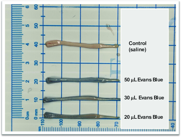
Figure 1: Visualization of Evans blue dye distribution following intrathecal injection in adult mice. Evans Blue Dye (0.5%) was administered via direct intrathecal (IT) injection in adult mice to qualitatively assess its spinal cord distribution. Three different dye volumes 20 μL, 30 μL, and 50 μL were delivered in separate experimental groups (n = 1 mouse per volume condition). Following injection, spinal cords were harvested and flushed to evaluate dye penetration as assessed by the staining intensity along the tissue. Images of the spinal cord tissue show the extent of dye dispersion associated with each injection volume, demonstrating a dose volume-dependent increase in coverage and staining intensity throughout the spinal cord. Please click here to view a larger version of this figure.
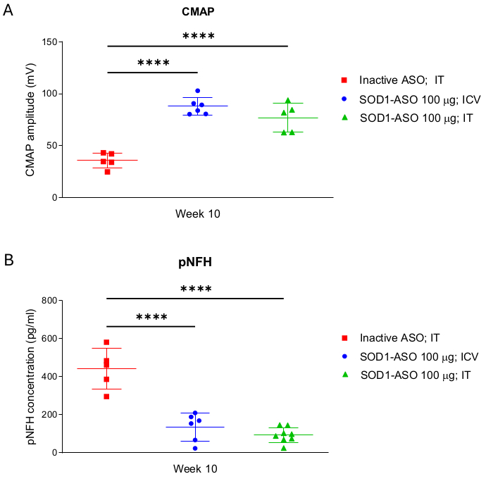
Figure 2: SOD1-targeting antisense oligonucleotide therapy restores motor function and reduces pathological neurofilament levels in mice. Adult SOD1-G93A transgenic mice received a single dose of either active SOD1-targeting antisense oligonucleotide (SOD1-ASO) via intrathecal (IT) or intracerebroventricular (ICV) injection at 5 weeks of age, or an inactive control ASO via IT injection. The therapeutic impact was evaluated at 10 weeks of age, corresponding to 5 weeks post-treatment. (A) Compound muscle action potential (CMAP) recordings were obtained to assess neuromuscular function in treated animals. SOD1-ASO-treated groups (n = 5 - 6 per group) showed a reversal in CMAP decline relative to the control group, indicating functional recovery. (B) Peripheral levels of phosphorylated neurofilament heavy chain (pNFH), a biomarker of neurodegeneration, were quantified from serum collected at week 10 (n = 5 - 8 per group). SOD1-ASO treatment led to a significant reduction in circulating pNFH levels compared to control, suggesting attenuation of disease-associated neuronal damage. All data are presented as mean ± SD. Statistical significance was determined using one-way analysis of variance (ANOVA) followed by Tukey's multiple comparisons test, with p-values ≤ 0.0001 indicating robust differences among treatment groups. Please click here to view a larger version of this figure.
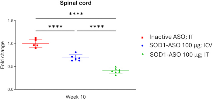
Figure 3: SOD1-ASO administration significantly suppresses human SOD1 mRNA expression in the transgenic SOD1-G93A mouse spinal cord. To evaluate the targeting efficiency of SOD1-targeting antisense oligonucleotide (SOD1-ASO) delivered through intrathecal (IT) or intracerebroventricular (ICV) injection, cervical/thoracic section of spinal cord tissue from SOD1-G93A transgenic mice euthanized at 10 weeks of age (5 weeks post-administration) was analyzed for human SOD1 mRNA expression using reverse transcription quantitative polymerase chain reaction (RT-qPCR). Human SOD1 transcript levels were quantified and normalized relative to expression in the inactive ASO-treated group. The figure depicts the fold change in human SOD1 mRNA (normalized to GAPDH) for each treatment group (n = 5-7 mice per group), highlighting a substantial downregulation in response to SOD1-ASO administration. Data are presented as mean ± SD. Statistical differences among groups were determined using one-way analysis of variance (ANOVA), followed by Tukey's multiple comparisons test, with p-values ≤ 0.0001 indicating robust differences among treatment groups. Please click here to view a larger version of this figure.
| Component | FW | Addition Amount (per L or kg) | Units |
| Sodium Phosphate Monobasic Dihydrate | 0.034 | g | |
| Sodium Phosphate Dibasic Anhydrous | 142 | 0.111 | g |
| Sodium Chloride | 58.44 | 8.766 | g |
| Potassium Chloride | 74.56 | 0.224 | g |
| Calcium Chloride Dihydrate | 147.02 | 0.206 | g |
| Magnesium Chloride Hexahydrate | 0.163 | g | |
| Water (Milli-Q) | 993 | g | |
| Poloxamer 188 | 0.01 | g |
Table 1: aCSF composition.
Supplementary Figure 1: Mouse positioning for lumbar intrathecal injection. A curved spatula or scoopula placed under the abdomen flexes the spine, widening the intervertebral space. The injection targets the L4-L5 or L5-L6 level, with proper alignment along the spine's medial axis as shown. Please click here to view a larger version of this figure.
Supplementary File 1: ASO sequence and chemistry. Please click here to download this file.
Discussion
The direct intrathecal (IT) injection technique presented here represents a minimally invasive yet highly effective method for delivering therapeutic agents into the central nervous system (CNS), particularly in small rodent models like mice. This blind-stick procedure is technically challenging but has proven to be both reproducible and clinically relevant when performed by a trained injector. This method utilizes precise anatomy and efficient technique to provide rapid CNS access without the need for catheterization, an important simplification with substantial benefits for expanding preclinical studies; however, its translational relevance will benefit from further quantitative assessment and standardized methodological validation5,8,10.
A successful IT injection depends on several key steps. Meticulous animal preparation is essential: anesthesia is first induced using a chamber and maintained with a nose cone, followed by shaving the lumbar region and administering analgesics to ensure animal comfort. Proper positioning of the mouse is achieved by placing a curved spatula under the abdomen, flexing the spine to widen the intervertebral space (Supplementary Figure 1). The goal of the intrathecal injection described here is to position the bevel of the needle within the lumbar cistern by advancing it between the laminae at the appropriate vertebral level. Accurate targeting of the intervertebral space between L4 and L5 laminae or, preferably, L5 and L6 laminae is critical for accessing the subarachnoid space5,10. The subarachnoid space, also referred to as the intrathecal space, lies between the arachnoid and pia mater39. Because the spinal cord is shorter than the vertebral canal, caudal spinal cord segments are located more rostrally relative to their corresponding vertebral levels. To avoid ambiguity, injection sites are designated by intervertebral space rather than spinal cord level. Notably, substantial variability exists in lumbar vertebral segmentation in mice, both within and across strains40. The injection must be performed along the midline of the spine to avoid lateral deviation, which can result in hindlimb paresis or, in severe cases, paralysis due to spinal cord injury. When carried out at the correct anatomical location, the risk of direct spinal cord injury is minimal. However, a small risk of nerve root irritation or damage remains, underscoring the importance of adequate operator training and procedural proficiency to minimize adverse outcomes. The tail-flick reflex serves as a helpful indicator of successful needle placement, allowing the injector to gauge depth and avoid superficial or excessively deep penetration. This technique takes practice to master, needing a steady control of depth, angle, and injection volume to avoid backflow and leakage into surrounding tissues. Although we recommend keeping the injection volume ≤10 μL, we have evaluated up to 20 μL without adverse effects. Previous studies have identified a 10 μL intrathecal injection volume as optimal for drug delivery38. Although this represents 25-30% of total CSF volume in mice41,42, the maximum tolerable intrathecal injection volume in adult mice has been reported to be 30 μL10. Injection volumes exceeding recommended limits may disrupt cerebrospinal fluid (CSF) dynamics, leading to altered CSF pressure and an increased risk of adverse effects. The total CSF volume in mice is estimated at 35-40 μL and turns over 12-13 times per day41,42, corresponding to a complete renewal roughly every 1.8 h42,43. Considering the rapid CSF turnover and empirical observations as well as previous studies demonstrating the safe use of intrathecal injection volumes up to 20-30 μL without complications, an injection volume of 10 μL is not expected to produce adverse effects10,44.
Scalability is a significant advantage of this technique. Once mastered, trained injectors can treat 10-12 animals per hour, facilitating larger preclinical studies without the logistical burdens of catheter implantation or more invasive surgical approaches. The procedure avoids complex instrumentation, reduces animal stress, and shortens recovery times compared with chronic catheterization, making it suitable for longitudinal studies requiring repeated dosing. To simulate clinical dosing regimens, we validated serial IT administrations in mice, delivering up to four doses spaced eight weeks apart. These injections were well tolerated, with no signs of distress, behavioral changes, or signs of illness, consistent with prior studies reporting no significant changes in CSF dynamics or spinal cord architecture following multiple IT treatments34,45. Thus, our studies confirm that repeated IT dosing is feasible with appropriate intervals, aligning with human treatment paradigms where sustained target engagement is necessary for therapeutic benefit. In our experience, these repeated IT injections were successfully performed across a broad age range of 5 to 29 weeks. While increasing age and body mass can slightly alter spinal cord accessibility due to greater subcutaneous tissue, successful administration remains achievable with a skilled operator who is proficient in the technique and familiar with the feel of needle placement. We note, however, that several factors beyond injection site and bolus volume, specifically injection velocity and the physical properties of the injected solution, such as density relative to CSF, can substantially influence the distribution of solutes within the CNS. While these parameters were not directly measured or manipulated in the present study, recent work by Linninger et al. (2023) demonstrates through a mechanistic pharmacokinetic model that such factors significantly affect intrathecal antisense oligonucleotide (ASO) biodistribution and tissue targeting46. Accordingly, we acknowledge this as a limitation of our study and highlight injection velocity and solution properties as important variables for future investigation to optimize CNS delivery and translational relevance. Together, these results highlight the reliability and feasibility of repeated IT dosing in multi-dose preclinical paradigms, supporting its use for sustained target engagement and translational modeling of human therapeutic regimens.
The IT approach also provides flexibility in therapeutic testing. It accommodates a broad range of modalities, including ASOs, small molecules, peptides, and viral vectors, enabling direct evaluation of CNS-targeted interventions. The ability to deliver precise, controlled volumes of therapeutics into the lumbar cistern ensures effective spinal cord and broader CNS exposure while limiting systemic spillover. The tail-flick reflex and other procedural cues allow real-time verification of successful intrathecal delivery, further enhancing reproducibility and reducing experimental failure rates5,6,8,47.
Previous studies in SOD1-G93A mice showed that the disease pathology prominently affects the spinal cord, especially the lumbar region, than in the cortex29,48. Therefore, this technique is especially relevant in the context of SOD1-ALS, a genetic form of ALS caused by mutations in the superoxide dismutase 1 (SOD1) gene. These mutations produce misfolded SOD1 protein aggregates that lead to a toxic gain of function within motor neurons, leading to progressive neuromuscular decline49,50. In this study, we employed SOD1-targeting ASOs to demonstrate successful engagement with target mRNA and functional recovery. RT-qPCR analysis revealed significant suppression of human SOD1 transcripts in the spinal cord following IT administration, while CMAP recordings indicated reversal of motor neuron dysfunction. Furthermore, serum analysis showed a decrease in phosphorylated neurofilament heavy chain (pNFH) levels, a biomarker of neurodegeneration, reinforcing the efficacy of the therapeutic strategy.
The clinical relevance of this model is emphasized by the recent FDA approval of tofersen (QALSODY®), an ASO designed to target SOD1 mRNA for the treatment of SOD1-ALS35. Tofersen, is administered intrathecally in people living with SOD1-ALS and has shown clear pharmacodynamic activity, including reductions in total CSF SOD1 protein levels (indirect marker of target engagement) and plasma NfL (marker of axonal injury and neurodegeneration). In integrated analyses of the phase 3 VALOR study and its OLE, earlier initiation of treatment with tofersen, was associated with reductions of decline in measures of clinical function, strength, and quality of life, as well as a reduction in the risk of death-equivalent events35. These data validate the intrathecal route as a powerful avenue for CNS-targeted ASO therapy and lend strong translational weight to preclinical studies employing similar delivery strategies.
In addition to its utility in ALS, the IT route has demonstrated success in spinal muscular atrophy (SMA)14,51,52, where the IT-administered ASO Nusinersen (Spinraza®) was the first approved therapy14,51,52. IT injections circumvent the blood-brain barrier and enable widespread CNS biodistribution53, which is critical for diseases characterized by diffuse neuronal pathology. While repeat dosing poses logistical and tolerability challenges, advancements in formulation and delivery techniques continue to make IT administration more viable for long-term treatment.
IT administration offers greater translational relevance to human clinical practice than ICV injections. ICV and IT injections in adult mice differ primarily in their target location within the CNS and the resulting distribution and effects of the administered substance. ICV injections deliver substances directly into the cerebrospinal fluid (CSF) within the lateral ventricles of the brain, primarily targeting supraspinal sites. In contrast, IT injections deliver substances into the CSF within the spinal subarachnoid space, primarily targeting the spinal cord. While both methods bypass the blood-brain barrier and allow for direct CNS delivery, IT administration can lead to a longer duration of action54 for some compounds and typically results in stronger transduction in the spinal cord, whereas ICV can achieve high local concentrations in brain regions adjacent to the lateral ventricle54. However, it is important to note that compounds administered into the spinal CSF can reach the brain, although the rate of transport is very slow1. These differences are also reflected in procedural risks. ICV injections carry a higher potential for contamination if sterility is compromised, while IT injections, though not risk-free, have fewer device-related vulnerabilities. Post-procedure, IT injections may induce transient discomfort or hindlimb-related reactions, whereas ICV injections are associated with less immediate discomfort, though local cranial irritation remains possible.
Future applications of this technique extend beyond current preclinical studies. IT delivery can be leveraged to evaluate next-generation CNS-targeted therapeutics, including gene-editing tools (e.g., CRISPR/Cas systems), RNA interference modalities, and viral vector-based gene therapies. The approach is adaptable for combination therapies targeting multiple CNS pathways, allowing real-time assessment of pharmacodynamic interactions55,56. Moreover, IT administration can be integrated with imaging modalities to track distribution and target engagement in vivo, accelerating translational pipelines. The technique also provides a platform for studying precision-medicine strategies across genetically defined mouse models of neurological disease, facilitating biomarker discovery, longitudinal functional assessments, and safety evaluations prior to clinical translation.
In conclusion, the direct lumbar IT injection technique using isoflurane anesthesia represents a minimally invasive, rapid, and clinically translatable method for delivering therapeutics into the murine CNS. Its successful application in SOD1-ALS models and compatibility with serial dosing provide a strong foundation for broader deployment in translational neuroscience research. As the field advances with growing emphasis on CNS gene modulation, RNA-targeted therapies, and precision medicine, the IT route will remain a key drug delivery strategy, bridging preclinical models with clinical success.
Disclosures
SM and S-C L are current employees of Biogen, Inc., while TC, DF, YL, JD, and MZ are former employees of the company. The antisense oligonucleotides discussed in this article were provided to the authors by Ionis Pharmaceuticals. Tofersen (QALSODY) is a jointly developed product of Biogen and Ionis Pharmaceuticals.
Acknowledgements
We would like to thank Ionis Pharmaceuticals for supplying the ASOs described in the article. We are grateful to Ryan Gamble, Steve Garafalo, Adria Martig, Isabel Isaza, Taras Tuczkewycz, and David Koske of Biogen for their expert contributions, which significantly enhanced the manuscript through their substantive revisions.
Materials
| Name | Company | Catalog Number | Comments |
|---|---|---|---|
| 0.3 mL 29 G, 1/2 inch insulin syringe for IT injection | Smiths Medical | 4429-3 | |
| 0.5 mm diameter sterile drill burr | Fine Science Tools (FST), or equivalent | 1900705 | |
| 5-0 Prolene Monofilament Suture | Ethicon | 8870H | |
| Alcohol swabsticks | Dynarex | 1203 | |
| Alcohol swabsticks | Dynarex | 1203 | |
| Analgesic- Buprenorphine Extended-release | Prescription required | ||
| Animal clipper | Wahl | 08787-450A | |
| Applied Biosystems High-Capacity cDNA Reverse Transcription Kit | Fisher Scientific | 43-688-14 | |
| Bead Ruptor 24 Homogenizer | Omni International, Inc. | 19-070 | |
| Blood collection and serum separation tubes | Beckton Dickinson (BD) | 365967 | |
| Calcium Chloride Dihydrate | JT Baker | 1335 | |
| Digital syringe pump | Harvard Apparatus | 70-4507 | |
| Ella Automated Immunoassay System | R&D Systems | 600-100 | |
| Eye Ointment-Puralube | Decra | 17033-211-38 | |
| Geno/Grinder | SPEX SamplePrep | 2010 | |
| Germinator 500 Dry sterilizer | Braintree Scientific, or equivalent | GER 5287-120V | |
| Hamilton syringe with needle (26s/2"/2 (point-style) | Hamilton | 80300 | 26s/2"/2: 26s: 26 G; 2" length; 2: point style |
| Hard Tissue Homogenizing Mix 2.8 mm Ceramic (2 mL Reinforced Tubes) (50/pk) | Fisher Scientific | 50-280-9286 | Supplier: Revvity Health Sciences Inc |
| Heating Pad | K&H Manufacturing | 1060 | |
| ICV platform | Stoelting | 502063 | |
| Isoflurane chamber with scavanging system | VetEquip | 901820 | |
| Magnesium Chloride Heahydrate | EMD Chemicals | 1.05832 | |
| Metzenbaum Scissors | Fine Science Tools (FST), or equivalent | 14016-14 | |
| Mouse GAPDH TaqMan Assay ID Mm99999915_g1 | ThermoFisher Scientific | 4331182 | |
| Needle Electrodes for CMAP, 28 G | Natus Medical (brand Teca) | #902-DMF25-TP | |
| NutraGel: Complete nutrition | Bio Serv | NGB-2 | |
| Olsen-Hegar Needle Holder | Fine Science Tools (FST), or equivalent | 12502-12 | |
| Poloxamer 188 | Mutchler | ||
| Potassium Chloride | JT Baker | 3045 | |
| Preamplifier (for CMAP) | World Precision Instruments (WPI) | SYS-DAM50 | |
| ProteinSimple pNFH kit | R&D Systems | SPCKB-PS-000519 | |
| Providone-Iodide swabsticks | Dynarex | 1201 | |
| QIAzol Lysis Reagent | Qiagen | 79306 | |
| QuantStudio 12K Flex system | Applied Biosystems | 4471087 | |
| Scoopula | Fisherbrand | 01-257-567 | |
| Signal v6.04 | Cambridge Electronic Design | Serial No. 061144 | CMAP Software |
| Small animal recovery chamber | Thermocare Portable Animal Intensive Care Units Model FW-1 | FW-1 | |
| Small animal stereotaxic frame with digital display console | KOPF | Model 940 | |
| SOD1 G93A mice | The Jackson Laboratory | Stock # 004435 | |
| Sodium Chloride | EMD Chemicals | 1.16224 | |
| Sodium Phosphate Dibasic Anhydrous | Sigma Aldrich | 55136 | |
| Sodium Phosphate Monobasic Dihydrate | Sigma Aldrich | 3819 | |
| Sterile disposable scalpel | Med Vet International, or equivalent | MIL4410 | |
| Sterile drape | Dynarex | 4410 | |
| Sterile gauze pads | Dynarex, or equivalent | DRX-3354 | |
| Sterile saline | ThermoFisher Scientific | 14190094 | |
| Sterile surgical gloves | Ansell, or equivalent | 20277290 | |
| Stimulus Isolator (for CMAP) | World Precision Instruments (WPI) | SYS-A365D | |
| Stoelting Micro Drill | Stoelting | 58610 | |
| Surgical curved forceps (Graefe Forceps - Titanium) | Fine Science Tools (FST), or equivalent | 11652-10 | |
| Surgical stright forceps (Graefe Forceps - Titanium) | Fine Science Tools (FST), or equivalent | 11650-10 | |
| Surgical tape | Dynarex, or equivalent | 3572 | |
| Syringe for subcutaneous injection of analgesic | BD | 309582 | |
| TaqMan Fast Advanced Master Mix | ThermoFisher Scientific | 4444556 | |
| Water (Milli-Q) | |||
| Weighing Scale | Mettler Toledo Model XSR2002S | 23-112795 | |
| Wild-type C57BL/6J mice | The Jackson Laboratory | Stock # 000664 |
References
- Hylden, J. L., Wilcox, G. L. Intrathecal morphine in mice: a new technique. Eur J Pharmacol. 67 (2-3), 313-316 (1980).
- Li, D., Li, Y., Tian, Y., Xu, Z., Guo, Y. Direct intrathecal injection of recombinant adeno-associated viruses in adult mice. J Vis Exp. (144), e58565(2019).
- Banks, W. A. Characteristics of compounds that cross the blood-brain barrier. BMC Neurol. 9 Suppl 1 (Suppl 1), S3(2009).
- Pardridge, W. M. The blood-brain barrier: bottleneck in brain drug development. NeuroRx. 2 (1), 3-14 (2005).
- Ajeeb, R., Clegg, J. R. Intrathecal delivery of macromolecules: clinical status and emerging technologies. Adv Drug Deliv Rev. 199, 114949(2023).
- Bennett, C. F., Swayze, E. E. RNA targeting therapeutics: molecular mechanisms of antisense oligonucleotides as a therapeutic platform. Annu Rev Pharmacol Toxicol. 50, 259-293 (2010).
- Corey, D. R. Nusinersen, an antisense oligonucleotide drug for spinal muscular atrophy. Nat Neurosci. 20 (4), 497-499 (2017).
- Bey, K., et al. Efficient CNS targeting in adult mice by intrathecal infusion of single-stranded AAV9-GFP for gene therapy of neurological disorders. Gene Ther. 24 (5), 325-332 (2017).
- Kennedy, Z., Gilbert, J. W., Godinho, B. Intrathecal delivery of therapeutic oligonucleotides for potent modulation of gene expression in the central nervous system. Methods Mol Biol. 2434, 345-353 (2022).
- Stavrou, M., Georgiou, E., Kleopa, K. A. Lumbar intrathecal injection in adult and neonatal mice. Curr Protoc. 4 (6), e1091(2024).
- Zou, Z. Y., Liu, C. Y., Che, C. H., Huang, H. P. Toward precision medicine in amyotrophic lateral sclerosis. Ann Transl Med. 4 (2), 27(2016).
- Quemener, A. M., et al. The powerful world of antisense oligonucleotides: from bench to bedside. Wiley Interdiscip Rev RNA. 11 (5), e1594(2020).
- Rook, M. E., Southwell, A. L. Antisense oligonucleotide therapy: from design to the Huntington disease clinic. BioDrugs. 36 (2), 105-119 (2022).
- Neil, E. E., Bisaccia, E. K. Nusinersen: a novel antisense oligonucleotide for the treatment of spinal muscular atrophy. J Pediatr Pharmacol Ther. 24 (3), 194-203 (2019).
- Amanat, M., Nemeth, C. L., Fine, A. S., Leung, D. G., Fatemi, A. Antisense oligonucleotide therapy for the nervous system: from bench to bedside with emphasis on pediatric neurology. Pharmaceutics. 14 (11), 2389(2022).
- Kingwell, K. Double setback for ASO trials in Huntington disease. Nat Rev Drug Discov. 20 (6), 412-413 (2021).
- Mead, R. J., Shan, N., Reiser, H. J., Marshall, F., Shaw, P. J. Amyotrophic lateral sclerosis: a neurodegenerative disorder poised for successful therapeutic translation. Nat Rev Drug Discov. 22 (3), 185-212 (2023).
- Gros-Louis, F., Gaspar, C., Rouleau, G. A. Genetics of familial and sporadic amyotrophic lateral sclerosis. Biochim Biophys Acta. 1762 (11-12), 956-972 (2006).
- Valentine, J. S., Hart, P. J. Misfolded CuZnSOD and amyotrophic lateral sclerosis. Proc Natl Acad Sci U S A. 100 (7), 3617-3622 (2003).
- Rotunno, M. S., Bosco, D. A. An emerging role for misfolded wild-type SOD1 in sporadic ALS pathogenesis. Front Cell Neurosci. 7, 253(2013).
- Winkler, D. D., et al. Structural and biophysical properties of the pathogenic SOD1 variant H46R/H48Q. Biochemistry. 48 (15), 3436-3447 (2009).
- Kaur, S. J., McKeown, S. R., Rashid, S. Mutant SOD1 mediated pathogenesis of amyotrophic lateral sclerosis. Gene. 577 (2), 109-118 (2016).
- Rosen, D. R., et al. Mutations in Cu/Zn superoxide dismutase gene are associated with familial amyotrophic lateral sclerosis. Nature. 362 (6415), 59-62 (1993).
- Bruijn, L. I., et al. Aggregation and motor neuron toxicity of an ALS-linked SOD1 mutant independent from wild-type SOD1. Science. 281 (5384), 1851-1854 (1998).
- Gurney, M. E., et al. Motor neuron degeneration in mice that express a human Cu, Zn superoxide dismutase mutation. Science. 264 (5166), 1772-1775 (1994).
- Peggion, C., et al. SOD1 in ALS: taking stock in pathogenic mechanisms and the role of glial and muscle cells. Antioxidants (Basel). 11 (4), 614(2022).
- Beers, D. R., et al. Endogenous regulatory T lymphocytes ameliorate amyotrophic lateral sclerosis in mice and correlate with disease progression in patients with amyotrophic lateral sclerosis. Brain. 134 (Pt 5), 1293-1314 (2011).
- Marchetto, M. C., et al. Non-cell-autonomous effect of human SOD1 G37R astrocytes on motor neurons derived from human embryonic stem cells. Cell Stem Cell. 3 (6), 649-657 (2008).
- Filipi, T., et al. Cortical glia in SOD1(G93A) mice are subtly affected by ALS-like pathology. Sci Rep. 13 (1), 6538(2023).
- Turner, B. J., Talbot, K. Transgenics, toxicity and therapeutics in rodent models of mutant SOD1-mediated familial ALS. Prog Neurobiol. 85 (1), 94-134 (2008).
- Smith, R. A., et al. Antisense oligonucleotide therapy for neurodegenerative disease. J Clin Invest. 116 (8), 2290-2296 (2006).
- Simonini, C., et al. Neurodegenerative and neuroinflammatory changes in SOD1-ALS patients receiving tofersen. Sci Rep. 15 (1), 11034(2025).
- Naseri Kouzehgarani, G., et al. Harnessing cerebrospinal fluid circulation for drug delivery to brain tissues. Adv Drug Deliv Rev. 173, 20-59 (2021).
- Duan, C., et al. Intrathecal administration of a novel siRNA modality extends survival and improves motor function in the SOD1(G93A) ALS mouse model. Mol Ther Nucleic Acids. 35 (1), 102147(2024).
- Miller, T. M., et al. Trial of antisense oligonucleotide tofersen for SOD1 ALS. N Engl J Med. 387 (12), 1099-1110 (2022).
- McCampbell, A., et al. Antisense oligonucleotides extend survival and reverse decrement in muscle response in ALS models. J Clin Invest. 128 (8), 3558-3567 (2018).
- DeVos, S. L., Miller, T. M. Direct intraventricular delivery of drugs to the rodent central nervous system. J Vis Exp. (75), e50326(2013).
- Bhusal, A., Suk, K. Protocol for delivering proteins, peptides, and shRNAs via the intrathecal route in the experimental allergic encephalomyelitis mouse model. STAR Protoc. 5 (3), 103219(2024).
- Rahman, M. M., Lee, J. Y., Kim, Y. H., Park, C. K. Epidural and intrathecal drug delivery in rats and mice for experimental research: fundamental concepts, techniques, precaution, and application. Biomedicines. 11 (5), 1413(2023).
- Rigaud, M., et al. Species and strain differences in rodent sciatic nerve anatomy: implications for studies of neuropathic pain. Pain. 136 (1-2), 188-201 (2008).
- Simon, M. J., Iliff, J. J. Regulation of cerebrospinal fluid (CSF) flow in neurodegenerative, neurovascular and neuroinflammatory disease. Biochim Biophys Acta. 1862 (3), 442-451 (2016).
- Law, V., et al. A murine Ommaya xenograft model to study direct-targeted therapy of leptomeningeal disease. J Vis Exp. (167), e62033(2021).
- Christensen, J., Li, C., Mychasiuk, R. Choroid plexus function in neurological homeostasis and disorders: the awakening of the circadian clocks and orexins. J Cereb Blood Flow Metab. 42 (7), 1163-1175 (2022).
- Wu, W., et al. In vivo imaging in mouse spinal cord reveals that microglia prevent degeneration of injured axons. Nat Commun. 15 (1), 8837(2024).
- Shi, L., et al. Repeated intrathecal administration of plasmid DNA complexed with polyethylene glycol-grafted polyethylenimine led to prolonged transgene expression in the spinal cord. Gene Ther. 10 (14), 1179-1188 (2003).
- Linninger, A. A., Barua, D., Hang, Y., Iadevaia, S., Vakilynejad, M. A mechanistic pharmacokinetic model for intrathecal administration of antisense oligonucleotides. Front Physiol. 14, 1130925(2023).
- Chen, Y., et al. Intrathecal delivery of antisense oligonucleotides in the rat central nervous system. J Vis Exp. 152, e60274(2019).
- Molnar-Kasza, A., et al. Evaluation of neuropathological features in the SOD1-G93A low copy number transgenic mouse model of amyotrophic lateral sclerosis. Front Mol Neurosci. 14, 681868(2021).
- Munch, C., O'Brien, J., Bertolotti, A. Prion-like propagation of mutant superoxide dismutase-1 misfolding in neuronal cells. Proc Natl Acad Sci U S A. 108 (9), 3548-3553 (2011).
- Une, M., et al. SOD1-interacting proteins: roles of aggregation cores and protein degradation systems. Neurosci Res. 170, 295-305 (2021).
- Gidaro, T., Servais, L. Nusinersen treatment of spinal muscular atrophy: current knowledge and existing gaps. Dev Med Child Neurol. 61 (1), 19-24 (2019).
- Hoy, S. M. Nusinersen: first global approval. Drugs. 77 (4), 473-479 (2017).
- Metz, T., et al. Biodistribution of radioactively labeled splice-modulating antisense oligonucleotides after intracerebroventricular and intrathecal injection in mice. Nucleic Acid Ther. 34 (1), 26-34 (2024).
- Adank, D. N., et al. Comparative intracerebroventricular and intrathecal administration of a nanomolar macrocyclic melanocortin receptor agonist MDE6-5-2c (c[Pro-His-DPhe-Arg-Trp-Dap-Ala-DPro]) decreases food intake in mice. ACS Chem Neurosci. 11 (19), 3051-3063 (2020).
- Sharma, A., et al. Multiple doses of cell therapy and neurorehabilitation in amyotrophic lateral sclerosis: a case report. Clin Pract. 10 (3), 1242(2020).
- Modol-Caballero, G., et al. Gene therapy overexpressing neuregulin 1 type I in combination with neuregulin 1 type III promotes functional improvement in the SOD1(G93A) ALS mice. Front Neurol. 12, 693309(2021).
Reprints and Permissions
