The article presents an easy and reproducible method for transurethral calcium electroporation using a newly developed endoscopic electrode that is compatible with standard urological equipment. The study is a significant advancement toward transurethral application of electroporation and paves the way for expanding innovative and minimally invasive treatment of urinary tumors.
Method Article
Integration of Electroporation in Urological Practice: Design and Evaluation of a New Transurethral Electrode
In This Article
Summary
Abstract
Bladder tumors constitute a substantial health challenge, yet advances in the treatment have been few. Electroporation is the use of brief electric pulses that permeabilize cell membranes with the purpose of a direct cytotoxic effect (irreversible electroporation) or for drug and gene delivery into cells (reversible electroporation). In this study, calcium was used (calcium electroporation). The use of electroporation in uro-oncology has been limited due to a lack of suitable instruments. The study focuses on the design and clinical usability of a new transurethral electrode and its technical performance in pigs. This fixed-geometry electrode consists of two opposing arrays of three needles each. The central needle is offset from an imaginary line between the two outermost needles. This ensures uniform, square-shaped electric field distribution. The electrode is compatible with a transurethral resectoscope and is an interchangeable working element through which the telescope inserts. The external pulse generator connects to the handle. Locking mechanisms ensure alignment of the equipment during assembly. The suitability was assessed in healthy porcine bladders, which resemble the human and allow the use of similar instruments. Under general anesthesia, the resectoscope was inserted transurethrally. Calcium was injected into the bladder wall with an endoscopic needle, the electrode was inserted, and the needles were placed in the area injected with calcium. Electric pulses were applied (8 pulses, 100 µs, 5000 Hz, and 400 V corresponding to 1000 V/cm voltage to electrode distance ratio). Afterwards the instruments were retracted. The handling of the instruments closely mirrors established urological techniques. The positioning of the electrode was visually managed, and the pulse delivery equaled standard transurethral procedures. Pulses were delivered successfully. The transurethral use and performance of the new electrode in the bladder aligns with existing urological instruments and techniques, enabling seamless adaptation by urologists.
Introduction
Tumors of the urinary bladder represent a significant global health burden. The high incidence and the tendency to recur positions the malignancy among the costliest types of cancers1,2. In addition, the incidence is expected to increase by approximately 70% by 20403. Despite the growing challenge, therapeutic innovations have remained few until recently4.
One emerging and promising treatment modality is electroporation, which includes the application of brief electric pulses to target tissues5. The pulses temporarily or permanently permeabilize cell membranes, enabling two distinct therapeutic mechanisms. Irreversible electroporation (IRE) that directly induces cytotoxicity6, and reversible electroporation that facilitates intracellular delivery of drugs or genes7. Electroporation has already been established as a treatment modality in other fields of medicine, such as cardiac ablation in arrhythmic heart diseases8 and electrochemotherapy (ECT) for various types of cancer9,10,11. However, the use of electroporation in uro-oncology has been limited.
Recent progress in electroporation therapy includes calcium electroporation (CaEP). CaEP uses the otherwise non-toxic calcium as the cytotoxic agent and has previously been shown to eradicate tumors of varying histologies12,13,14. In cutaneous tumors, fixed-geometry needle electrodes or plates are commonly used14,15,16. In a phase I observational study of CaEP on recurrent mucosal head and neck tumors, a flexible finger electrode with linear array needles, a hexagonal or a linear array electrode on a handle was used17. In studies of colorectal and esophageal cancers, CaEP was performed with electrodes compatible with the endoscopes (colonoscope or gastroscope)13,18,19. However, intravesical CaEP has been hindered by the absence of suitable instruments for the transurethral approach, and to date, only a single report documents the effect of CaEP treatment on metastases from urothelial carcinomas in humans20.
The current treatment of bladder tumors in primis transitional cell (urothelial) carcinomas is transurethral resection (TURBT) that employs a resectoscope inserted through urethra under visual guidance, irrigation, and instrument exchange facilitated by a protective sheath placed in the urethra. The tumors are removed with a bipolar resection loop capable of cutting and coagulating the tissue. Given the procedural similarities between TURBT and electroporation with a transurethral electrode, CaEP would be a major step forward as a feasible and minimally invasive approach for the treatment of urinary bladder cancer.
The new “OpField electrode” (Figure 1) by our group is designed to integrate the familiar setup in TURBT with electroporation directly in the urinary bladder and enables a simple and innovative therapeutic option for both non-muscle-invasive and muscle-invasive bladder tumors. In the present article, we describe the design, assembly, and in vivo manipulation of the first transurethral electrode for electroporation in the urinary bladder with direct endoscopic visualization.
The electrode is a single-use, fixed-geometry electrode, which consists of two opposing arrays of each three needles. The needles measure 4 mm in length, and the central needle is offset ~0.8 mm from an imaginary line between the two outermost needles. The offset central needle ensures a uniform, square-shaped electric field (Figure 2). The electrode array assembly comprises a distal end with a bipolar construction, with two laser-cut fork-like electrodes each having 3 needle tips. Each electrode pole is mounted in a precision stainless steel tube and overmolded with medical-grade UV glue. Each electrode leg is insulated using a medical-grade polyolefin-based heat shrink. The handle of the device is 3D printed using a medical-grade resin. This sub-assembly undergoes 100% visual inspection and is cleaned and disinfected before entering the final assembly in the cleanroom. The fully assembled device is packaged in a heat sealed Tyvek sterilization pouch and sterilized using E-beam radiation. The electrode is compatible with a transurethral resectoscope used for TURBT and works as an interchangeable working element through which the telescope inserts. The external pulse generator connects to the handle and locking mechanisms ensure alignment of the equipment during assembly.
Another very important advantage of CaEP compared with ECT is the use of the non-toxic calcium as the therapeutic agent in the benefit of both patients and medical staff21.
In vivo the potential clinical adoption, user experience and practical properties of the electrode are being evaluated, and its compatibility and performance with standard urological instruments for transurethral and endoscopic use are judged. This part is carried out in pigs that are similar in size to humans, and therefore, the same instruments will be usable. A schematic diagram of the procedure is shown in Figure 3.
Protocol
Use four- to five-month-old healthy pigs weighing 50–80 kg (Sus scrofa domesticus, females), as their urinary bladders resemble those of humans. The size of the pigs allows for the use of the same transurethral endoscopic instruments as in humans. Anesthetize the pigs according to institutional guidelines (i.m. ketamine (7-10 mg·kg -1), dexmedetomidine (20-40 µg·kg -1), and butorphanol (0.1-0.2 mg·kg -1) as preanesthetic, and i.v. propofol (0.3-0.5 mg·kg -1) and sevoflurane in 60% oxygen for a tidal volume of 10 mL·kg -1 as anesthesia. Verify adequate anesthetic depth prior to initiation of the procedure. Apply eye lube, provide thermal support and monitor the perioperative vitals throughout the procedure.
Ethics statement
The animal studies are conducted in compliance with the authorization from the Animal Experiments Inspectorate (permit no. 2024-15-0201-01759) and follow the guidelines of the European Convention for the Protection of Vertebrate Animals used for Experimental and Other Scientific purposes.
1. Preparation of the instruments and equipment
NOTE: The sterile resectoscope is assembled by personnel wearing sterile gowns and gloves on a table with sterile drapes. For the treatment, two doctors skilled in transurethral procedures are present. One handles the insertion and manipulation of the instruments in the pig; the other acts as an assistant.
- Insert the telescope into a bridge with a working channel.
- Insert the instruments into the sheath.
- Connect saline to the irrigation system on the sheath with an intravenous tubing set.
- Open the tubing set, fill the tube with saline, and displace the air.
- Attach the camera head and lighting cable to the telescope base.
- Connect the sterile transurethral equipment with the clean visualization platform to produce a real-time image on the connected screen.
NOTE: This is carried out with the assistance of non-sterile personnel. Before the procedure, the image is white balanced to ensure that white objects recorded by the camera appear white. All locking mechanisms must be fully secured to prevent misalignment or disconnection and ensure proper irrigation during the procedure.
2. Preparation of the transurethral electrode
- Remove the electrode from its sterile package.
- Connect the electrode to the external pulse generator by connecting the sterile insulated cable at the electrode’s handle to the clean insulated cable at the external pulse generator.
3. External pulse generator
- Choose the linear configuration electrode setting on the display, as this is compatible with the transurethral electrode.
- Set 8 pulses of 100 µs, frequency 5000 Hz, and voltage 400 V, correlating to 1000 V/cm voltage to electrode distance ratio in the designated fields on the screen.
4. Injection needle and calcium gluconate
CAUTION: Handle calcium gluconate in accordance with institutional safety regulations. Dispose of calcium gluconate as dangerous waste.
- Remove the cystoscopy needle from the sterile package and adjust the needle-tip length to 2 mm.
- Acquire a bottle of calcium gluconate 100 mg·mL-1.
- Fill one or more syringes with the needed calcium gluconate.
- Connect the syringe (preferably with luer-lock-type syringe) to the distal end of the cystoscopy needle.
NOTE: 2.2.-4.2. are carried out with the assistance of non-sterile personnel. Use of other calcium compounds with the same molarity of calcium is possible22.
5. Preparation of the pig
- Place the pig in a dorsal lithotomy position and the buttocks aligned with the edge of the operating table.
- Secure a fluid collection pouch with drainage used for transurethral procedures underneath the pig.
- Clean the vulva with water and soap. Install gel in the urethra for disinfection and analgesia.
6. Calcium electroporation procedure
- Open the irrigation system on the resectoscope and confirm saline flow.
- Insert the resectoscope, visually guided through the urethra into the urinary bladder.
- Stop the saline flow once the resectoscope is in place. Empty the bladder of residual urine.
- Resume the saline water influx until the urinary bladder is filled.
- Perform a cystoscopy and inspect the entire bladder.
- Insert the cystoscopy needle through the working channel of the bridge and visualize the tip of the needle.
- Inject 0.3 mL calcium gluconate 100 mg·mL -1 into the bladder wall to produce a vesicle in the bladder wall.
NOTE: Calcium injection is repeated as needed to ensure complete coverage of the electroporation area. Throughout the procedure, the irrigation is continuously adjusted to secure an optimal view of the bladder wall. - Remove the cystoscopy needle.
- Remove the telescope. Leave the outer protective sheath in the urethra.
- Remove the bridge.
- Insert the telescope into the transurethral electrode.
- Place the telescope and the electrode in the sheath to the bladder under visual guidance.
- Place the electrode needles in the tissue covering the areas injected with calcium gluconate.
- Apply the electric pulses either by the surgeon using the foot pedal, or by a non-sterile assistant operating the external pulse generator. First, use the “charge” button followed by the “pulse” button.
- Confirm completion of the electric pulses and successful pulse delivery by reading the pulses generated by the instrument (Voltage) and the pulses delivered to the tissue (Current) shown on the external pulse generator.
NOTE: Mild muscle contractions may appear during pulse delivery. As a safety precaution, muscle relaxants can be administered during the procedure. Electroporation should be performed immediately after calcium injection. During treatment of multiple/larger tumors, alternating between calcium injection and electroporation is recommended. In theory, the protocol can be paused after treatment of CaEP, but this is not advisable as the treatment is performed under general anesthesia.
7. Post-procedure regiment
- Remove the telescope and electrode when every area injected with calcium gluconate has been electroporated.
- Empty the bladder.
- Carefully remove the resectoscope with the outer sheath using an obturator.
- Finish the general anesthesia and medication.
- Let the pig wake up in accordance with institutional guidelines.
- Manage post-procedural pain when needed in accordance with institutional guidelines (e.g., meloxicam, 0.4 mg/kg, i.m).
- Dispose of the electrode as sharp waste.
Results
A preliminary study in 3 pigs weighing 30-40 kg resulted in successful electroporation, but 2 of the pigs had to be euthanized post-operatively, one because of urethral bleeding and the other because of unintentional intraperitoneal injection of ink meant to mark treatment sites. Therefore, in the principal study, larger pigs weighing approximately 80 kg were used, and the injection of ink was considered unnecessary. A total of 27 areas in 7 pigs were treated, including 5 lesions injected with calcium gluconate without electroporation, and 5 lesions with electroporation only. A total of 176 pulses were given. The applied voltage was 400 V, and the current ranged from 3–4 A.
The treatment was performed by a consultant urologist specialized in transurethral surgery. The insertion and manipulation of the electrode closely mirror established urological techniques. Specifically, the manipulation of instruments was intuitive for the surgical team and reflected familiarity of the movements required during procedures such as TURBT. The electrode was positioned within the bladder wall under continuous visual guidance to ensure accurate placement in the tissue. The pulse and current delivery (8 pulses of 100 µs, 5000 Hz, and 400 V correlating to 1000 V/cm voltage-to-electrode distance ratio) was successful, constant, and stable in all cases according to the readings on the external pulse generator; and the procedure paralleled that of conventional resection loops. The study confirmed the high degree of familiarity and ease of adoption for urologists for clinical integration.
Cystoscopic examinations of the bladder 5 and 7 days after CaEP showed thickened mucosa with edema, erythema, and small hemorrhages. In all cases, the bladder wall was intact without perforations. After euthanasia and evisceration, the bladders were filled with fluid, the walls were still intact, and calcifications could be seen through pellucid walls. Similar changes were observed in the bladder treated with calcium injections alone, whereas electroporation alone showed minimal hemorrhage and no other changes. All other abdominal organs were normal.
CONCLUSION:
The application of the new transurethral electrode within the urinary bladder demonstrates alignment with established urological instruments and techniques. The procedural similarity to conventional transurethral surgery and the stability of pulse and current delivery support rapid and seamless translation to clinical urology. The clinical implementation is planned to begin with a first-in-human trial, where CaEP and the transurethral electrode will be evaluated in patients with advanced bladder cancer.
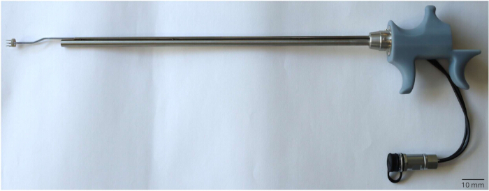
Figure 1. Prototype of the transurethral electrode. The electrode is compatible with a transurethral resectoscope used for TURBT. The electrode is an interchangeable working element. The telescope is inserted into the electrode, allowing the visually guided placement of the needles in the bladder wall. The external pulse generator is cabled to the electrode handle. Locking mechanisms ensure the equipment is aligned during assembly. Please click here to view a larger version of this figure.
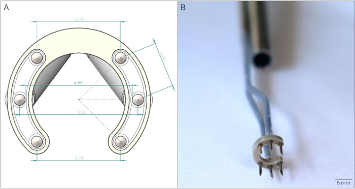
Figure 2. (A) A schematic illustration of the active part of the electrode. The fixed-geometry electrode consists of two opposing arrays of three needles each. The central needle in each array is offset from an imaginary line between the two outermost needles. The arrangement of the needles ensures a uniform, square-shaped electric field distribution. The measurements are in mm. (B) The active part of the electrode. In a horseshoe configuration, the two arrays of needles are placed with the offset central needle. The telescope channel is visible at the top of the photo. Please click here to view a larger version of this figure.
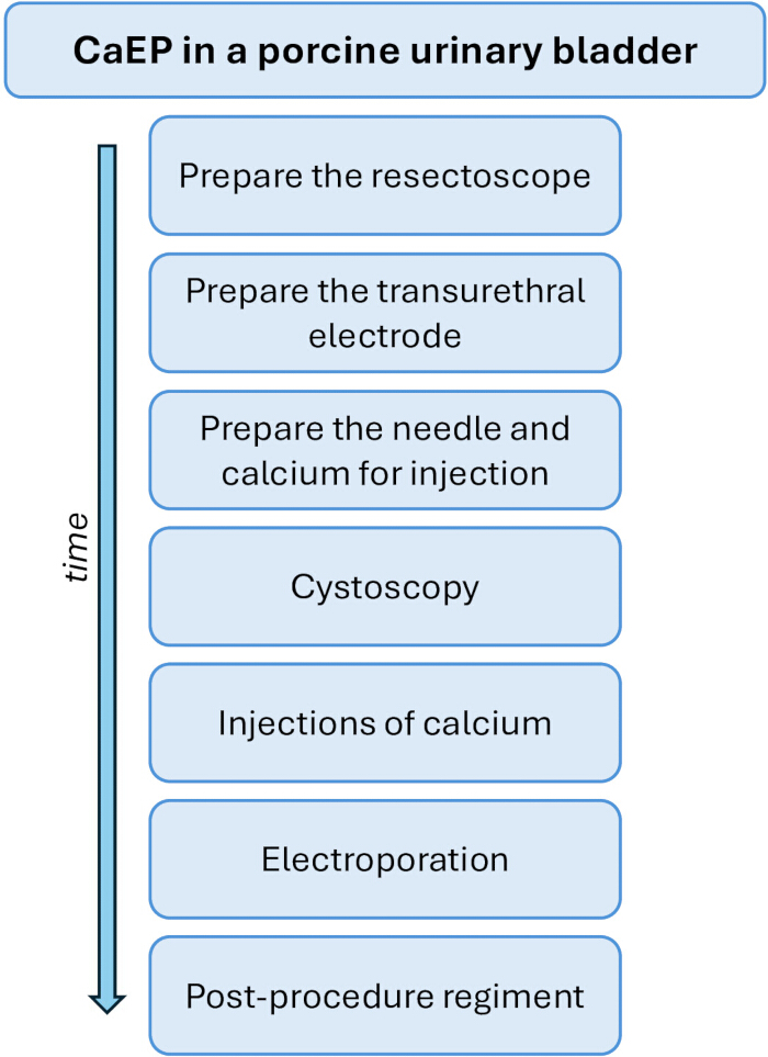
Figure 3. A schematic illustration of the CaEP procedure, highlighting the main procedural phases. Please click here to view a larger version of this figure.
Discussion
The protocol details the use of a new transurethral OpField electrode for CaEP in the porcine urinary bladder and presents a significant advance in the application of electroporation in urology23. One strength of the method is its seamless integration into standard urological equipment. The compatibility allows a close comparison to TURBT and supports immediate adoption in the clinical setting due to procedural similarity. The duration of CaEP and TURBT depends on the size of the tumor, but the length of the procedures is similar. The fixed-geometry design of the electrode ensures a homogeneous electric field distribution that is essential for consistent electroporation efficacy.
Several steps in the protocol are important to ensure successful execution. This includes proper irrigation to keep optimal visibility during the procedure. A proper assembly and locking of the components of the resectoscope and electrode is critical for stability and safety. Poor visibility, any misalignment, or disconnection could compromise the accuracy of needle placement and the delivery of pulses. Successful treatment also depends on the precise visual placement of the electrode needles into the area injected with calcium. Adjustments to the protocol may be necessary depending on tumor size and characteristics of the bladder. Changes in the calcium volume may be required for complete coverage of the tumors. The irrigation system may be adapted if visibility is poor. In the event of failed pulse delivery, the feedback system of the external generator provides immediate notice and allows prompt identification of connection, contact, or placement issues. If pulse delivery fails, ensure correct connection of the cables and sufficient contact of the electrode to the tissue. Reconnect the cables of the electrode to the external pulse generator or reposition the needles in the tissue, if necessary.
The use of muscle relaxants in humans is deemed important to prevent strong pelvic contractions during pulse delivery, and omission thereof may cause muscle contractions, potential bladder wall perforation, postoperative pain, and unnecessary procedural difficulties24,25. In the present study, the pigs did not show any signs of discomfort in relation to the urinary system; and perforation, thermal effects, bleeding, or urethral trauma were not seen as a result of electroporation.
Moreover, unlike ECT, which requires toxic drugs, CaEP uses a non-toxic and readily available agent, and the complexity of handling chemotherapeutics during surgery is avoided21. In case, surgical complications like bleeding or perforations will be similar to those met in TURBT and will be handled accordingly25.
Despite more advantages, the method has certain limitations. The placement of the electrode might be problematic in poorly accessible tumors in difficult anatomic areas. In the porcine study, the posterior wall can be difficult to reach because of the oblong shape of the porcine bladder. This happened twice during the study and was alleviated by emptying the bladder, reducing distention, and applying pressure to the pig's abdomen with a hand (by the surgeon). The electrode delivers pulses only to tissue directly penetrated by the needles, and large or multifocal tumors may require repositioning and/or multiple insertions.
In addition to its use in CaEP, the new electrode could be adapted for other modalities such as IRE, gene therapy, and ECT16,23. Also, it may be an alternative to extensive surgical options such as cystectomy.
CaEP might also be cost-efficient. A single treatment with CaEP will potentially be sufficient and spare repeated installations of chemotherapeutics or immunotherapy1,26. Finally, the electrode may provide a mean for translational studies into electroporation-mediated immunogenic cell therapy, cell death, and systemic immune memory – perhaps in combination with immunotherapies11,12,20.
In summary, the protocol introduces a novel, reproducible, and clinically adaptable method for endoscopic electroporation in the urinary bladder. The newly designed electrode addresses longstanding barriers by offering a tool that is technically aligned with standard equipment and easy to use. While certain limitations remain, the design offers broad potential for clinical application in minimally invasive cancer therapies, and the results pave the way for the first-in-human clinical trial planned to evaluate the safety and effect of transurethral CaEP in patients with advanced bladder cancer.
Disclosures
The authors disclose the following conflicts of interest. JLV serves on the advisory boards of AMBU, Lina Medical, Photocure, MSD, Janssen, and Olympus, and has been an invited speaker for Olympus and Medac. JLV and JG are co-inventors of the patented OpField electrode: An electrode assembly for improved electric field distribution (PCT/EP2019/080198). JG holds a patent regarding calcium electroporation: Therapeutic applications of calcium electroporation to effectively induce tumor necrosis (PCT/DK2012/050496). No other authors have a conflict of interest to declare.
Acknowledgements
The study is funded by grants from Innoexplorer (Grant No. 5287-00042B), and Beta. Health (ID: 2023-1486 and ID: 2024-1834), as well as by Region Sjællands Forskningsfond, Agnethe Løvgreens Fond and Christian Larsen og Dommer Ellen Larsens Legat.
Materials
| Name | Company | Catalog Number | Comments |
|---|---|---|---|
| Calcium gluconate 100 mg/mL | B.Braun (Germany) | ||
| Catheterization gel | Medac (Faxe, Denmark) | Instillagel | |
| Clean insulated cable for the external pulse generator | IGEA (Carpi, Italy) | ||
| Cystoscopy needle | Laborie (NH, USA) | InjeTAK | |
| Disposable plastic sterile camera cover | |||
| Drapes for transurethral procedures with fluid collection pouch with drainage | |||
| External pulse generator | IGEA (Carpi, Italy) | Cliniporator EPS01;IGEA, incl. foot pedal if necessary | |
| Intravenous tubing set | |||
| Loop resection working element | Olympus (Tokyo, Japan) | ||
| Resectoscope ch27 | Olympus (Tokyo, Japan) | Incl. telescope, bridge with working channel, inner and outer sheath, obturator | |
| Saline in a 3 L bag | |||
| Sterile drapes for at table | |||
| Sterile gloves | |||
| Sterile surgery gown for the assistant | |||
| Syringe | Preferably with Luer-Lock | ||
| The OpField electrode | Soltech (Måløv, Denmark) | Endoscopic electrode | |
| Urologist sterile surgeon gown | |||
| Visualization platform | Olympus (Tokyo, Japan) | Incl. lightning cable, camera head |
References
- Scilipoti, P., et al. The financial burden of localized and metastatic bladder cancer. Eur Urol. 87 (5), 536-550 (2025).
- Cookson, M. S. The economics of bladder cancer in the us: Increasing costs and unsustainable patterns. Urol Times. 53 (02), 37(2025).
- Zhang, Y., et al. The global landscape of bladder cancer incidence and mortality in 2020 and projections to 2040. J Glob Health. 13, 04109(2023).
- Patel, V. G., Oh, W. K., Galsky, M. D. Treatment of muscle-invasive and advanced bladder cancer in 2020. CA Cancer J Clin. 70 (5), 404-423 (2020).
- Hoejholt, K. L., et al. Calcium electroporation and electrochemotherapy for cancer treatment: Importance of cell membrane composition investigated by lipidomics, calorimetry and in vitro efficacy. Sci Rep. 9 (1), 4758(2019).
- Tasu, J. P., Tougeron, D., Rols, M. P. Irreversible electroporation and electrochemotherapy in oncology: State of the art. Diagn Interv Imaging. 103 (11), 499-509 (2022).
- Campana, L. G., et al. Pulsed electric fields in oncology: A snapshot of current clinical practices and research directions from the 4th world congress of electroporation. Cancers (Basel). 15 (13), (2023).
- Jacobs, E. J., Rubinsky, B., Davalos, R. V. Pulsed field ablation in medicine: Irreversible electroporation and electropermeabilization theory and applications. Radiol Oncol. 59 (1), 1-22 (2025).
- Bastrup, F. A., Vissing, M., Gehl, J. Electrochemotherapy with intravenous bleomycin for patients with cutaneous malignancies, across tumour histology: A systematic review. Acta Oncol. 61 (9), 1093-1104 (2022).
- Bertino, G., et al. Electrochemotherapy for the treatment of cutaneous squamous cell carcinoma: The inspect experience (2008-2020). Front Oncol. 12, 951662(2022).
- Falk, H., et al. Electrochemotherapy and calcium electroporation inducing a systemic immune response with local and distant remission of tumors in a patient with malignant melanoma - a case report. Acta Oncol. 56 (8), 1126-1131 (2017).
- Falk, H., et al. Calcium electroporation induces tumor eradication, long-lasting immunity and cytokine responses in the ct26 colon cancer mouse model. Oncoimmunology. 6 (5), e1301332(2017).
- Broholm, M., et al. Endoscopic calcium electroporation for colorectal cancer: A phase i study. Endosc Int Open. 11 (5), E451-E459 (2023).
- Falk, H., Matthiessen, L. W., Wooler, G., Gehl, J. Calcium electroporation for treatment of cutaneous metastases; a randomized double-blinded phase ii study, comparing the effect of calcium electroporation with electrochemotherapy. Acta Oncol. 57 (3), 311-319 (2018).
- Frandsen, S. K., Vissing, M., Gehl, J. A comprehensive review of calcium electroporation -a novel cancer treatment modality. Cancers (Basel). 12 (2), (2020).
- Gehl, J., et al. Updated standard operating procedures for electrochemotherapy of cutaneous tumours and skin metastases. Acta Oncol. 57 (7), 874-882 (2018).
- Plaschke, C. C., et al. Calcium electroporation for recurrent head and neck cancer: A clinical phase i study. Laryngoscope Investig Otolaryngol. 4 (1), 49-56 (2019).
- Broholm, M., et al. Neoadjuvant calcium electroporation for potentially curable colorectal cancer. Surg Endosc. 38 (2), 697-705 (2024).
- Egeland, C., Baeksgaard, L., Gehl, J., Gogenur, I., Achiam, M. P. Palliative treatment of esophageal cancer using calcium electroporation. Cancers (Basel). 14 (21), (2022).
- Vissing, M., et al. Calcium electroporation as an adjunct therapy for pembrolizumab-resistant bladder cancer: A case report. Acta Oncol. 62 (6), 614-620 (2023).
- Shiwani, T., Singh Dhesi, S., Wah, T. M. Reversible electroporation for cancer therapy. Br J Radiol. 98 (1167), 313-320 (2025).
- Frandsen, S. K., Gissel, H., Hojman, P., Eriksen, J., Gehl, J. Calcium electroporation in three cell lines: A comparison of bleomycin and calcium, calcium compounds, and pulsing conditions. Biochim Biophys Acta. 1840 (3), 1204-1208 (2014).
- Kielbik, A., Szlasa, W., Saczko, J., Kulbacka, J. Electroporation-based treatments in urology. Cancers (Basel). 12 (8), 2208(2020).
- Panagoda, P. I., Vasdev, N., Gowrie-Mohan, S. Avoiding the obturator jerk during TURBT. Curr Urol. 12 (1), 1-5 (2018).
- Osman, Y., et al. Bladder perforation as a complication of transurethral resection of bladder tumors: The predictors, management, and its impact in a series of 1570 at a tertiary urology institute. Int Urol Nephrol. 55 (9), 2161-2167 (2023).
- Szewczyk, A., et al. Advancing cancer therapy: Mechanisms, efficacy, and limitations of calcium electroporation. Biochim Biophys Acta Rev Cancer. 1880 (3), 189319(2025).
Reprints and Permissions
