The in vitro bioactivity of Terfezia claveryi extracts depended on the biophysical extraction method and solvent polarity. The water extract from maceration-assisted extraction (MAE) exhibited the strongest antioxidant activity, whereas the Soxhlet extraction (SE) extract induced greater cytotoxicity in SH-SY5Y cells. These results demonstrate clear method-dependent differences in the in vitro bioactivity of Terfezia claveryi extracts.
A subscription to JoVE is required to view this content. Sign in or start your free trial.
Method Article
Impact of Different Extraction Methods on the In Vitro Bioactivity Profiles of Terfezia Claveryi
In This Article
Summary
Abstract
Terfezia claveryi is an edible desert truffle traditionally consumed in arid regions. In recent years, it has attracted increasing scientific interest due to its potential antioxidant, antimicrobial, and therapeutic properties, suggesting possible applications in food and health-related fields. However, despite growing evidence of its biological potential, the influence of different extraction strategies on the recovery of bioactive compounds and the resulting in vitro bioactivity profile of Terfezia claveryi has not yet been clarified in the literature. In this study, the in vitro bioactivity of Terfezia claveryi extracts obtained using different extraction methods and solvents with varying polarity was evaluated. Extraction yield was determined, and the obtained extracts were assessed for antioxidant, antimicrobial, antibiofilm, and cytotoxic activities using established testing systems. Antioxidant capacity was measured using 2,2-diphenyl-1-picrylhydrazyl (DPPH), 2,2′-azino-bis(3-ethylbenzothiazoline-6-sulfonic acid) (ABTS), and ferric reducing antioxidant power (FRAP) tests, while cytotoxic effects were investigated in SH-SY5Y neuroblastoma cells. Extraction yield varied depending on both method and solvent polarity, with the highest yield obtained from the aqueous extract prepared by the ultrasound-assisted extraction (UAE) method (18.40%). It was determined that antioxidant activity showed significant differences depending on the extraction method, and aqueous extracts obtained by the maceration-assisted extraction (MAE) method exhibited a profile rich in phenolic compounds along with the strongest radical scavenging capacity. LC-MS/MS analysis of this extract revealed the presence of nine phenolic compounds; quinic acid and chlorogenic acid were identified as the dominant components. In contrast, Soxhlet-derived extracts induced greater cytotoxic effects in SH-SY5Y cells. Antimicrobial and antibiofilm responses displayed relatively limited variation among extraction methods, indicating a weaker dependence on extraction strategy for these activities. Overall, these findings demonstrate that the biophysical extraction method and solvent selection are critical determinants of the in vitro bioactivity profile of Terfezia claveryi extracts, with pronounced effects observed for antioxidant and cytotoxic activities.
Introduction
The use of various mushroom species to improve health and to prevent and treat diseases dates back to ancient times1,2,3. Macrofungi are generally classified into two groups: Epigeous (mushrooms) and hypogeous (truffles)4. Known as the desert truffle, Terfezia claveryi (T. claveryi) is a world-renowned and edible truffle species belonging to the genus Terfezia5. T. claveryi is commonly distributed in arid and semi-arid regions where Helianthemum species are prevalent6,7,8,9,10,11. In recent years, the global burden of chronic diseases such as cancer and neurodegenerative disorders has increased significantly, highlighting the urgent need for new, safe, and effective therapeutic agents. According to global health reports, cancer remains one of the leading causes of death worldwide, while neurodegenerative diseases continue to increase alongside an ageing population. Given this growing public health issue, the investigation of natural products derived from fungi and medicinal plants as potential sources of bioactive compounds has become an important research priority12.
In parallel, the rapid emergence of antimicrobial resistance poses another major threat to global health. Drug resistance has significantly reduced the effectiveness of traditional antibiotics, creating an urgent need for alternative antimicrobial strategies13. Secondary metabolites derived from natural sources, particularly phenolic and polyphenolic compounds, are attracting increasing interest due to their diverse antimicrobial mechanisms and low tendency to develop resistance14. Oxidative stress is a fundamental mechanistic factor underlying many chronic diseases, including cancer, neurodegenerative disorders, and metabolic syndromes. Excessive production of reactive oxygen species (ROS) can ultimately disrupt cellular homeostasis by causing DNA damage, lipid peroxidation, and protein oxidation. In this context, natural antioxidants that can scavenge free radicals and regulate redox balance are considered promising agents for mitigating oxidative damage and disease progression15.
Truffles are rich in unsaturated fatty acids, proteins, minerals, vitamins, amino acids, phenolics, and polyphenols, components that have been associated with antimicrobial, antioxidant, and anticancer properties16,17,18,19,20,21,22,23,24. Their antioxidant capacity has been linked to their effectiveness in scavenging free radicals25,26,27. Among truffle species, T. claveryi is particularly noteworthy due to its edible structure, regional economic value, and reported richness in phenolic compounds23. Despite emerging evidence regarding its traditional consumption and biological activity, comprehensive studies investigating how extraction strategies affect its chemical composition and biological effects remain limited.
The recovery of biologically active compounds from truffles is strongly influenced by extraction parameters, including the applied extraction technique and solvent polarity. Biophysical extraction methods such as ultrasound-assisted extraction (UAE), maceration-assisted extraction (MAE), and Soxhlet extraction (SE) show significant differences in terms of energy input, extraction efficiency, and selectivity for polar compounds28. However, most research has relied on a single extraction method or limited solvent systems, and studies that comparatively address the effects of these methods on the in vitro biological activity profile of T. claveryi are limited. Therefore, this study systematically compares multiple biophysical extraction methods using solvents of varying polarity to evaluate the in vitro biological activities of T. claveryi extracts. It was hypothesized that the extraction method and solvent polarity would lead to method-dependent differences in biological activities. Accordingly, the aims of the study are: (i) to compare extraction yields between different methods and solvents, (ii) to evaluate method-dependent variation in antioxidant and cytotoxic activities, and (iii) to examine antimicrobial and antibiofilm responses, considering their sensitivity to the extraction strategy.
Access restricted. Please log in or start a trial to view this content.
Protocol
NOTE: The overall experimental workflow for the preparation and analysis of T. claveryi extracts is schematically summarized in Figure 1. All materials used in the study are described in the Table of Materials.
1. Fungal material authentication and preparation
- Collection and processing of material
NOTE: Fresh T.claveryi truffles were collected in May 2023 from steppe and sandy, uncultivated (non-agricultural) lands in Kırsehir Province, Central Anatolia, Türkiye. Sampling was carried out at three distinct locations: Dedeli Village (38°54′15.8″ N, 34°06′40.3″ E; ~1050 m above sea level), Buyukkayapa Village (38°53′45.1″ N, 34°19′37.5″ E; ~1020 m above sea level), and Akcaagıl Village (39°01′39.9″ N, 34°12′39.8″ E; ~1100 m above sea level). The collection sites are characterized by a semi-arid continental climate, with cold winters, hot and dry summers, and an average annual precipitation of approximately 380–420 mm. All samples were harvested during the natural fruiting season from areas with no recent agricultural activity to minimize potential anthropogenic contamination.- Select clean and undamaged truffles, peel them, and wash three times with distilled water.
- Slice the samples into small pieces and shade-dry until a constant weight is achieved (approximately 20 days).
- Pulverize the dried material using a mechanical grinder and pass through a 328-micron (50 mesh) sieve.
- Store the processed samples in zip-lock bags in a cool, dry, and dark environment at room temperature until further analyses.
- Molecular identification of T. claveryi
- Isolate genomic DNA from dried T. claveryi samples for molecular identification.
- Assess the DNA quantity and purity spectrophotometrically.
- Perform species identification polymerase chain reaction (PCR) by amplifying the internal transcribed spacer (ITS) region using universal primers ITS1 (5′-TCCGTAGGTGAACCTGCGG-3′) and ITS4 (5′-TCCTCCGCTTATTGATATGC-3′).
- Resolve PCR amplification products by electrophoresis on a 1.5% agarose gel prepared in 1x TAE buffer and visualized under UV illumination following ethidium bromide staining.
- Purify single-band PCR products according to the manufacturer’s instructions29. Subject purified amplicons to Sanger sequencing.
- Assemble forward and reverse sequence reads into consensus sequences using the contig assembly programme (CAP) algorithm.
- Species identity was confirmed by comparison of the consensus ITS sequence with reference sequences available in public databases.
NOTE: A voucher specimen of T. claveryi was retained in the laboratory collection for future reference. Species identification was confirmed by ITS-based molecular analysis (Supplementary File 1).
2. Fungal material preparation and extraction
- Preparation of T. Claveryi extracts
- Weigh approximately 30 g of dried T. claveryi powder.
- Add 150 mL of solvent (methanol, acetone, n-hexane, ethyl acetate, or distilled water) to maintain a constant solid–liquid ratio across all extraction methods.
- Use analytical-grade solvents for all extraction procedures.
- Filter each extract under reduced pressure using qualitative filter paper.
- Concentrate the filtrates under reduced pressure at 30–45 °C using a rotary evaporator.
- Freeze-dry (lyophilize) the concentrated extracts until complete solvent removal is achieved.
- Store the dried crude extracts at −20 °C until further analysis.
NOTE: Apply identical post-extraction processing steps to all samples to ensure methodological consistency and comparability.
- Biophysical extraction methods
- Maceration-assisted extraction (MAE)
- Place the powdered T. claveryi sample into a closed extraction container.
- Add the selected solvent while maintaining a constant solvent-to-solid ratio.
- Incubate the mixture at room temperature under dark conditions for 72 h.
- Keep the containers closed to prevent solvent evaporation.
- Do not apply external agitation during the maceration period.
- Allow the mixture to settle at the end of the extraction period.
- Filter the extract prior to further processing.
- Soxhlet extraction (SE)
NOTE: Ensure proper condenser water circulation and avoid dry heating during reflux.- Place the powdered sample into a cellulose extraction thimble.
- Position the thimble inside the Soxhlet extraction chamber connected to a round-bottom flask.
- Add the selected solvent to the flask while maintaining a constant solvent-to-solid ratio.
- Heat the solvent to reflux using a thermostatically controlled heating mantle.
- Maintain continuous extraction for 24 h.
- Maintain the extraction temperature at the boiling point of each solvent (n-hexane: 68.7 °C; ethyl acetate: 77.1 °C; methanol: 64.7 °C; acetone: 56 °C; distilled water: 100 °C).
- Ultrasound-assisted extraction (UAE)
NOTE: Monitor temperature continuously to prevent overheating during sonication.- Place the powdered T. claveryi sample into the extraction vessel.
- Add the selected solvent while maintaining a constant solvent-to-solid ratio.
- Sonicate the mixture at a fixed frequency of 35 kHz and an output power of 520 W for 25 min.
- Maintain the extraction temperature at approximately 30 °C.
- Add ice at 5-minute intervals to keep the temperature within 25–28 °C during sonication.
- Maceration-assisted extraction (MAE)
- Extraction efficiency
- Weigh the dried fungal material (Wp) prior to extraction.
- Weigh the dried crude extract (Wext) after complete solvent removal.
- Calculate the extraction efficiency using Equation (1).
 (1)
(1) - Re-dissolve each dried crude extract in its respective solvent to prepare a stock solution at a standardized concentration of 10 mg/mL.
- Prepare stock solutions freshly prior to each in vitro bioactivity experiment.
NOTE: Standardize all extract concentrations to ensure comparable dosing across subsequent bioactivity assays.
3. Antioxidant activity determination
- Perform antioxidant activity analysis using DPPH, ABTS, and FRAP assays.
- Prepare five different extract concentrations ranging from 0.25 to 1.0 mg/mL from freshly prepared stock solutions.
- Conduct all measurements in triplicate to ensure reproducibility.
- Record absorbance values spectrophotometrically using a microplate reader.
NOTE: Perform all assays according to established literature protocols with minor modifications. - DPPH radical scavenging activity
NOTE: The DPPH radical scavenging activity of T. claveryi extracts was evaluated according to the method described in the literature, with minor modifications30. Protect DPPH solution and reaction mixtures from light exposure.- Prepare a DPPH solution in 70% methanol.
- Adjust the DPPH solution to an absorbance of 1.00 ± 0.02 at 517 nm.
- Pipette 50 µL of extract at different concentrations into a microplate well.
- Add 250 µL of DPPH solution to each well.
- Incubate the reaction mixture in the dark at room temperature for 30 min.
- Measure absorbance at 517 nm using a microplate spectrophotometer.
- Use methanol as a negative control and ascorbic acid as a positive control.
- Construct dose–response curves and calculate IC₅₀ values to quantify radical scavenging activity.
- ABTS cation radical scavenging activity
NOTE: Perform the ABTS radical scavenging assay according to the method described in the literature, with minor modifications31. Prepare the ABTS⁺ radical solution at least 16 h before use and protect it from light.- Generate the ABTS radical solution by incubating the reaction mixture in the dark at room temperature for 16 h.
- Dilute 1 mL of ABTS solution with 80 mL of ethanol prior to analysis.
- Adjust the absorbance of the diluted ABTS solution to 0.80 ± 0.02 at 734 nm.
- Pipette 50 µL of extract at different concentrations into a microplate well.
- Add 250 µL of ABTS solution to each well.
- Incubate the mixture at room temperature for 10 min.
- Measure absorbance at 734 nm using a microplate spectrophotometer.
- Construct dose–response curves and calculate IC₅₀ values.
- Ferric reducing antioxidant power (FRAP) assay
NOTE: Perform the FRAP assay according to the method proposed in the literature, with minor modifications32. Prepare the FRAP reagent freshly before use and protect the reaction mixture from light.- Prepare the FRAP reagent according to the referenced protocol.
- Pipette 20 µL of extract at different concentrations into a microplate well.
- Add 280 µL of freshly prepared FRAP reagent.
- Incubate the reaction mixture at room temperature in the dark for 45 min.
- Measure absorbance at 593 nm using a microplate spectrophotometer.
- Generate a calibration curve using Trolox as a standard.
- Express antioxidant capacity as mg Trolox equivalents (TE) per gram of extract.
4. Total phenolic content (TPC) analysis
NOTE: Determine the total phenolic content (TPC) of T. claveryi extracts using the Folin–Ciocalteu method as described in the literature, with minor modifications33. Protect the reaction mixtures from light during incubation. Prepare the sodium carbonate solution freshly before use.
- Prepare extract solutions at a concentration of 1 mg/mL from freshly prepared stock solutions.
- Pipette 25 µL of extract into a microplate well, add 75 µL of distilled water, and then add 25 µL of Folin–Ciocalteu reagent.
- Incubate the mixture at room temperature for 6 min.
- Add 100 µL of 7% (w/v) sodium carbonate solution to each well.
- Incubate the reaction mixture at room temperature in the dark for 90 min.
- Measure absorbance at 665 nm using a microplate spectrophotometer.
- Prepare a calibration curve using gallic acid as a standard and calculate total phenolic content, expressing the results as mg gallic acid equivalents (GAE) per mL of extract.
5. Phytochemical analysis using LC-MS/MS
NOTE: Perform LC-MS/MS analysis according to previously published chromatographic methods with minor modifications34. Filter all samples and mobile phases through 0.22 µm membrane filters and degas the mobile phases prior to analysis.
- Select the extract exhibiting the highest total phenolic content for quantitative phytochemical profiling.
- Perform chromatographic separation using a reversed-phase C18 column maintained at 30°C.
- Prepare the mobile phase consisting of (A) 0.1% formic acid in deionized water and (B) 0.1% formic acid in acetonitrile, set the flow rate to 0.4 mL/min, adjust the injection volume to 5 µL, and set the total run time to 17 min.
- Conduct mass spectrometric detection under electrospray ionization (ESI) conditions using nitrogen as the drying, sheath, and nebulizing gas.
- Set the drying gas temperature to 350 °C with a flow rate of 12 L/min, set the sheath gas temperature to 250 °C with a flow rate of 5 L/min, and adjust the nebulizer pressure to 55 psi.
- Prepare external calibration curves for each phenolic standard and quantify the analytes using the corresponding calibration curves.
6. Antimicrobial activity test
NOTE: Evaluate antibacterial activity using the agar well diffusion method according to established microbiological protocols. Perform all microbiological procedures under aseptic conditions in a biosafety cabinet. Sterilize all materials before and after use.
- Select the following type strains for antimicrobial testing: Pseudomonas aeruginosa ATCC 27853, Escherichia coli ATCC 25922, Staphylococcus aureus ATCC 25923, Enterococcus faecalis ATCC 29212, Bacillus cereus ATCC 14579, Staphylococcus epidermidis ATCC 12228, Listeria monocytogenes ATCC 19115, Acinetobacter baumannii ATCC 17978, Shigella dysenteriae ATCC 13313, Klebsiella pneumoniae ATCC 13883, and Enterobacter aerogenes ATCC 13048.
- Inoculate each bacterial strain into tryptone soy broth (TSB) and incubate at the optimal growth temperature until reaching an optical density of OD₆₀₀ = 0.7.
- Spread the bacterial cultures uniformly onto tryptone soy agar (TSA) plates using a sterile swab.
- Punch wells aseptically into the agar using a sterile cork borer.
- Add 100 µL of each extract (100 µg/mL) into the corresponding wells.
- Incubate the plates at 37 °C for 18 h.
- Measure the diameters of the inhibition zones surrounding each well to assess antimicrobial activity.
- Perform all experiments in triplicate and express the results as mean values.
7. Biofilm inhibition
NOTE: Evaluate anti-biofilm activity using a crystal violet staining assay against reference bacterial strains. Perform all procedures under aseptic conditions and avoid disturbing adhered biofilms during washing steps.
- Select Escherichia coli ATCC 25922 and Pseudomonas aeruginosa ATCC 27853 as reference strains and culture them in TSB at 37 °C until reaching the logarithmic growth phase.
- Adjust the bacterial suspensions to an optical density of OD₆₀₀ = 0.132.
- Pipette 100 µL of extract solutions (0.625–10 mg/mL) into sterile 96-well polystyrene microtiter plates, add 100 µL of bacterial suspension to each well, and mix gently.
- Incubate the plates at 37 °C for 24 h. Use wells containing TSB alone as blanks and wells containing bacterial suspension without extract as controls.
- Remove non-adherent cells carefully, wash the wells twice with distilled water, and allow the plates to air-dry.
- Add 200 µL of 0.4% (w/v) crystal violet solution to each well and incubate for 30 min.
- Remove excess stain, rinse the wells twice with distilled water, and allow the plates to dry completely.
- Add ethanol to solubilize the bound dye and measure absorbance at 595 nm using a microplate reader.
- Calculate antibiofilm activity using Equation (2).
 (2)
(2)
8. Cell culture and cytotoxic activity analysis (MTT assay)
NOTE: Evaluate cytotoxic effects using the MTT assay according to standard cell viability protocols. Perform all procedures under sterile conditions in a biosafety cabinet and pre-warm media and reagents to 37 °C before use.
- Obtain the human neuroblastoma cell line SH-SY5Y from an accredited cell repository and culture the cells in Dulbecco’s modified Eagle medium (DMEM) supplemented with 10% fetal bovine serum (FBS), 1% penicillin/streptomycin, and 1% L-glutamine in 25 cm2 culture flasks.
- Maintain the cultures at 37 °C in a humidified atmosphere containing 5% CO2 and grow the cells to approximately 80% confluence.
- Detach the cells by trypsinization, seed them into 96-well plates at a density of 2 × 10⁴ cells per well, and incubate for 24 h to allow attachment.
- Treat the cells with T. claveryi extracts at concentrations of 150, 300, 600, 1200, and 2000 µg/mL and incubate for 24 h.
- Remove the culture medium, add 100 µL of fresh medium containing 10 µL of MTT solution to each well, and incubate for 4 h at 37 °C.
- Aspirate the medium, dissolve the formazan crystals by adding 100 µL of dimethyl sulfoxide (DMSO) to each well, and measure absorbance at 570 nm using a microplate spectrophotometer.
- Perform all experiments in triplicate.
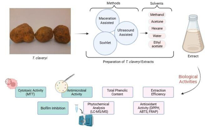
Figure 1: Schematic overview of the experimental workflow. Please click here to view a larger version of this figure.
9. Statistical analysis
- Perform statistical analyses using appropriate statistical software packages and compare control and treatment groups using Student’s t-test or two-way analysis of variance (ANOVA), as appropriate.
- Express all data as mean ± standard error of the mean (SEM).
- Define statistical significance at p < 0.05.
- Indicate statistical significance in figures using asterisks as follows: **** for p < 0.0001, *** for p < 0.001, ** for p < 0.01, and * for p < 0.05, and use “ns” to denote non-significance.
Access restricted. Please log in or start a trial to view this content.
Results
Molecular identification of T. claveryi
PCR amplification of the ITS region was performed using the universal primers ITS1 and ITS4 for molecular identification of T. claveryi. The resulting sequences were analyzed and compared against reference sequences in the NCBI database using the BLAST algorithm, allowing definitive species identification (Supplementary file 1).
Extraction efficiency
Extraction efficiencies vari...
Access restricted. Please log in or start a trial to view this content.
Discussion
The present study establishes a comparative and standardized extraction–bioactivity platform for T. claveryi, demonstrating that the extraction strategy is a decisive determinant of both chemical composition and biological functionality. The primary methodological strength of this protocol lies in the strict control of extraction parameters, particularly the fixed solid–liquid ratio, standardized solvent volume, identical post-extraction processing, and uniform stock solution preparation prior to bio...
Access restricted. Please log in or start a trial to view this content.
Disclosures
The authors declare no competing interests. All data supporting the findings of this study are available within the main manuscript and the supplementary information files. The datasets generated and analyzed during the current study are available from the corresponding author upon reasonable request.
Acknowledgements
The authors declared that this study has received no financial support.
Access restricted. Please log in or start a trial to view this content.
Materials
| Name | Company | Catalog Number | Comments |
|---|---|---|---|
| 96-well polystyrene microtiter plates | Nest Scientific | 701001 | Cell culture / assay plates |
| ABTS (2,2'-azino-bis(3-ethylbenzothiazoline-6-sulfonic acid)) | Sigma-Aldrich | A3219 | Cation radical scavenging assay |
| Acetone (Analytical grade) | Sigma-Aldrich | 179124 | Extraction solvent |
| Acetonitrile (LC-MS grade) | Sigma-Aldrich | 34851 | Organic mobile phase |
| Acinetobacter baumannii | ATCC | Manassas, VA, USA | ATCC 17978 |
| Triple Quad LC-MS/MS with 1290 Infinity UPLC system | Agilent Technologies | 6460 | Quantitative analysis |
| Analytical column ( C18, 4.6 × 100 mm, 3.5 μm) | Agilent Technologies (Zorbax) | 959990-902 | Reversed-phase column |
| Antioxidant standards (Ascorbic acid / Trolox) | Sigma-Aldrich | A7506 / 238813 | Positive controls |
| Applied Biosystems 3730xl DNA Analyzers | Applied Biosystems | 3730xl DNA Analyzers | DNA Sequencing System |
| Applied Biosystems™ BigDye™ Terminator v3.1 Cycle Sequencing Kit | Applied Biosystems | 4337455 | Sanger Sequencing Kit |
| Bacillus cereus | ATCC | Manassas, VA, USA | ATCC 14579 |
| Bioinformatics Analysis, BioEdit software, Algorithm used: CAP contig assembly | Ibis Biosciences | NA | Sequence Assembly Software |
| Cellulose extraction thimble | Fisherbrand™ | 11724043 | Used for Soxhlet extraction |
| Dimethylsulfoxide (DMSO) | Merck | 102952 | Solvent |
| DNA isolation kit (Plant&Fungi DNA isolation kit (Poland) ) | EurX GeneMATRIX | E3595 | DNA isolation |
| DPPH (2,2-diphenyl-1-picrylhydrazyl) | Sigma-Aldrich | D9132 | Radical scavenging assay |
| Dulbecco's Modified Eagle Medium | Capricorn, | DMEM-HA | |
| Enterobacter aerogenes | ATCC | Manassas, VA, USA | ATCC 13048 |
| Enterococcus faecalis | ATCC | Manassas, VA, USA | ATCC 29212 |
| Escherichia coli | ATCC | Manassas, VA, USA | ATCC 25922 |
| Ethanol | Merck | 100983 | Solvent |
| Ethyl acetate | Merck | 109623 | Extraction Solvent |
| Fetal Bovine Serum (FBS) | Capricorn Scientific | FBS-16A | |
| BigDyeTerminator v3.1 Cycle Sequencing Kit | ThermoFisher | 4337455 | Sequencing Kit |
| Folin-Ciocalteu reagent | Sigma-Aldrich | 47641 | Total phenolic content |
| Formic acid | Sigma-Aldrich | 33015 | Mobile phase additive |
| Gallic acid | Carlo Erba | 406335 | Calibration curve |
| Gene MATRIX Plant&Fungi DNA isolation kit | EurX | E3595 | DNA isolation kit |
| GraphPad Prism (v9) | Data visualization and statistical analysis | N/A | Statistical analysis |
| Heating mantle | Electrothermal | EME30500 | Used to maintain solvent reflux |
| HighPrep™ PCR Clean-upSystem | MAGBIO | AC-60005 | |
| Ice | Laboratory grade | — | Temperature control during UAE |
| Klebsiella pneumoniae | ATCC | Manassas, VA, USA | ATCC 13883 |
| L-glutamine | Capricorn Scientific | Germany | |
| Listeria monocytogenes | ATCC | Manassas, VA, USA | ATCC 19115 |
| Lyophilizer | Scientz | N/A | Freeze-drying of extracts |
| Magnetic bead–based PCR clean-up system | MAGBIO | AC-60005 | PCR |
| Methanol | Merck | 106009 | Extraction solvent |
| n-Hexane | Merck | 103701 | Extraction solvent |
| Penicillin/Streptomycin | Capricorn Scientific | Germany | |
| Pseudomonas aeruginosa | ATCC | Manassas, VA, USA | ATCC 27853 |
| Filter paper (No. 541) | Cytiva | 1541-125 V | In the extract filtration process |
| Rotary evaporator | Buchi R100 | 11100V101 | Solvent removal |
| Shigella dysenteriae | ATCC | Manassas, VA, USA | ATCC 13313 |
| Sodium carbonate | Merck | 106392 | Total phenolic analysis |
| SPECTROstar Nano Microplate Reader | BMG Labtech | Absorbance measurements | |
| Staphylococcus aureus | ATCC | Manassas, VA, USA | ATCC 25923 |
| Staphylococcus epidermidis | ATCC | Manassas, VA, USA | ATCC 12228 |
| Statistical analysis software (SPSS, v23) | Data analysis | NA | |
| Thermo Scientific Nanodrop 2000 | Thermo Fisher Scientific | ND-2000 | |
| Tryptone Soy Agar (TSA) | Merck | Germany | — |
| Tryptone Soy Broth (TSB) | Merck | Germany | — |
| Ultrasonic homogenizer | Scientz | SCIENTZ-650L | Ultrasound-assisted extraction |
References
- Wasser, S. P., Weis, A. L. Medicinal properties of substances occurring in higher basidiomycetes mushrooms: current perspectives (Review). Int. J. Med. Mushrooms. 1 (1), 31-62 (1999).
- Wasser, S. P. Current findings, future trends, and unsolved problems in studies of medicinal mushrooms. Appl. Microbiol. Biotechnol. 89 (5), 1323-1332 (2011).
- Badalyan, S. Medicinal aspects of edible ectomycorrhizal mushrooms. Edible Ectomycorrhizal Mushrooms: Current Knowledge and Future Prospects. Zambonelli, A., Bonito, G. M. , Springer. Berlin, Heidelberg. 317-334 (2012).
- El Enshasy, H. A., Hatti-Kaul, R. Mushroom immunomodulators: unique molecules with unlimited applications. Trends Biotechnol. 31 (12), 668-677 (2013).
- Morte, A., Kagan-Zur, V., Navarro-Ródenas, A., Sitrit, Y. Cultivation of desert truffles—A crop suitable for arid and semi-arid zones. Agronomy. 11 (8), 1462(2021).
- Gutiérrez, A., Morte, A., Honrubia, M. Morphological characterization of the mycorrhiza formed by Helianthemum almeriense Pau with Terfezia claveryi Chatin and Picoa lefebvrei (Pat.) Maire. Mycorrhiza. 13 (6), 299-307 (2003).
- Loizides, M., Hobart, C., Konstandinides, G., Yiangou, Y. Desert truffles: the mysterious jewels of antiquity. Field Mycology. 13 (1), 17-21 (2012).
- Atila, F., Kazankaya, A. Terfezia species natural distributed in Türkiye and their ecologies. Mantar Dergisi. 13 (3), 119-130 (2022).
- Türkoğlu, A., Castellano, M., Trappe, J., Yaratanakul Güngör, M. Turkish truffles I: 18 new records for Turkey. Turkish J. Bot. 39, 359-376 (2015).
- Doğan, H., Kurt, F. New macrofungi records from Turkey and macrofungal diversity of Pozantı-Adana. Turkish J. Bot. 40, 209-217 (2016).
- İnci, Ş, Kırbağ, S. Nutritional content, antioxidant and antimicrobial activity of Terfezia claveryi Chatin. Artvin Çoruh Univ. J. For. Fac. 19 (2), 138-143 (2018).
- Sung, H., et al. Global cancer statistics 2020: GLOBOCAN estimates of incidence and mortality worldwide for 36 cancers in 185 countries. CA Cancer J. Clin. 71 (3), 209-249 (2021).
- Ventola, C. L. The antibiotic resistance crisis: part 1: causes and threats. P T. 40 (4), 277-283 (2015).
- Cushnie, T. P. T., Lamb, A. J. Antimicrobial activity of flavonoids. Int. J. Antimicrob. Agents. 26 (5), 343-356 (2005).
- Valko, M., et al. Free radicals and antioxidants in normal physiological functions and human disease. Int. J. Biochem. Cell Biol. 39 (1), 44-84 (2007).
- Morte, A., Honrubia, M., Gutiérrez, A. Biotechnology and cultivation of desert truffles. Mycorrhiza: State of the Art. Varma, A. , 467-483 (2008).
- Corrêa, R. C. G., et al. Biotechnological, nutritional and therapeutic uses of Pleurotus spp. related with its chemical composition: a review. Trends Food Sci Technol. 50, 103-117 (2016).
- Ma, G., et al. A critical review on the health promoting effects of mushrooms nutraceuticals. Food Sci. Hum. Wellness. 7 (2), 125-133 (2018).
- Reis, F. S., et al. Functional foods based on extracts or compounds derived from mushrooms. Trends Food Sci. Technol. 66, 48-62 (2017).
- Mandeel, Q. A., Al-Laith, A. A. Ethnomycological aspects of the desert truffle among native Bahraini and non-Bahraini peoples. J. Ethnopharmacol. 110 (1), 118-129 (2007).
- Patel, S. Food, health and agricultural importance of truffles: a review. Curr. Trends Biotechnol. Pharm. 6, 15-27 (2012).
- Janakat, S., Nassar, M. Hepatoprotective activity of desert truffle (Terfezia claveryi) compared with Nigella sativa in rats. Pak. J. Nutr. 9, 52-56 (2010).
- Al-Laith, A. A. A. Antioxidant components and activities of desert truffle (Tirmania nivea). J. Food Compos. Anal. 23 (1), 15-22 (2010).
- Shavit, E., Shavit, E. The medicinal value of desert truffles. Desert Truffles. Kagan-Zur, V., Roth-Bejerano, N., Sitrit, Y., Morte, A. 38, 323-340 (2014).
- Patel, S., et al. Potential health benefits of natural products derived from truffles. Trends Food Sci. Technol. 70, 1-8 (2017).
- Beara, I. N., et al. Phenolic profile, antioxidant, anti-inflammatory and cytotoxic activities of truffles. Food Chem. 165, 460-466 (2014).
- Dahham, S. S., et al. Antioxidant, anticancer and apoptosis properties of Terfezia claveryi. Saudi J. Biol. Sci. 25 (8), 1524-1534 (2018).
- Azmir, J., et al. Techniques for extraction of bioactive compounds: a review. J. Food Eng. 117 (4), 426-436 (2013).
- Kumar, M., Shukla, P. K. PCR targeting of ITS regions for rapid diagnosis of mycotic keratitis. J. Clin. Microbiol. 43 (2), 662-668 (2005).
- Bondet, V., Brand-Williams, W., Berset, C. Kinetics and mechanisms of antioxidant activity using the DPPH method. LWT Food Sci. Technol. 30, 609-615 (1997).
- Re, R., et al. Antioxidant activity applying an improved ABTS assay. Free Radic. Biol. Med. 26 (9-10), 1231-1237 (1999).
- Đorđević, T. M., et al. Effect of fermentation on antioxidant properties of cereals. Food Chem. 119 (3), 957-963 (2010).
- Herald, T. J., et al. High-throughput micro plate assays for screening flavonoid content and DPPH-scavenging activity in sorghum bran and flour. Sci. Food Agric. 92 (11), 2326-2331 (2012).
- Kısa, D., et al. Evaluation of Biological Potency of two Endemic Species Integrated with in vitro and in silico Approches: LC-MS/MS Analysis of the Plants. Chem. Biodivers. 21 (3), e202301351(2024).
- Kumoro, A., Singh Hasan, Effects of solvent properties on the soxhlet extraction of diterpenoid lactones from andrographis paniculata leaves. ScienceAsia. 35, 306-309 (2009).
- Ng, Z. X., Samsuri, S. N., Yong, P. H. The antioxidant index and chemometric analysis of tannin, flavonoid, and total phenolic extracted from medicinal plant foods with the solvents of different polarities. Food Process. Preserv. 44 (9), e14680(2020).
- Kıvrak, İ Analytical methods applied to assess chemical composition, nutritional value and ın vitro bioactivities of Terfezia olbiensis and Terfezia claveryi from Turkey. Food Anal. Methods. 8 (5), 1279-1293 (2015).
- Neggaz, S., et al. In vitro evaluation of antioxidant, antibacterial and antifungal activities of Terfezia claveryi Chatin. Phytothérapie. 13, 1-7 (2015).
- Monteiro, M., et al. Effect of extraction method and solvent system on the phenolic content and antioxidant activity of selected macro- and microalgae extracts. J. Appl. Phycol. 32 (1), 349-362 (2020).
- Pingret, D., et al. Degradation during application of ultrasound in food processing: A review. Food Control. 31, 593-606 (2013).
- Şahin, A., et al. Total phenol, antioxidant activity, fatty acid composition and mineral contents of the species of fungi known as domalan. J. Agroalimentary Process. Technol. 26 (3), 149-154 (2020).
- Fidan, M., et al. cytotoxic and protective effects of truffles. Anal. Biochem. 641, 114566(2022).
- Awika, J. M., et al. Screening methods to measure antioxidant activity of sorghum (sorghum bicolor) and sorghum products. J. Agric. Food Chem. 51 (23), 6657-6662 (2003).
- Gursoy, N., et al. Antioxidant activities, metal contents, total phenolics and flavonoids of seven Morchella species. Food Chem. Toxicol. 47 (9), 2381-2388 (2009).
- Elsayed, E., et al. Truffles: A potential source of bioactive compounds. Bioactive Compounds in Food. , CRC Press. 163-174 (2021).
- Elsayed, E. A., et al. Mushrooms: a potential natural source of anti-inflammatory compounds for medical applications. Mediators Inflamm. 2014, 805841(2014).
- Stanikunaite, R., et al. Cyclooxygenase-2 inhibitory and antioxidant compounds from the truffle Elaphomyces granulatus. Phytother. Res. 23 (4), 575-578 (2009).
- Doğan, H. H., Aydın, S. Determination of antimicrobial effect, antioxidant activity and phenolic contents of desert truffle in Turkey. Afr. J. Tradit. Complement. Altern. Med. 10 (4), 52-58 (2013).
- Abu-Odeh, A., et al. In vitro and In vivo Antidiabetic Activity, phenolic content and microscopical characterization of Terfezia claveryi. Molecules. 27 (15), 4843(2022).
- Nguyen, V., et al. Chlorogenic Acid: A systematic review on the biological functions, mechanistic actions, and therapeutic potentials. Nutrients. 16 (7), 924(2024).
- Suhag, R., et al. Mapping antioxidant mechanisms in plant extracts via reactivity kinetics inhibiting oil autoxidation. Appl. Food Res. 5 (2), 101476(2025).
- Harir, M., et al. Evaluation of antimicrobial activity of Terfezia arenaria extracts collected from Saharan desert against bacteria and filamentous fungi. 3 Biotech. 9 (7), 281(2019).
- Hamza, A., et al. Nutraceutical potential, antioxidant and antibacterial activities of Terfezia boudieri Chatin, a wild edible desert truffle from Tunisia arid zone. Arab. J. Chem. 9 (3), 383-389 (2016).
- Neggaz, S., et al. In vitro evaluation of antioxidant, antibacterial and antifungal activities of Terfezia claveryi Chatin. Phytothérapie. 13, 1-7 (2020).
- Dib, S., Fortas, Z. Antimicrobial activities of desert truffle extracts and their chemical ıdentification. J. Pharm. Pharmacol. 7, 1-7 (2019).
- Gargano, M. B., et al. Antimicrobial activity of the extracts of Terfezia Claveryi and Tirmania Pinoyi against gram-positive and gram-negative bacteria causal agent of diseases in tomato. Chem. Eng. Trans. 58, 73-78 (2017).
- Khojah, H. M. J., et al. Antimicrobial efficacy of extracts of Saudi Arabian desert Terfezia claveryi truffles. Saudi J. Biol. Sci. 29 (11), 103462(2022).
- Akyüz, M., et al. Cytotoxicity and DNA protective effects of the Terfezia and Picoa species from the eastern region of Turkey. Istanbul J. Pharm. 51 (2), 198-203 (2021).
- Al Obaydi, M. F., et al. Terfezia boudieri: A Desert Truffle With Anticancer and Immunomodulatory Activities. Front. Nutr. 7, 38(2020).
- Pakrashi, S., et al. Quercetin alleviates 6-OHDA-caused apoptosis in SH-SY5Y cells. Toxicol. Res. 13 (4), tfae117(2024).
Access restricted. Please log in or start a trial to view this content.