This protocol evaluates and compares the clinical efficacy, safety, and patient-reported outcomes of ab externo canaloplasty performed alone or in combination with phacoemulsification in eyes with advanced primary open-angle glaucoma, providing a standardized approach for assessing long-term intraocular pressure control, structural preservation, and quality-of-life improvement.
A subscription to JoVE is required to view this content. Sign in or start your free trial.
Method Article
Ab Externo Canaloplasty Alone or Combined with Phacoemulsification for Advanced Primary Open-Angle Glaucoma
* These authors contributed equally
In This Article
Summary
Abstract
Advanced primary open-angle glaucoma (POAG) requires surgical strategies that achieve effective reduction of intraocular pressure (IOP) while preserving optic nerve structure and function. This study aims to compare the long-term clinical performance of ab externo canaloplasty (AC) alone versus AC combined with phacoemulsification (AC + phaco) in patients with advanced POAG. In this retrospective comparative study, 140 patients with advanced POAG were assigned to AC (n = 70) or AC + phaco (n = 70). Over 24 months of follow-up, both procedures significantly reduced IOP and medication use. However, AC + phaco achieved greater mean IOP reduction (45.5% vs. 36.1%; p < 0.001), higher retinal nerve fiber layer thickness (62.55 ± 6.03 µm vs. 60.82 ± 6.51 µm; p = 0.047), larger neuroretinal rim area (0.91 ± 0.12 mm² vs. 0.85 ± 0.12 mm²; p = 0.009), and improved visual field stability. Quality-of-life measures favored AC + phaco, with improved patient-reported visual function and fewer vision-related limitations. These findings demonstrate that both AC and AC + phaco are safe and effective surgical options for advanced POAG. AC + phaco may be preferentially selected in patients with coexisting cataract or in cases requiring enhanced long-term IOP control and structural preservation, without increasing surgical risk.
Introduction
Primary open-angle glaucoma (POAG) is a progressive optic neuropathy and the second leading cause of irreversible blindness worldwide1. It is characterized by the degeneration of retinal ganglion cells and optic nerve fibers, resulting in progressive visual field loss and eventual visual disability. Due to its insidious onset and largely asymptomatic early course, POAG is often diagnosed only after substantial structural and functional damage has occurred. With increasing global life expectancy, urbanization, and population aging, the burden of POAG continues to rise, particularly in low- and middle-income regions where access to early screening and sustained care remains limited2. In addition to visual impairment, POAG imposes significant socioeconomic and quality-of-life burdens, highlighting the need for surgical strategies that achieve durable intraocular pressure (IOP) control while preserving optic nerve structure and functional vision.
Conventional glaucoma surgeries, including trabeculectomy and glaucoma drainage device implantation, have long been considered the standard surgical options for eyes with uncontrolled IOP. Although effective in lowering pressure, these procedures are associated with well-recognized complications such as hypotony, bleb leakage, infection, and long-term bleb-related morbidity3. The reliance on subconjunctival filtration also necessitates intensive postoperative management and may compromise ocular surface integrity over time. To overcome these limitations, Schlemm's canal-based procedures and minimally invasive glaucoma surgeries have been developed to restore physiologic aqueous humor outflow while minimizing tissue disruption4. These approaches preserve conjunctival integrity, reduce complication rates, and allow faster visual recovery, making them increasingly relevant across the glaucoma severity spectrum5,6.
Ab externo canaloplasty (AC) is a bleb-independent Schlemm's canal-based procedure designed to enhance trabecular outflow by circumferential dilation of Schlemm's canal under a watertight scleral flap7,8,9. By avoiding subconjunctival filtration, this technique reduces bleb-related complications, simplifies postoperative care, and preserves future surgical options10,11,12,13. These features have supported its use primarily in early and moderate POAG; however, its application in advanced disease remains an area of active investigation14. Importantly, AC maintains the native aqueous outflow pathway, offering a physiologic alternative to filtering surgery in appropriately selected patients.
Combining AC with phacoemulsification (AC + phaco) offers potential additional benefits, particularly in older patients with coexisting cataract. Cataract extraction alone is known to lower IOP modestly by widening the anterior chamber angle and improving aqueous outflow dynamics15,16,17. When combined with AC, phacoemulsification may enhance outflow facility while simultaneously restoring visual acuity, thereby improving both functional vision and quality of life18,19,20,21,22. This combined approach may be especially advantageous in patients with visually significant lens opacity, advanced glaucoma, and a need for sustained IOP reduction with minimal postoperative risk.
Despite these advantages, concerns persist regarding the safety and efficacy of combining cataract surgery with Schlemm's canal-based procedures in eyes with advanced optic nerve damage, given the potential inflammatory and hemodynamic stresses associated with phacoemulsification23,24. However, emerging evidence from longitudinal studies, matched cohort analyses, and multicenter registries suggests that AC combined with phacoemulsification may provide superior preservation of retinal nerve fiber layer thickness, neuroretinal rim area, and visual field stability compared with canaloplasty alone25,26,27,28,29. Additional studies have demonstrated improved patient-reported quality-of-life outcomes, reduced dependence on anti-glaucoma medications, and durable IOP control extending beyond two years30,31. These findings underscore the importance of integrating structural, functional, and patient-centered metrics when evaluating surgical options for advanced POAG32,33,34,35,36,37.
Both procedures may be less suitable in patients with angle-closure glaucoma, extensive peripheral anterior synechiae, or secondary glaucomas such as neovascular or active uveitic glaucoma, where trabecular outflow resistance is not the primary mechanism of pressure elevation. Eyes requiring very low target IOP, particularly in the low teens or single-digit range due to rapidly progressive disease, may be better managed with filtering surgery or drainage devices. Prior conjunctival or scleral surgery, distorted Schlemm's canal anatomy, or ocular comorbidities that limit reliable structural and functional assessment may also reduce the feasibility or interpretability of outcomes with this approach.
The overall goal of the present method is to provide a reproducible surgical approach to evaluate the long-term efficacy, safety, and functional impact of AC performed alone or in combination with phacoemulsification in advanced POAG.
Access restricted. Please log in or start a trial to view this content.
Protocol
All procedures described in this protocol were reviewed and approved by the Institutional Human Research Ethics Committee of the Third People's Hospital of Dalian University of Technology (Approval No. 2025-176-001). The study was conducted in accordance with the ethical principles outlined in the Declaration of Helsinki. Informed consent was obtained from all participants prior to enrollment.
1. Patient recruitment and screening
NOTE: Patients were recruited consecutively and concurrently from the glaucoma outpatient clinic of the Third People's Hospital of Dalian University of Technology between January 2020 and December 2022.
- Confirm eligibility using the following inclusion criteria:
- Ensure patient age is between 50 and 80 years.
- Confirm diagnosis of advanced primary open-angle glaucoma (POAG) defined by a visual field mean deviation (MD) ≤−12 dB, cup-to-disc ratio ≥0.8, and significant retinal nerve fiber layer (RNFL) thinning on optical coherence tomography (OCT).
- Verify uncontrolled IOP (≥21 mmHg) despite maximal tolerated anti-glaucoma medical therapy.
- Confirm the patient's ability to provide written informed consent and comply with scheduled postoperative follow-up visits.
- Exclude patients with a history of glaucoma filtering or cyclodestructive surgery, secondary glaucomas, active ocular infection or inflammation, advanced systemic illness, visually significant corneal opacity or retinal pathology, or pseudophakic/aphakic eyes at baseline.
- Screen all eligible patients during the same enrollment period to minimize temporal bias related to surgical experience or procedural refinement.
2. Group allocation
- After excluding individuals who do not meet eligibility requirements or decline participation, enroll eligible patients with advanced primary open-angle glaucoma.
- Allocate them in comparable numbers to either AC alone or AC + phaco based on clinical indication and lens status (Figure 1).
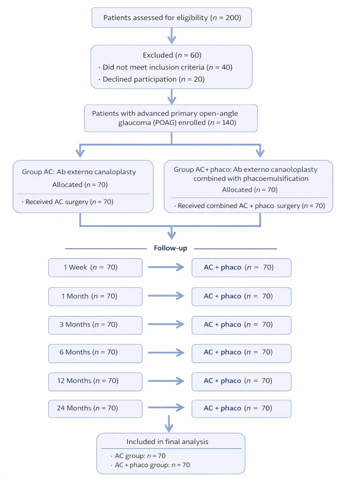
Figure 1: Patient enrollment and group allocation flow diagram. Flow diagram illustrating patient screening, eligibility assessment, exclusions, and final allocation to the AC group or the AC + phaco group. Abbreviations: AC = ab externo canaloplasty; AC + phaco = ab externo canaloplasty combined with phacoemulsification; POAG = primary open-angle glaucoma. Please click here to view a larger version of this figure.
3. Preoperative evaluation
- Measure Best Corrected Visual Acuity (BCVA) using a Snellen chart under standardized lighting.
- Record IOP using Goldmann applanation tonometry. Take three consecutive readings and calculate the mean.
- Perform gonioscopy to assess the anterior chamber angle.
- Conduct slit-lamp biomicroscopy to examine anterior segment structures.
- Use spectral-domain OCT to measure RNFL thickness and neuroretinal rim area.
- Capture fundus photographs of the optic disc for baseline documentation.
- Perform automated perimetry (24-2 SITA-Standard protocol) to evaluate visual fields.
4. Surgical intervention
- General surgical conditions
- Perform all surgical procedures by the same experienced glaucoma surgeon throughout the study period to minimize inter-operator variability.
- Perform all procedures under local anesthesia using standard sterile ophthalmic technique.
- Position the patient supine and prepare and drape the operative field in the usual manner.
- Ab externo canaloplasty alone (AC group)
- Create a fornix-based conjunctival peritomy in the superior quadrant.
- Dissect Tenon's capsule to expose bare sclera and achieve meticulous hemostasis.
- Fashion a superficial scleral flap measuring approximately 4 × 4 mm, followed by a deep scleral flap to expose and unroof Schlemm's canal.
NOTE: Accurate identification and exposure of Schlemm's canal is critical, as incorrect flap depth or canal misidentification is the most frequent source of technical failure. - Identify the two ostia of Schlemm's canal and confirm correct localization.
- Introduce a flexible microcatheter into Schlemm's canal and advance it circumferentially through 360°.
NOTE: Confirm complete circumferential passage before proceeding. Recognize incomplete canal cannulation by resistance to catheter advancement or failure to visualize the catheter tip, and reassess canal localization if this occurs. - Inject viscoelastic material incrementally during catheter withdrawal to achieve uniform canal dilation.
NOTE: Identify inadequate viscodilation by minimal early postoperative IOP reduction. - Place a 10-0 Prolene suture within Schlemm's canal and apply moderate, evenly distributed tension to obtain circumferential canal distension.
NOTE: Avoid under- or over-distension. Recognize over-tensioning by early postoperative IOP spikes or patient discomfort. - Reposition the superficial scleral flap and secure it with interrupted 10-0 nylon sutures.
- Close Tenon's capsule and conjunctiva with interrupted 10-0 nylon sutures to prevent wound leakage and postoperative hypotony.
NOTE: Mild hyphema and transient IOP fluctuations in the early postoperative period are common and typically self-limiting, reflecting restoration of physiologic outflow rather than surgical failure.
- Combined Ab externo canaloplasty with phacoemulsification (AC + phaco group)
- Perform the standard clear corneal phacoemulsification using the divide-and-conquer technique.
- Remove the crystalline lens and implant a foldable acrylic intraocular lens into the capsular bag. Hydrate corneal incisions and confirm watertight wound closure.
- Proceed with AC as described for the AC group.
- Create a superior conjunctival peritomy, fashion superficial and deep scleral flaps, identify Schlemm's canal, and perform circumferential microcatheterization with incremental viscoelastic injection.
- Insert and tension a 10-0 Prolene suture to achieve uniform canal distension. Reposition and secure the scleral flap and close the conjunctiva with interrupted 10-0 nylon sutures.
5. Postoperative evaluation and follow-up
- 5.1.Conduct follow-up assessments at 1 week, 1 month, 3 months, 6 months, 12 months, and 24 months.
- At each visit, repeat BCVA, IOP measurement, slit-lamp examination, and gonioscopy.
- Perform OCT imaging and fundus photography annually.
- Assess visual fields at 6, 12, and 24 months.
- Document postoperative complications, dividing into early (<30 days) or late (>30 days). Include hyphema, IOP spikes, shallow anterior chamber, infection, and need for reintervention.
- Record medication burden by counting topical anti-glaucoma agents used at each follow-up.
6. Quality-of-life assessment
- Administer the National Eye Institute Visual Function Questionnaire-25 (NEI VFQ-25) to assess vision-related quality of life.
NOTE: The NEI VFQ-25 assesses vision-related functional status across multiple domains, including general vision, near and distance activities, social functioning, mental health, and role limitations. - Administer EuroQol-5D (EQ-5D) to evaluate general health-related quality of life.
NOTE: The EQ-5D evaluates general health-related quality of life across five dimensions: mobility, self-care, usual activities, pain/discomfort, and anxiety/depression. - Administer Glaucoma Quality of Life-15 (GQL-15) to measure glaucoma-specific disability.
NOTE: The GQL-15 specifically measures glaucoma-related disability, focusing on difficulties with central and peripheral vision, dark adaptation, glare, and outdoor mobility. - Conduct all questionnaires in the patient's preferred language with trained personnel.
7. Outcome measures
- Record primary outcomes:
- IOP reduction at each follow-up relative to baseline.
- Stability of RNFL thickness and neuroretinal rim area on OCT.
NOTE: The change in retinal nerve fiber layer (RNFL) thickness is analyzed as the difference from baseline (ΔRNFL) at each follow-up time point to reflect longitudinal structural preservation. - Quality-of-life scores from NEI VFQ-25, EQ-5D, and GQL-15.
- Complete success, defined as IOP ≤18 mmHg without medications
- Qualified success, defined as IOP ≤18 mmHg with medications
- Record secondary outcomes:
- Change in visual field indices.
- Reduction in medication burden.
- Frequency and severity of complications.
8. Statistical analysis
- Enter all data into SPSS software (version 26.0; IBM Corp., Armonk, NY, USA) for statistical analysis.
- Assess the normality of continuous variables using the Shapiro-Wilk test. Present normally distributed data as mean ± standard deviation (SD) and non-normally distributed data as median (interquartile range).
- Perform between-group comparisons using the independent-samples t-test for normally distributed continuous variables and the Mann-Whitney U test for non-normally distributed variables. Use repeated-measures ANOVA to evaluate within-group longitudinal changes across follow-up time points.
- Analyze categorical variables using the chi-square test or Fisher's exact test, as appropriate.
- Consider a p value ≤ 0.05 as statistically significant for all analyses.
- Perform Kaplan-Meier survival analysis to evaluate long-term surgical success according to predefined IOP control thresholds, with group differences assessed using the log-rank test.
Access restricted. Please log in or start a trial to view this content.
Results
A total of 200 patients were assessed for eligibility. After excluding patients who did not meet the inclusion criteria or declined to participate, 140 patients with advanced POAG were enrolled and allocated into two groups: AC (n = 70) and AC + phaco (n = 70) (Figure 1).
Baseline clinical characteristics were comparable between the AC group and the AC + phaco group (Table 1). No statistically significant differences were observed between groups i...
Access restricted. Please log in or start a trial to view this content.
Discussion
The efficacy of Schlemm’s canal–based strategies in slowing the progression of POAG is demonstrated by the consistent IOP reduction pattern observed in both groups. However, statistical analysis showed that the AC + phaco group achieved superior pressure-reducing effects at all postoperative time points. These results are supported by Zhang and Wang38 and Seethe et al.39, who reported significantly greater IOP reduction with combined canaloplasty and phacoemulsi...
Access restricted. Please log in or start a trial to view this content.
Disclosures
The authors have no conflicts of interest to declare.
Acknowledgements
The authors acknowledge the support of the Scientific Research Start-up Fund Project of Dalian Third People's Hospital (2023ky006) and the Medical Science Research Program Project of Dalian Municipal (2111007). The authors also thank the clinical and nursing staff of the Glaucoma Service for their assistance with patient care and data collection.
Access restricted. Please log in or start a trial to view this content.
Materials
| Name | Company | Catalog Number | Comments |
|---|---|---|---|
| Equipment | |||
| Automated perimeter (HFA 24-2 SITA-Standard) | Carl Zeiss Meditec | ZEISS-HFA3-860 | Visual field indices: MD, PSD at 6, 12, 24 months. |
| Canaloplasty microcatheter | Nova Eye Medical (iTrack) | NOVA-ITRACK-250 | 360° Schlemm’s canal catheterization for viscodilation. |
| Fundus camera / Imaging system | ZEISS (VISUCAM) | ZEISS-VISC-524 | Optic disc and posterior pole documentation. |
| Goldmann applanation tonometer | Haag-Streit AG | HS-GAT-870 | Standard IOP measurement; three consecutive readings averaged. |
| Gonioscopy lens (4-mirror) | Volk Optical | VOLK-G4 | Pre-/post-operative angle assessment. |
| Ophthalmic operating microscope | ZEISS (OPMI Lumera series) | ZEISS-LUMERA-T | Microsurgical visualization. |
| Phacoemulsification system | Alcon (CENTURION) | ALC-CENT-PHACO | Divide-and-conquer technique; used only in PCP+Phaco group. |
| Slit-lamp biomicroscope | Topcon | TOP-SLD701 | Anterior segment examination and postoperative monitoring. |
| Spectral-domain OCT (RNFL/GCC) | Heidelberg Engineering (SPECTRALIS) | HE-SDOCT-SPC | Peripapillary RNFL thickness and neuroretinal rim area acquisition. |
| Consumable | |||
| Balanced salt solution (BSS) | Alcon | ALC-BSS-500 | Irrigation during anterior segment surgery. |
| Foldable posterior chamber IOL | Alcon (AcrySof IQ) | ALC-SN60WF | IOL power individualized; acrylic lens implantation. |
| Microsurgical sutures, Nylon 10-0 | Ethicon | ETH-NYL-10-0 | Watertight scleral flap closure. |
| Ophthalmic viscosurgical device (OVD), 1.0% Sodium hyaluronate | Johnson & Johnson Vision (Healon) | JJ-HEALON-10 | Maintains anterior chamber; viscodilation support. |
| Viscodilation cannula (27G) | Nova Eye Medical | NOVA-VD-27G | Controlled viscoelastic delivery into Schlemm’s canal. |
| Drug | |||
| Moxifloxacin 0.5% Ophthalmic solution | Alcon (Vigamox) | ALC-VIG-05 | Postoperative topical antibiotic. |
| Prednisolone acetate 1% Ophthalmic suspension | Allergan (Pred Forte) | AGN-PF-1 | Postoperative topical steroid taper. |
| Timolol 0.5% / Brimonidine 0.2% / Dorzolamide 2% / Latanoprost 0.005% | Various (Generic/Branded) | GLC-RESCUE-SET | IOP-lowering agents as needed per protocol. |
| Software/Instrument | |||
| EQ-5D Instrument (License) | EuroQol Research Foundation | EQ-5D-LIC | General health-related quality of life. |
| GQL-15 Questionnaire (Permission) | Instrument Owner/Publisher | GQL-15-PERM | Glaucoma-specific disability index. |
| NEI VFQ-25 Questionnaire (License/Permission) | National Eye Institute | NEI-VFQ25-LIC | Vision-related quality of life assessment. |
| SPSS Statistics v26 | IBM | IBM-SPSS-26 | Statistical analysis: t-tests, ANOVA, repeated measures, Kaplan–Meier. |
References
- Lavia, C., Dallorto, L., Maule, M., Ceccarelli, M., Fea, A. M. Minimally invasive glaucoma surgeries (MIGS) for open angle glaucoma: a systematic review and meta-analysis. PLoS One. 12 (8), e0183142(2017).
- Koerber, N., Ondrejka, S. Six-year efficacy and safety of iTrack ab interno canaloplasty as a stand-alone procedure and combined with cataract surgery in primary open angle and pseudoexfoliative glaucoma. J Glaucoma. 33 (3), 176-182 (2024).
- Dorairaj, S., Radcliffe, N. M., Grover, D. S., Brubaker, J. W., Williamson, B. K. A review of excisional goniotomy performed with the Kahook Dual Blade for glaucoma management. J Curr Glaucoma Pract. 16 (1), 59-64 (2022).
- Radcliffe, N. M., Harris, J., Garcia, K., Zwick, E., Chang, R. T., Mbagwu, M. Standalone canaloplasty and trabeculotomy using the OMNI surgical system in eyes with primary open angle glaucoma: a 36-month analysis from the AAO IRIS registry. Am J Ophthalmol. 271, 436-444 (2025).
- Rhee, D. J., Lau, S., Sozeri, Y. G. Glaucoma staging and progression risk factors. Rev Ophthalmol. , (2022).
- Streamline Surgical System. Instructions for Use. , New World Medical. Rancho Cucamonga, CA. (2021).
- Ahmed, I. I. K., et al. COMPARE study: Hydrus versus iStent microinvasive glaucoma surgery implants for standalone treatment of open angle glaucoma. Ophthalmology. 127 (1), 52-61 (2020).
- Samuelson, T. W., et al. Prospective randomized controlled trial of ab interno trabecular micro-bypass in primary open angle glaucoma and cataract: two-year results. Ophthalmology. 126 (6), 811-821 (2019).
- Samuelson, T. W., et al. HORIZON study: Schlemm canal microstent for intraocular pressure reduction in primary open angle glaucoma and cataract. Ophthalmology. 126 (1), 29-37 (2019).
- Hughes, T., Traynor, M. Clinical results of ab interno canaloplasty in patients with open angle glaucoma. Clin Ophthalmol. 14, 3641-3650 (2020).
- Ondrejka, S., Koerber, N. 360° ab interno Schlemm's canal viscodilation in primary open angle glaucoma. Clin Ophthalmol. 13, 1235-1246 (2019).
- Tracer, N., Dickerson, J. E., Radcliffe, N. M. Circumferential viscodilation ab interno plus phacoemulsification: 12-month outcomes. Clin Ophthalmol. 14, 1357-1364 (2020).
- Gillmann, K., Aref, A., Niegowski, L. J., Baumgartner, J. M. Combined ab interno canaloplasty in open angle glaucoma: 12-month outcomes. Int Ophthalmol. 41, 3295-3301 (2021).
- Kazerounian, S., et al. Canaloplasty ab interno (ABiC): two-year results in minimally invasive glaucoma surgery technique. Klin Monbl Augenheilkd. 238 (10), 1113-1119 (2021).
- Davids, A. M., et al. ABiC: 12-month results of a new MIGS. Graefes Arch Clin Exp Ophthalmol. 257 (9), 1947-1953 (2019).
- Gallardo, M. J., Supnet, R. A., Ahmed, I. I. K. Circumferential viscodilation of Schlemm's canal in open angle glaucoma: ab interno versus ab externo with tensioning suture. Clin Ophthalmol. 12, 2493-2498 (2018).
- Gallardo, M. J., Supnet, R. A., Ahmed, I. I. K. Viscodilation of Schlemm's canal via ab interno approach. Clin Ophthalmol. 12, 2149-2155 (2018).
- Gallardo, M. J. 24-month efficacy of viscodilation with iTrack ab interno canaloplasty. Clin Ophthalmol. 15, 86-89 (2021).
- Gallardo, M. J. 36-month effectiveness of ab interno canaloplasty with or without cataract surgery. Ophthalmol Glaucoma. 5 (5), 476-482 (2022).
- Koerber, N., Ondrejka, S. Four-year efficacy and safety of iTrack ab interno canaloplasty with or without phacoemulsification. Klin Monbl Augenheilkd. 240, 1394-1404 (2022).
- Lazcano Gomez, G., Garg, S. J., Yeu, E., Kahook, M. Y. Interim analysis of Streamline outcomes in glaucoma eyes. Clin Ophthalmol. 16, 1313-1320 (2022).
- Lazcano Gomez, G., Antzoulatos, G. L., Kahook, M. Y. Combined phacoemulsification and Streamline in Hispanic patients with mild to moderate glaucoma. Clin Ophthalmol. 17, 1911-1918 (2023).
- Patel, V. N., Smith, A. L., Nguyen, D. T. EQ-5D and NEI VFQ-25 outcomes in canaloplasty with versus without cataract extraction in severe primary open angle glaucoma. Am J Ophthalmol. 240, 160-168 (2022).
- Kumar, R. S., et al. Glaucoma Quality of Life (GQL-15) following canaloplasty versus phacocanaloplasty in end-stage primary open angle glaucoma: 24-month prospective trial. Qual Life Res. 31 (6), 1579-1589 (2022).
- Verma, S., et al. Longitudinal neuroretinal rim preservation in canaloplasty versus combined surgery: OCT RNFL analysis over three years. Graefes Arch Clin Exp Ophthalmol. 259 (7), 1765-1774 (2021).
- Huang, J., et al. Comparative hemodynamics and rim preservation after ab interno canaloplasty with or without phacoemulsification: Doppler study. Clin Exp Ophthalmol. 49 (5), 472-479 (2021).
- Fernandez, R., et al. Patient-reported outcomes and visual field stability after canal-based MIGS with or without cataract surgery: a multicenter registry analysis. Ophthalmol Glaucoma. 4 (2), 123-132 (2021).
- Johnson, C. E., et al. Phacoemulsification and canaloplasty versus canaloplasty alone in pseudoexfoliation and advanced primary open angle glaucoma: three-year multicenter randomized controlled trial. Int J Ophthalmol. 44 (3), 401-409 (2021).
- Moreno, P., et al. Neuroretinal rim area and macular thickness preservation in canaloplasty with or without phacoemulsification in Chinese primary open angle glaucoma patients: a 24-month observational study. Eye. 34, 2238-2246 (2020).
- Singh, A., et al. Quality of life and medication reduction after phacocanaloplasty versus canaloplasty in advanced glaucoma: a matched cohort 18-month study. Br J Ophthalmol. 104 (12), 1646-1654 (2020).
- Alvarado, J. A., et al. Longitudinal study comparing surgical outcomes and quality of life in ab interno canaloplasty with or without cataract in advanced primary open angle glaucoma. Clin Ophthalmol. 14, 2895-2903 (2020).
- Steinmetz, J. D., et al. Causes of blindness and trends in vision impairment: Global Burden of Disease 2020. Lancet Glob Health. 9 (2), e144-e160 (2021).
- Riva, I., et al. Canaloplasty in open angle glaucoma: patient selection and outcomes review. Adv Ther. 36 (1), 31-43 (2019).
- Xin, C., et al. Mechanism of reconstruction of aqueous outflow drainage. Sci China Life Sci. 61 (5), 534-540 (2018).
- Khaimi, M. A., Dvorak, J. D., Ding, K. Three-year outcomes following canaloplasty in primary open angle glaucoma. J Ophthalmol. 2017, 1-7 (2017).
- Garris, W., Le, C., Zurakowski, D., Ayyala, R. Canaloplasty versus trabeculectomy with mitomycin C: two-year outcomes. Indian J Ophthalmol. 66 (1), 66-71 (2018).
- Konopińska, J., Mariak, Z., Rękas, M. Safety profile of canaloplasty and phacocanaloplasty: complication review. J Ophthalmol. 2020, 1-6 (2020).
- Zhang, J., Wang, N. L. Progression on canaloplasty for primary open angle glaucoma. Int. J Ophthalmol. 12 (10), 1629-1633 (2019).
- Seuthe, A. M., Szurman, P., Januschowski, K. Canaloplasty with suprachoroidal drainage in pseudoexfoliation glaucoma: four-year results. Curr. Eye Res. 46 (2), 217-223 (2021).
- Vastardis, I., et al. Comparison of two microcatheter systems in ab externo canaloplasty. Eur J Ophthalmol. 30 (3), 487-493 (2020).
- Holste, G., et al. Longitudinal transformer model for primary open angle glaucoma prognosis using serial fundus imagery: survival analysis. arXiv. , (2024).
- Ng, J. C., et al. Quality of life and neuroretinal rim outcomes in advanced primary open angle glaucoma: canaloplasty versus canaloplasty plus phacoemulsification. J Glaucoma. 32 (8), e227-e233 (2023).
- López-García, G., et al. Comparative analysis of peripapillary rim changes in primary open angle glaucoma after canal modification with or without phacoemulsification. Int Ophthalmol. 43 (3), 1121-1129 (2023).
- Kicińska, A. K., et al. Safety and efficacy of canaloplasty variants with phacoemulsification: 12-month follow-up. J Clin Med. 11 (21), 6501(2022).
- Grover, D. S., et al. Two-year outcomes of phacoemulsification plus GATT versus standalone GATT in moderate to severe primary open angle glaucoma: a retrospective comparative cohort study. Diagnostics (Basel). 15 (5), 542(2024).
- Brusini, P., Papa, V., Zeppieri, M. Canaloplasty in pseudoexfoliation glaucoma: a good choice. J Clin Med. 11 (9), 2532(2022).
- Beres, H., Scarioth, G. B. Canaloplasty in the spotlight: alternatives and future perspectives. Rom J Ophthalmol. 66 (3), 225-232 (2022).
- Kodomskoi, L., Kotliar, K., Schröder, A. C., Bertram, B., Kasmann-Kellner, B. S. Suture probe canaloplasty as an alternative to iTrack. J Glaucoma. 28 (9), 811-817 (2019).
Access restricted. Please log in or start a trial to view this content.