This protocol describes a two-phase acid–solvent extraction method for isolating short-chain fatty acids from low-input mouse fecal samples without derivatization.
A subscription to JoVE is required to view this content. Sign in or start your free trial.
Method Article
Extraction of Short-chain Fatty Acids from Mouse Feces Using A Two-phase Acid–Solvent Method
In This Article
Summary
Abstract
This article describes a protocol for extracting short-chain fatty acids (SCFAs) from low-input mouse fecal samples using a two-phase acid–solvent extraction strategy. The method is designed for individual fecal inputs of approximately 20–50 mg and employs dilute hydrochloric acid and ethyl acetate to enable phase separation without derivatization. The protocol consists of fecal homogenization, acidification, organic solvent partitioning, and low-temperature centrifugation, and can be performed using standard laboratory equipment commonly available in research laboratories. Following extraction, SCFAs are suitable for downstream analysis by routine gas chromatography–based platforms. Successful execution of the protocol is indicated by the detection of clearly resolved SCFA peaks at characteristic retention times above background noise, as demonstrated for major fecal SCFAs including acetate, propionate, and butyrate from individual mouse samples. This protocol addresses a common procedural challenge in small-animal studies by providing a standardized workflow for fecal SCFA extraction from limited sample material while maintaining compatibility with routine analytical workflows.
Introduction
This protocol was developed to enable the extraction of short-chain fatty acids (SCFAs) from low-input mouse fecal samples in a format compatible with routine gas chromatography (GC)–based analysis. SCFAs are volatile organic fatty acids with fewer than six carbon atoms, primarily including acetic acid, propionic acid, and butyric acid, which are produced by gut microbiota through the fermentation of indigestible dietary fibers1. Quantifying these metabolites from individual mice is frequently required in small-animal studies where fecal material is limited, and samples are linked to behavioral, metabolic, or physiological phenotypes2,3,4,5,6. A practical, low-input extraction workflow is therefore necessary to support experimental designs that rely on per-animal SCFA measurements.
Existing approaches for fecal SCFA analysis present several methodological constraints. Quantifying SCFAs in fecal samples remains analytically challenging due to their volatility, low abundance, and the complexity of biological matrices7. Standard workflows often require derivatization steps, large sample volumes, and specialized equipment such as automated liquid handling, specialized homogenization platforms, or solid-phase extraction columns8,9,10,11. These requirements can limit accessibility and scalability, particularly in mouse studies where fecal output per animal is minimal and longitudinal or genotype-specific sampling is common12,13,14. As a result, there remains a methodological gap for protocols that allow SCFA extraction from small fecal inputs without derivatization while remaining compatible with standard gas chromatography workflows.
The protocol described here addresses this gap by employing a two-phase acid–solvent extraction strategy using dilute hydrochloric acid and ethyl acetate. The workflow consists of fecal homogenization, acidification, organic solvent partitioning, and low-temperature centrifugation, and is designed to operate with small fecal input masses. Unlike derivatization-based workflows that require additional chemical modification steps or specialized sample preparation platforms, this protocol enables direct extraction of SCFAs in a format compatible with routine GC–based analysis using standard laboratory equipment2.
This method is intended for studies that require SCFA extraction from limited amounts of mouse fecal material, particularly when individual-level measurements are needed or when sample availability constrains experimental design. It is suitable for GC-based analysis of common SCFAs (C2–C6) in fecal samples but is not designed for applications requiring derivatization-dependent detection, ultra-trace quantification beyond GC sensitivity, or assessment of systemic SCFA concentrations.
Access restricted. Please log in or start a trial to view this content.
Protocol
The Standing Committee on Animals at Mass General Brigham (MGB), Boston, MA, USA, approved the preceding animal protocol, which was designed to minimize the number of animals used. This protocol abides by the National Institutes of Health guidelines and regulations. This manuscript was formed according to ARRIVE guidelines. The protocol diagram is shown in Figure 1, and the required materials are listed in the Table of Materials.
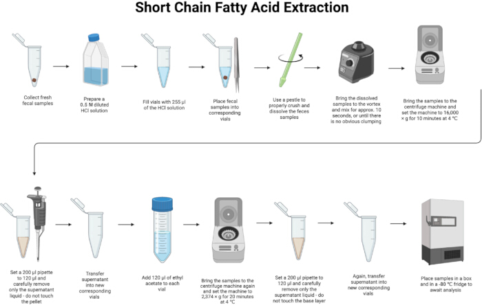
Figure 1: Diagram of protocol. Please click here to view a larger version of this figure.
1. Fecal sample collection and preparation
- Collect fresh fecal (~30 mg) samples in separate 1.5 mL microcentrifuge tubes. Record the exact fecal input mass for each sample to allow accurate normalization of SCFA concentrations.
NOTE: Ensure retrieval includes clean vials, gloves, and tweezers for each sample. All glassware and instruments must be autoclaved prior to use. This protocol has been validated for fecal input masses of approximately 20–50 mg per sample, with ~30 mg used as a representative input. Extraction volumes are kept constant within this range. - Place and label the necessary number of centrifuge vials corresponding to the samples.
- Prepare diluted hydrochloric acid (0.5 M; pH 1–2) by slowly adding concentrated HCl (12 M, ACS reagent grade) to ultrapure water.
CAUTION: HCl is corrosive; it can cause severe burns to skin, eyes, and respiratory tract. Dangerous to inhale. - Thaw samples at room temperature for 30–45 min, or until completely thawed.
2. Sample homogenization and acidification
- Add 255 µL of the 0.5 M HCl solution to each centrifuge vial.
- Take a new vial and fill it with 70% ethanol to use for cleaning tweezers. Clean the tweezers by dipping them into the ethanol and then drying them on a clean paper towel.
CAUTION: Ethanol is flammable and it can irritate skin and eyes; inhalation of vapors may cause dizziness. - Set up an ethanol bath for the pestles by placing pestles into a container and filling the container with 70% ethanol until all pestles are completely submerged.
- Retrieve the correct sample from the sample box. Take the lid off the vial and remove one sample (~ 30 mg) of feces.
- Place the fecal samples into the corresponding centrifuge vials.
- Once all the samples are placed into the correct centrifuge vials, remove a pestle from the ethanol bath. Dry the pestle with a clean paper towel.
NOTE: Use a new pestle for each sample. - Use the pestles to properly crush and dissolve the feces samples, see Figure 2. Grind the fecal sample until a visually homogeneous suspension is obtained, with no visible solid fragments.
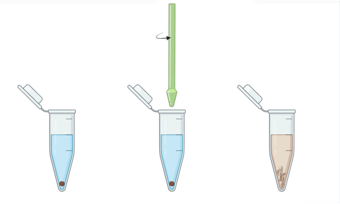
Figure 2: Grinding of fecal samples using a pestle. Freshly collected feces are ground to achieve a uniform consistency for SCFA analysis. Please click here to view a larger version of this figure.
- Place the used pestles in an empty box to be washed later.
- Vortex the samples using a pulse-type vortex mixer at maximum speed for 10 s, with tubes tightly capped.
NOTE: Ensure that the centrifuge vials are tightly covered to prevent spilling the mixture.
3. SCFA extraction and phase separation
- Centrifuge the samples at 16,000 × g for 10 min at 4 °C, ensuring proper balancing of the centrifuge.
- After starting the centrifuge, label the new centrifuge vials corresponding to the samples. Remove the samples from the centrifuge once centrifugation is complete.
- Set the 200 µL pipette to 120 µL. Carefully collect 120 µL of the aqueous supernatant after centrifugation, avoiding disturbance of the pellet. Transfer the supernatant into the newly labeled centrifuge vials and discard the pellet.
- Add an equal amount (120 µL) of ethyl acetate into the newly labeled vials with the supernatant liquid. Repeat this for all samples.
CAUTION: Ethyl acetate is flammable and can irritate skin, eyes, and respiratory system. Avoid inhaling vapors and prolonged contact. - Bring the samples to the centrifuge again. Ensure the centrifuge is properly balanced and the rotor lid is secure. Spin at 2,374 × g for 20 min at 4 °C.
- After centrifugation, carefully collect 120 µL of the upper organic phase without disturbing the lower aqueous phase. Transfer the extracted SCFA-containing supernatant into new, labeled centrifuge vials.
NOTE: The collected volume may be less than 120 µL. If an emulsion is present at the interface, extend centrifugation or chill the samples on ice until clear phase separation is achieved. Procedural blank samples processed in parallel without fecal material may be included as an optional quality-control step to assess background levels of SCFAs introduced by reagents or handling. - Examine previous vials to ensure that the bottom layer was not accidentally drawn up.
NOTE: Steps involving acid addition and organic solvent handling should be performed rapidly, with tubes kept capped whenever possible, to minimize volatilization or evaporation losses.
4. Extract handling and storage
- Locate and properly label an extraction box, then place the extracted samples within it.
NOTE: Samples may be safely paused after organic phase collection by storing the extracts at −20 °C for up to 24 h in tightly capped tubes, then transfer to −80 °C for longer-term storage. - Place the box into a -80 °C freezer to await SCFA analysis.
NOTE: Because both ethyl acetate and SCFA are highly volatile, extracted samples should be analyzed promptly after storage at −80 °C. Extended long-term storage may compromise sample integrity.
5. Gas chromatography analysis
- Analyze the extracted SCFAs by GC using a fused-silica capillary column with a free fatty acid stationary phase at flow rates of 30, 300, and 20 mL/min for hydrogen, air, and nitrogen, respectively. Inject a 1 µL aliquot of the organic extract for analysis.
- For quantitative applications, incorporate internal standards and calibration procedures (e.g., 2-ethylbutyric acid) as described previously2.
NOTE: Detailed instrument-specific settings are provided in the referenced methods14.
Access restricted. Please log in or start a trial to view this content.
Results
The above protocol will have yielded extracted SCFA aliquots of 120 µL corresponding with the number of fecal samples from which they were derived. Successful execution of the extraction protocol is indicated by the presence of clearly resolved SCFA peaks at characteristic retention times with signal intensity above background noise. When performed as described, the protocol yields fecal SCFA profiles suitable for downstream quantitative comparison across samples. We quantified eight SCFAs: acetic acid, propionic acid, b...
Access restricted. Please log in or start a trial to view this content.
Discussion
This protocol contains several critical steps that ensure efficient and reproducible extraction of SCFAs from small-volume mouse fecal samples. The protocol employs a two-phase acid–solvent extraction system using defined volumes of diluted hydrochloric acid and ethyl acetate, which is tailored to the physicochemical properties of SCFAs as low-molecular-weight, weak organic acids. Acidification shifts SCFAs into their protonated form, reducing ionic interactions with the fecal matrix and facilitating their partitio...
Access restricted. Please log in or start a trial to view this content.
Disclosures
The authors have no conflicts of interest to declare.
Acknowledgements
We gratefully acknowledge the funding support for this study provided by the National Institutes of Health through R21AG065606 and R21AG08176 awarded to Yiying Zhang, as well as support from the Borten Family Foundation and the Tang Family gift to Yiying Zhang.
Access restricted. Please log in or start a trial to view this content.
Materials
| Name | Company | Catalog Number | Comments |
|---|---|---|---|
| Centrifuge | ThermoFisher | 75003424 | "Sorvall Legend Micro 21R Microcentrifuge" |
| Centrifuge vials | Fisher Scientific | 05-408-129 | |
| Microtube Rack | Millipore Sigma | HS29025F-5EA | |
| Ethyl acetate | ThermoFisher | 022912.K2 | HPLC Grade, ≥99.5% |
| Green pestles | Fisher Scientific | 50-189-9195 | |
| HCl | Fisher Scientific | SA49 | ACS Certified (10 N, stock solution); diluted to 0.5 M for use |
| 1000 μL pipette | USA Scientific | 7110-1000 | |
| 1000 μL pipette tips | USA Scientific | 1111-2821 | |
| Ethanol | Fisher Scientific | BP82011 | Molecular Biology Grade, 70% solution |
| 200 μL pipette | USA Scientific | 7100-2200 | |
| 200 μL pipette tips | USA Scientific | 1110-1800 | |
| Tweezers | ThermoFisher | 046638.KT | |
| Vortex | Fisher scientific | 50-136-7909 | "Research Products International Corp VORNADO Miniature Vortex Mixer, Purple Cup" |
References
- Koh, A., De Vadder, F., Kovatcheva-Datchary, P., Bäckhed, F. From dietary fiber to host physiology: short-chain fatty acids as key bacterial metabolites. Cell. 165 (6), 1332-1345 (2016).
- Zhang, Y., et al. Transmission of Alzheimer's disease-associated microbiota dysbiosis and its impact on cognitive function: evidence from mice and patients. Mol Psychiatry. 28 (10), 4421-4437 (2023).
- Sun, Y., et al. PYY-mediated appetite control and obesity alleviation through short-chain fatty acid-driven gut-brain axis modulation by Lacticaseibacillus rhamnosus HF01 isolated from Qula. J Dairy Sci. 108 (8), 7960-7978 (2025).
- He, J., et al. Short-chain fatty acids and their association with signalling pathways in inflammation, glucose and lipid metabolism. Int J Mol Sci. 21 (17), 6356(2020).
- Fock, E., Parnova, R. Mechanisms of blood-brain barrier protection by microbiota-derived short-chain fatty acids. Cells. 12 (4), 657(2023).
- Kachoueiyan, F., et al. Butyrate: a key mediator of gut-brain communication in Alzheimer's disease. Metab Brain Dis. 40 (5), 189(2025).
- Kim, K. S., Lee, Y., Chae, W., Cho, J. Y. An improved method to quantify short-chain fatty acids in biological samples using gas chromatography-mass spectrometry. Metabolites. 12 (6), 525(2022).
- Ueyama, J., et al. Freeze-drying enables homogeneous and stable sample preparation for determination of fecal short-chain fatty acids. Anal Biochem. 589, 113508(2020).
- Papanicolas, L. E., et al. Bacterial viability in faecal transplants: which bacteria survive. EBioMedicine. 41, 509-516 (2019).
- Yao, L., et al. Quantitative analysis of short-chain fatty acids in human plasma and serum by GC-MS. Anal Bioanal Chem. 414 (15), 4391-4399 (2022).
- Zhang, C., Liu, A., Zhang, T., Li, Y., Zhao, H. Gas chromatography detection protocol of short-chain fatty acids in mice feces. Bio Protoc. 10 (13), e3672(2020).
- Zha, X., et al. Microbiota-derived lysophosphatidylcholine alleviates Alzheimer's disease pathology via suppressing ferroptosis. Cell Metab. 37 (1), 169-186.e9 (2025).
- Li, L. J., et al. Neuronal double-stranded DNA accumulation induced by DNase II deficiency drives tau phosphorylation and neurodegeneration. Transl Neurodegener. 13 (1), 39(2024).
- Zhou, H., et al. Glia-to-neuron conversion by CRISPR-CasRx alleviates symptoms of neurological disease in mice. Cell. 181 (3), 590-603.e16 (2020).
- Zhao, G., Nyman, M., Jönsson, J. A. Rapid determination of short-chain fatty acids in colonic contents and faeces of humans and rats by acidified water-extraction and direct-injection gas chromatography. Biomed Chromatogr. 20 (8), 674-682 (2006).
Access restricted. Please log in or start a trial to view this content.
Reprints and Permissions
