This paper examines current literature surrounding the use of peroxisome proliferator-activated receptor γ (PPAR-γ) agonists in the treatment of cutaneous fibrosis. The rationale for repurposing pioglitazone is discussed, with a focus on the drug's dual anti-inflammatory and anti-fibrotic mechanisms. The literature on this topic encourages further clinical studies in this area.
A subscription to JoVE is required to view this content. Sign in or start your free trial.
Review Article
Pioglitazone in Skin Fibrosis: Mechanistic Rationale and Therapeutic Potential of Pioglitazone for Scarring Dermatoses
* These authors contributed equally
In This Article
Summary
Abstract
Cutaneous fibrosis – encompassing keloids, hypertrophic scars, localized scleroderma (morphea), and scarring alopecias – remains fundamentally undertreated, with conventional interventions frequently yielding incomplete therapeutic responses and high rates of recurrence. Pioglitazone, a Food and Drug Administration (FDA)-approved peroxisome proliferator-activated receptor γ (PPAR-γ) agonist traditionally utilized for glycemic control in type 2 diabetes mellitus, represents a highly compelling candidate for dermatologic repurposing. Beyond its canonical metabolic functions, robust target engagement of PPAR-γ by pioglitazone suppresses NF-κB–driven cytokine cascades, directly inhibits NOD-like receptor pyrin domain-containing protein 3 (NLRP3) inflammasome assembly, promotes macrophage polarization toward tissue-reparative phenotypes, and disrupts profibrotic TGF-β/SMAD signaling architectures. Consequently, the drug exerts a dual mechanism of action, simultaneously attenuating chronic inflammation while inhibiting pathological fibrogenesis. This review expands upon prior analyses by integrating recently published clinical and scientific data, specifically highlighting new translational milestones, including human registry data in progeroid syndromes and randomized controlled trials in cicatricial alopecia. A distinct focus is placed on advancements in localized delivery strategies, such as nanostructured lipid carriers and niosomes, that offer the potential to overcome pioglitazone's inherent physicochemical constraints and thereby maximize dermal target engagement while avoiding systemic toxicities like heart failure and bladder cancer. Furthermore, this analysis synthesizes recent molecular insights connecting fibrosis and inflammation via target engagement biomarkers (e.g., FABP4, CD36) and proposes actionable, biomarker-driven trial designs, including adaptive basket trials, to bridge current gaps in dermatology-specific translation. Demonstrating localized anti-fibrotic and anti-inflammatory efficacy with minimal systemic risk could establish pioglitazone as a mechanism-based, disease-modifying therapy for a broad spectrum of fibrotic and scarring dermatologic disorders.
Introduction
Cutaneous fibrosis represents a complex, heterogeneous spectrum of dermatologic disorders in which aberrant, sustained wound healing responses lead to the excessive deposition of Extracellular Matrix (ECM) proteins, profound architectural distortion, and functional impairment. Conditions such as keloids, hypertrophic scars, and localized scleroderma (morphea), alongside chronic inflammatory dermatoses like Hidradenitis Suppurativa (HS) and primary scarring alopecias, currently lack consistently reliable, disease-modifying, and target-specific therapies1. Conventional therapeutic options for keloids and hypertrophic scars – ranging from intralesional corticosteroid injections and aggressive surgical excision to silicone sheeting and localized pressure therapy – often yield highly variable clinical outcomes. Recurrence rates following these standard modalities can easily exceed 50% and are heavily dependent upon the specific anatomical site and the distinct intervention utilized1. Similarly, the standard therapeutic arsenal for morphea, which frequently relies upon ultraviolet phototherapy and broad systemic immunosuppressive agents like methotrexate or systemic corticosteroids, frequently leaves patients with residual sclerosis, permanent functional deficits, and a high propensity for clinical relapse2. Within the context of HS, the therapeutic landscape has recently expanded beyond long-term antibiotic administration and aggressive surgical debridement to include targeted monoclonal antibodies (biologics); however, even with these advanced, highly specific immunotherapies, a substantial demographic of patients fails to achieve satisfactory, long-term clinical remission, underscoring the significant, unmet clinical need for novel therapeutic approaches3. Furthermore, scarring alopecias, including lichen planopilaris (LPP) and frontal fibrosing alopecia (FFA), represent additional disorders characterized by chronic, destructive perifollicular inflammation and irreversible scarring that may theoretically benefit from metabolic and immunomodulatory interventions like pioglitazone therapy. Early case series and retrospective analyses involving LPP and FFA have demonstrated variable clinical benefits from the systemic administration of pioglitazone, highlighting the need for highly controlled, localized interventional trials to standardize these outcomes4.
Collectively, these varied dermatologic conditions share highly conserved pathogenic features, primarily characterized by Transforming Growth Factor-Beta (TGF-β)-driven myofibroblast activation, pathological ECM accumulation, and self-perpetuating chronic inflammatory signaling networks5,6,7. This shared biological architecture makes interventions capable of both limiting inflammatory cascades and fundamentally reprogramming fibrogenesis an appealing focus for advanced pharmacological study. Thiazolidinediones (TZDs), which act as potent Peroxisome Proliferator-Activated Receptor Gamma (PPAR-γ) agonists, are particularly attractive therapeutic candidates due to their proven capacity to intersect and downregulate both inflammatory and fibrogenic intracellular pathways simultaneously2,7,8,9. A continually expanding body of molecular evidence implicates PPAR-γ as a central regulator of fibroblast activation, collagen synthesis kinetics, and pro-inflammatory cytokine production2,5,10,11. Thus, targeting the PPAR-γ receptor with specific glitazones—compounds originally developed and optimized as insulin sensitizers for endocrinology—offers a highly rational, mechanism-based strategy to modulate the fibrotic cascade while simultaneously attenuating destructive inflammation across a wide variety of skin diseases.
By reviewing the available literature for new advances, insights, and clinical findings, we provide a comprehensive and up-to-date report on the potential use of pioglitazone in dermatologic conditions, with specific highlights on advancements in localized delivery strategies designed to overcome pioglitazone's physicochemical constraints12,13. We also highlight new findings demonstrating how pioglitazone's anti-fibrotic and anti-inflammatory activity can be actively monitored through the measurement of cutaneous PPAR-γ target engagement biomarkers such as Fatty Acid Binding Protein 4 (FABP4) and Cluster of Differentiation 36 (CD36), while also outlining proposed biomarker-driven, adaptive basket trial designs that may facilitate future study in this area14,15. These novel approaches to therapeutic assessment and study design offer a rational method to address persistent gaps in dermatology-specific translation through appropriately designed clinical trials. Finally, this analysis updates the risk-benefit paradigm by contextualizing recent human trial data against the systemic risks of bladder cancer and heart failure, emphasizing the benefits of non-systemic administration.
The practical applications of these findings are substantial and highly relevant to modern dermatologic practice. If successfully formulated for localized, transdermal delivery, topical pioglitazone could serve as an effective, non-immunosuppressive, and cost-conscious therapeutic option for a variety of fibrosing skin diseases, such as keloids, morphea, and scarring alopecias16,17. Though therapeutic limitations do exist, primarily related to the inherent physicochemical challenges of formulating the highly lipophilic, crystalline form of pioglitazone into an effective topical matrix, recent advances in biopharmaceutical engineering show promise that such formations are achievable18,19. And while epidemiology studies have demonstrated a small but significant risk of fluid retention, congestive heart failure, and dose-dependent bladder cancer associated with systemic TZD exposure, recent clinical trials have demonstrated a favorable safety profile for pioglitazone when used in the treatment of cutaneous diseases20,21.
Access restricted. Please log in or start a trial to view this content.
Review and Perspective
Drug profile
Pioglitazone, alongside structurally related glitazone compounds such as rosiglitazone and troglitazone, belongs to the thiazolidinedione (TZD) class of orally active PPAR-γ agonists initially developed and approved for the management of type 2 diabetes mellitus. In the context of metabolic disease, these agents act to enhance systemic insulin sensitivity by modulating the transcriptional activity of numerous genes involved in adipogenesis, cellular lipid transport, and systemic glucose metabolism. However, the pharmacological influence of PPAR-γ activation extends far beyond isolated glycemic control. Activation of this nuclear hormone receptor heavily antagonizes the nuclear factor kappa-light-chain-enhancer of activated B cells (NF-κB) signaling cascade, systematically suppresses the assembly of the NLR family pyrin domain containing 3 (NLRP3) inflammasome, and directs macrophage polarization toward tissue-reparative, anti-inflammatory M2 phenotypes2,22,23.
Early in vitro laboratory investigations and preclinical animal models have demonstrated that PPAR-γ agonists significantly suppress TGF-β–driven profibrotic gene transcription programs and actively reduce dermal thickness and collagen deposition in bleomycin-induced murine models of scleroderma8,9,24,25. In isolated human keloid fibroblasts, earlier iterations of TZDs, such as troglitazone, were shown to inhibit AXL receptor tyrosine kinase signaling, leading to significantly reduced collagen-related outputs and decreased cellular proliferation7. Clinically, systemic pioglitazone has demonstrated substantial efficacy signals in inflammatory dermatoses, with meta-analyses of randomized controlled trials (RCTs) showing statistically significant improvements in the Psoriasis Area and Severity Index (PASI) scores of treated patients3,20. Furthermore, early clinical case series have reported variable, yet promising, clinical benefits in the management of severe scarring alopecias like LPP21,26. This data forms the foundational anti-fibrotic rationale, collectively supporting the premise that glitazones can successfully modulate key pathogenic drivers of dermal fibrosis.
Mechanistic hypothesis
Anti-inflammatory pathways
The anti-inflammatory properties of PPAR-γ agonists are mediated through their interaction with multiple intersecting intracellular pathways governing both the innate and adaptive immune responses (Figure 1). The primary mechanism of action involves direct interference with the NF-κB signaling axis. By physically transrepressing NF-κB, PPAR-γ activation leads to the transcriptional repression of key pro-inflammatory cytokines, including Tumor Necrosis Factor-alpha (TNF-α), Interleukin-1 beta (IL-1β), and Interleukin-6 (IL-6)2,22,23. Within innate immune cells, particularly tissue-resident macrophages, pioglitazone effectively inhibits both the priming and subsequent activation steps of the NLRP3 inflammasome complex. This inhibition directly reduces the processing and extracellular secretion of IL-1β and interleukin-18 (IL-18), while simultaneously suppressing the activation of caspase-1, thereby breaking the cycle of chronic autoinflammation22,23.
Furthermore, recent molecular profiling indicates that PPAR-γ activation is heavily reliant on the induction of lipid chaperone proteins. The upregulation of FABP4 and the scavenger receptor CD36 facilitates organized intracellular lipid uptake, driving macrophages away from a classically activated, tissue-destructive M1 state and promoting an M2-like reparative phenotype. These M2 macrophages are characterized by the secretion of key anti-inflammatory mediators, such as Interleukin-10 (IL-10) and Transforming Growth Factor β3 (TGF-β3), which actively support the resolution of acute tissue injury and halt chronic inflammatory cycling2,22,23.
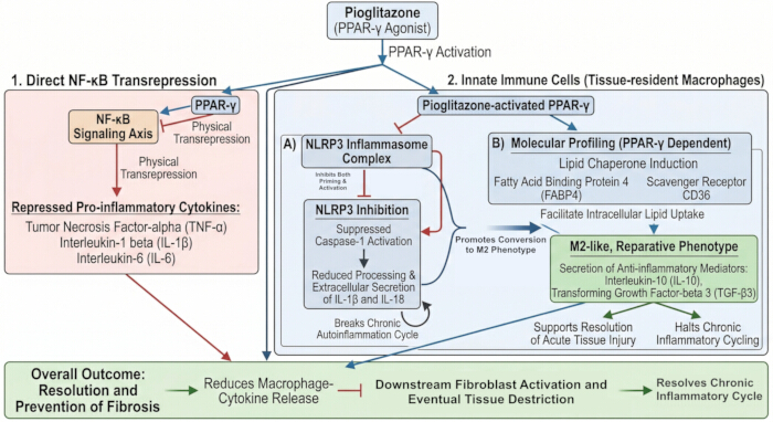
Figure 1: Mechanistic map of anti-inflammatory pathways induced by PPAR-γ receptor activation. The various inflammatory pathways activated by PPAR-γ agonists are shown, with specific focus on the role of PPAR-γ in NF-κB transrepression and the induction of cutaneous macrophages into M2-like reparative phenotypes. Please click here to view a larger version of this figure.
Anti-fibrotic pathways
PPAR-γ agonists also possess direct anti-fibrotic properties by intercepting various signaling cascades responsible for the pathological development of fibrosis. A principal mechanism by which pioglitazone inhibits fibrogenesis is through the direct and indirect interference of TGF-β signaling, encompassing both SMAD-dependent and SMAD-independent intracellular routes (Figure 2). In TGF-β/SMAD signaling, the binding of TGF-β to its cognate cell surface receptors induces the immediate phosphorylation of SMAD family member 2 (SMAD2) and SMAD family member 3 (SMAD3), which subsequently form heterocomplexes with SMAD family member 4 (SMAD4) and translocate to the nucleus to drive the transcription of profibrotic genes10,27,28. PPAR-γ agonists act to disrupt this signaling pathway by both actively competing for essential SMAD binding partners and upregulating the expression of SMAD family member 7 (SMAD7), an inhibitory SMAD protein that provides negative feedback to directly antagonize further SMAD2/3 phosphorylation28. Furthermore, PPAR-γ agonists also possess the ability to inhibit the recruitment of the transcriptional coactivator p300 to the DNA-bound complex, effectively neutralizing TGF-β-mediated fibroblast activation29.
Recent literature has also identified the vital role of PPAR-γ in modulating SMAD-independent pathways, most notably by disrupting the crosstalk between TGF-β and the Janus kinase and signal transducer and activator of transcription (JAK/STAT) signaling axis2,6,30,31. The attenuation of these combined signaling modalities ultimately curtails fibroblast-to-myofibroblast differentiation, resulting in a decrease in alpha-smooth muscle actin (α-SMA) expression and reduced cellular contractility. Consequently, pioglitazone inhibits the synthesis of Type I and Type III collagen at the transcriptional level, thereby limiting excessive ECM accumulation2,5,6,7,9,25. Moreover, emerging molecular evidence suggests that PPAR-γ plays a key role in modulating the bidirectional mechanical and paracrine crosstalk between dermal white adipose tissue and the overlying dermal fibroblasts, with recent data suggesting that the targeted activation of PPAR-γ stimulates localized adipogenesis within the deeper dermis, which inherently antagonizes and attenuates the uncontrolled proliferation of fibroblasts and the subsequent deposition of fibrotic tissue. This results in a tissue-regenerative effect rather than mere biochemical suppression (Figure 2)11,32,33,34.
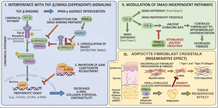
Figure 2: Mechanistic map of anti-fibrotic actions of PPAR-γ agonists. The anti-fibrotic activity of PPAR-γ agonists is shown, summarizing the three main anti-fibrotic outcomes resulting from PPAR-γ activation (interference with TGF-β/SMAD-dependent signaling, modulation of SMAD-independent pathways, and facilitation of adipocyte-fibroblast crosstalk). Please click here to view a larger version of this figure.
Skin-specific implications
In addition to its well-documented systemic effects on deep dermal fibroblasts and infiltrating immune cells, PPAR-γ activation also carries beneficial implications for the epidermis and its associated structures (Figure 3). Within the epidermis, PPAR-γ agonists have demonstrated the potential to enhance terminal differentiation of keratinocytes and actively restore the expression of barrier-related genes, specifically those encoding filaggrin, loricrin, and involucrin35,36,37. A functional epidermal barrier is essential for skin homeostasis as it helps limit water loss and protects against environmental irritants, pathogens, and allergens38,39. The epidermal barrier of the skin is largely dependent on the integrity of the cornified envelope, a crosslinked structure composed of key barrier proteins. Deficiencies in these proteins are a hallmark of various inflammatory skin diseases, and in vivo models have demonstrated that topical PPAR-γ activators stimulate a robust expression of these proteins35,36,37,38,39,40. Additionally, murine models have shown that the damaging effects of topical glucocorticoids, which are known to cause skin atrophy and impair barrier function, may be attenuated with the co-application of a PPAR-γ agonist, which acts to normalize the expression of filaggrin, loricrin, and involucrin36.
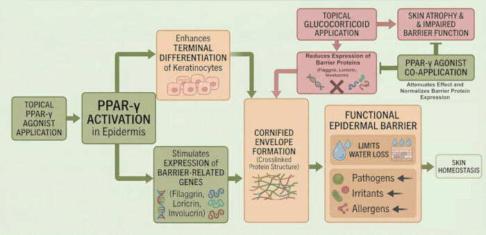
Figure 3: Mechanistic map of PPAR-γ activation in the epidermis. An overview of PPAR-γ activity in the epidermis is provided, summarizing the ability of PPAR-γ agonists to enhance terminal keratinocyte differentiation, attenuate the harmful effects of topical glucocorticoid application, and ultimately improve epidermal barrier function. Please click here to view a larger version of this figure.
Within the hair follicle microenvironment, physiological PPAR-γ activation is required to preserve immune privilege, reduce spontaneous follicular keratinocyte apoptosis, and dampen local, destructive cytokine production (Figure 4)4,41,42. The anatomical bulge region, which houses the epithelial hair follicle stem cell (eHFSC) population, is recognized as a site of relative immune privilege within hair follicles and is characterized by the downregulation of major histocompatibility complex (MHC) molecules and the local production of immunosuppressive factors41,42. The collapse of this localized immune privilege appears to be the central initiating event in the pathogenesis of LPP, FFA, and other primary scarring alopecias4,41,43. Advanced murine models featuring a targeted, keratinocyte-specific deletion of PPAR-γ within the hair follicle bulge unequivocally developed progressive skin pathology phenotypically mirroring human LPP43,44. The mechanism by which PPAR-γ preserves immune privilege appears to be indirect but critical. Defective PPAR-γ signaling leads to impaired lipid metabolism and peroxisome biogenesis within the sebaceous gland, resulting in the accumulation of pro-inflammatory lipids (Figure 4). These lipids may act as endogenous "danger signals" that trigger an inflammatory response and lead to the collapse of immune privilege4,43. Therefore, PPAR-γ activation does not create immune privilege directly but rather preserves it by maintaining the metabolic health and homeostasis of the pilosebaceous unit4,41. Furthermore, the broad anti-inflammatory effects exerted by PPAR-γ agonists help reduce follicular keratinocyte apoptosis and dampen cytokine production. These processes represent the downstream effector mechanisms that execute the destruction of the hair follicle following the collapse of immune privilege4,41,42. The inflammatory microenvironment that develops in scarring alopecia is a primary driver of apoptosis in the eHFSCs. By suppressing the production of these inflammatory mediators, PPAR-γ activation removes the principal extrinsic signal for apoptosis4,45.
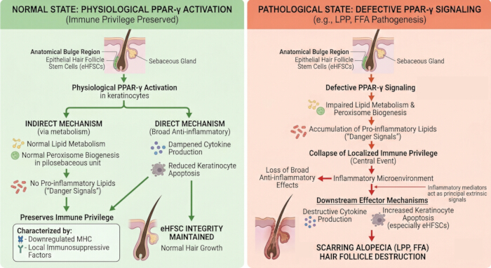
Figure 4: Role of PPAR- γ within hair follicle microenvironment. The normal hair follicle state, wherein immune privilege is preserved, is compared to the pathological state seen in scarring alopecia, demonstrating the critical role PPAR-γ plays in maintaining homeostasis within the hair follicle microenvironment. Please click here to view a larger version of this figure.
Target indications
Translational evidence and pathophysiological context
The translation of this preclinical data to the clinical dermatology setting requires rigorous scrutiny, particularly because much of the foundational anti-fibrotic data originates from internal organ models, such as hepatic or pulmonary fibrosis. Extrapolating therapeutic efficacy from hepatic internal organ models directly to dermal fibroblasts carries inherent limitations due to the vastly different biomechanical forces, the unique presence of dermal white adipose tissue, and the complex, stratified barrier dynamics specific to human skin46. However, new data has come to light that may help bridge this gap and move the focus from preclinical studies to real world clinical application.
In a 2024 analysis of the Japanese Werner Syndrome Registry – a rare, autosomal recessive progeroid syndrome characterized by severe, refractory skin ulcers, dermal sclerosis, and premature aging – patients receiving systemic pioglitazone demonstrated a statistically significant reduction in the development and persistence of severe fibrotic skin ulcers, independent of age and blood pressure variables47. This study offers robust data to support the hypothesis that pioglitazone actively enhances wound healing and suppresses fibrotic architectural breakdown in deeply compromised human skin. Furthermore, a 2022 clinical trial comparing oral pioglitazone to highly potent topical clobetasol lotion in 40 patients with Lichen Planopilaris (LPP) confirmed that pioglitazone safely and effectively reduced the Lichen Planopilaris Activity Index (LPPAI) over six months21. While not proven statistically superior to the high-potency topical steroid, pioglitazone was shown to possess biological activity in halting a primary human scarring dermatosis without the atrophogenic side effects of chronic steroid use.
Though the anti-fibrotic and anti-inflammatory properties of pioglitazone make it an appealing investigational drug for many dermatologic indications, the level of evidence for these indications varies greatly (Table 1). To appropriately contextualize these indications, we employed the Oxford Centre for Evidence-Based Medicine (OCEBM) 2011 Levels of Evidence framework to proportion clinical claims to the currently available data48.
| Category | Specific Indication | Rationale for Pioglitazone Use | OCEBM Level of Evidence |
| Fibrotic | Hypertrophic scars / keloids | PPAR-γ agonists (e.g., troglitazone, pioglitazone) suppress canonical TGF-β/SMAD-mediated collagen I/III synthesis, inhibit p300 coactivator recruitment, and reduce α-SMA expression and myofibroblast proliferation in isolated human keloid fibroblasts7,24. | Level 4/5 - In vitro mechanistic data and isolated clinical case reports7,24. |
| Fibrotic | Localized scleroderma (morphea) | Localized delivery of pioglitazone via PLGA nanoparticles or gel matrices severely attenuated dermal thickness and fibrosis in bleomycin-induced murine models. Mechanistically, it restores essential adipocyte–fibroblast crosstalk and limits ECM expansion8,11,32–34 | Level 5 - Preclinical animal models and mechanism-based reasoning. |
| Fibrotic | Refractory skin ulcers (e.g., Werner Syndrome) | Registry data indicates pioglitazone modulates inflammation, promotes cellular reparative phenotypes, and significantly reduces the incidence of severe, fibrotic skin ulcers in progeroid human subjects48. | Level 3/4 - Non-randomized registry/cohort data. |
| Inflammatory with scarring | Lichen planopilaris (LPP) / FFA / CCCA | PPAR-γ dysfunction is the central recognized trigger for the collapse of follicular immune privilege. A randomized clinical trial demonstrated pioglitazone effectively lowers disease activity indices (LPPAI) comparably to conventional topical corticosteroids4,21,41-45. | Level 2 - Randomized controlled trial and robust mechanistic murine knock-out models. |
| Inflammatory with scarring | Hidradenitis suppurativa (HS) | Pioglitazone strictly modulates macrophage polarization toward an M2 reparative state and inhibits the NF-κB and NLRP3 inflammasome. pathways, which are the central biological drivers of chronic, deep-tissue inflammatory signaling in HS3,22,23. | Level 5 - Mechanism-based reasoning derived from deep immune profiling of the disease state. |
| Inflammatory (no primary fibrosis) | Psoriasis | Meta-analysis of RCTs demonstrate that systemic pioglitazone significantly improves PASI scores. Immunohistochemistry of treated skin demonstrates a marked reduction in pro-inflammatory macrophage infiltration and cytokine burden20,51,52. | Level 1 - Systematic review and meta-analysis of randomized controlled trials. |
Table 1: Target indications and evidence framework for specific dermatoses. Potential dermatologic targets for pioglitazone repurposing are listed and grouped according to their key pathological characteristics, with brief summaries provided for the mechanistic rationale of each target indication. The OCEBM 2011 Level of Evidence is also given for each condition, with a brief justification for the assigned level.
Expanded rationales
Hypertrophic scars and keloids (OCEBM Level 4/5): The distinct pathophysiology of keloids is driven by an unremitting, exaggerated inflammatory response to acute cutaneous injury coupled with hyperactive TGF-β signaling. This results in highly disorganized collagen deposition extending beyond the original wound margins49. Cellular evidence indicates that TZDs suppress the pathological differentiation of fibroblasts by antagonizing the canonical TGF-β/SMAD pathway and interfering with AXL receptor tyrosine kinase signaling50. While large-scale clinical trial data for pioglitazone in human keloids remains sparse, the strong mechanistic suppression of collagen synthesis observed in isolated human keloid tissues provides a compelling theoretical basis for utilizing locally delivered pioglitazone formulations as an adjuvant to surgical excision to prevent high-rate recurrences7,24.
Localized scleroderma/morphea (OCEBM Level 5): Morphea is clinically characterized by an early, intense inflammatory phase driven largely by T helper 1 (TH1) and T helper 17 (TH17) cells, which subsequently shifts to a T helper 2 (TH2)-driven profibrotic phase featuring collagen crosslinking, tissue hardening, and the complete loss of dermal appendages16. Preclinical investigations utilizing nanoparticle-mediated localized delivery of pioglitazone in bleomycin-induced mouse models demonstrated a highly significant suppression of myofibroblast differentiation and a measurable reduction in dermal sclerosis8. Furthermore, pioglitazone's unique ability to stimulate localized adipogenesis directly counteracts the significant loss of subcutaneous fat often observed in deep and pansclerotic morphea32,33,34.
Scarring alopecias (OCEBM Level 2): In conditions such as LPP, FFA, and Central Centrifugal Cicatricial Alopecia (CCCA), the destruction of the eHFSC niche leads to permanent hair loss4,41,42,43. Human clinical trial data have shown that daily oral pioglitazone can effectively reduce the clinical signs of inflammation and patient-reported symptoms in LPP, operating with comparable efficacy to high-potency topical clobetasol21. By directly correcting the underlying lipid metabolism defects that disrupt follicular immune privilege, pioglitazone offers a disease-modifying intervention without the risks associated with high-potency corticosteroids.
Hidradenitis suppurativa (OCEBM Level 5): HS involves intense, painful, recurrent deep dermal nodules and tunneling sinus tracts driven by follicular occlusion and hyperactive innate immune signaling, predominantly mediated via the NLRP3 inflammasome and the overproduction of IL-1β. Because pioglitazone is a recognized inhibitor of NLRP3 assembly and actively drives the switch of macrophages from a tissue-destructive M1 to a tissue-reparative M2 state, it theoretically intersects the inflammatory axes responsible for HS pathogenesis2,22,23.
Psoriasis (OCEBM Level 1): Psoriasis is a chronic inflammatory skin disease characterized by the hyperproliferation and aberrant differentiation of epidermal keratinocytes and is often associated with comorbid systemic metabolic derangements and inflammatory disorders, such as metabolic syndrome and insulin resistance. PPAR-γ receptors are prominently expressed on epidermal keratinocytes, where their activation normalizes aberrant cell differentiation and suppresses key pro-inflammatory cytokines that directly drive psoriasis pathogenesis. Multiple meta-analyses of randomized controlled trials have concluded that daily doses of systemic pioglitazone yield a statistically significant reduction in Psoriasis Area and Severity Index (PASI) scores compared to placebo, without a significant increase in total adverse events20,51,52.
Therapeutic modalities and delivery
Physicochemical constraints of pioglitazone relevant to skin penetration
While the mechanistic rationale for deploying pioglitazone in cutaneous fibrosis is strong, significant hurdles must be overcome to transform the raw active pharmaceutical ingredient into an effective topical therapeutic formulation. Pioglitazone hydrochloride is defined by low aqueous solubility paired with high membrane permeability53,54. The molecule is highly lipophilic, possessing an unfavorable partition coefficient (LogP), and exists natively as a rigid, highly stable crystalline structure with a high melting point53,54. In the context of topical drug application, the stratum corneum acts as a selectively permeable biological barrier that must be overcome to achieve effective drug delivery. Ideal topical drug candidates typically require a LogP between 1 and 3, low molecular weight, and sufficient aqueous solubility to easily partition out of the delivery vehicle and traverse the alternating hydrophilic/lipophilic layers of the epidermis55. Pioglitazone's significant hydrophobicity and poor dissolution kinetics ultimately result in low-efficiency skin permeation when conventionally incorporated into standard creams or ointments56.
Realistic formulation approaches for localized delivery
To successfully circumvent these physicochemical constraints, recent advancements in pharmaceutical engineering have led to the development of several innovative nanoparticle-mediated formulation approaches applicable to pioglitazone13.
Niosomes: Niosomes are non-ionic surfactant-based synthetic lipid vesicles that securely encapsulate highly hydrophobic drugs within their structured lipid bilayers57. Recent formulation utilizing a precise structural matrix successfully generated niosomal suspensions of pioglitazone with an ideal, uniform nanoparticle size, a strong electronegative zeta potential, and a high maximum drug entrapment efficiency, which facilitates a favorable drug release profile for transdermal delivery13. The drug delivery profile plays a pivotal role in determining the success of topical formulations by directly influencing skin penetration. Profiles that demonstrate a controlled or sustained release allow the drug to maintain a consistent concentration gradient across skin layers, which is the driving force for passive diffusion. The resulting gradient facilitates deep skin penetration and prolonged therapeutic action13.
Nanostructured lipid carriers (NLCs) and solid lipid nanoparticles (SLNs): These advanced delivery systems utilize a highly calibrated blend of solid and liquid lipids to create an imperfect crystal lattice. This architectural irregularity vastly improves overall drug loading capacity and protects the encapsulated drug from premature chemical degradation12. Pioglitazone-loaded NLCs have demonstrated highly favorable biphasic release kinetics, with an initial rapid burst to saturate the epidermal barrier followed by prolonged, controlled release, thereby drastically enhancing transdermal bioavailability compared to crude free drug suspensions12.
Nanoemulsions and poly(lactic-co-glycolic acid) (PLGA) nanoparticles: Nanoemulsions, utilizing customized oil-in-water pseudo-ternary systems, drastically lower the surface tension of the skin, maximizing thermodynamic activity and transdermal drug flux58. Furthermore, the single emulsion-solvent evaporation method has been highly effective in encapsulating pioglitazone within biodegradable PLGA nanospheres, which have been shown in murine models to safely deposit the active drug into the deep dermis following localized subcutaneous injection8.
Human tolerability and safety of lipid-based nanoparticles
The safety and tolerability of any novel therapeutic approach must be carefully examined before transitioning to the clinical setting. While ongoing evaluation is required to fully establish the safety profile of these advanced nanoformulations, the molecular composition and early data strongly suggest a safe risk profile for human use. Most lipid-based nanosystems are formulated utilizing specific excipients that hold official "Generally Recognized As Safe" (GRAS) status from regulatory agencies, and in vitro safety assessments utilizing immortalized human keratinocytes and primary human fibroblasts have repeatedly demonstrated high cellular viability and a lack of severe cytotoxicity when directly exposed to these nanocarriers59. Furthermore, the specific nanosystems discussed above do not utilize Polyethylene Glycolated (PEGylated) lipids, which have been associated with significant adverse events, including hypersensitivity and anaphylactoid reactions. PEGylated lipids are typically added to intravenous liposomes to improve immune evasion and thereby increase systemic half-life. However, topical NLCs, SLNs, and niosomes utilize different surfactants aimed at improving topical drug delivery rather than increasing drug half-life59,60. Nevertheless, there remains a notable lack of long-term human tolerability data regarding the continuous application of such nanocarriers. As such, additional study in this area is required before a comprehensive risk assessment can be made.
Practicality, safety, and systemic exposure risks of proposed modalities
The value of developing an effective topical formulation is not limited to a theoretical higher local efficacy, either, but may also serve to limit the risk of systemic toxicities associated with oral pioglitazone. Although clinical trials examining systemic pioglitazone in the treatment of cutaneous diseases such as psoriasis and LPP did not find a significant increase in total adverse events, the long-term management often required for fibrosing dermatoses may expose otherwise metabolically healthy dermatologic patients to unnecessary oncologic and cardiovascular risks if treated with oral pioglitazone61,62,63,64,65,66. Epidemiologic studies have indicated a modest, but statistically significant increase in bladder cancer risk associated with chronic use, particularly in patients exposed to cumulative doses exceeding 28,000 mg or utilizing the systemic drug continuously for more than 1 to 2 years62,63,64,65,66. However, data regarding bladder cancer risk is conflicting; much of the evidence suggesting increased risk comes from meta-analyses of clinical trial data, whereas many large, long-term observational studies have failed to find statistically significant associations62,63,64,65,66.
Biomarker integration and standardized pharmacokinetic (PK) and pharmacodynamic (PD) endpoints
Though a localized approach offers an attractive solution to minimize the risks of systemic exposure, a reliable method of measuring both therapeutic activity and systemic spill over must be implemented into human-level trials to ensure topical formulations achieve therapeutic effect while mitigating possible systemic side effects. To account for this, early phase human skin studies should employ a rigorous PK and PD biomarker framework as part of their study design. Several methodologies have been developed that facilitate this process, such as tape-stripping (dermatopharmacokinetics), which allows for an accurate quantification of stratum corneum drug partitioning, and dermal open flow microperfusion (dOFM), which can directly measure unbound, pharmacologically active pioglitazone concentrations within the dermal interstitial fluid over continuous time points67,68,69. Peripheral blood sampling may also be implemented to ensure that systemic plasma concentrations remain negligible70.
Furthermore, the implementation of validated PD endpoints offers an effective method to determine whether topically therapy successfully activates the PPAR-γ receptor inside target cells. Current, state-of-the-art molecular profiling dictates that specific downstream genetic targets of PPAR-γ activation, including the expression of FABP4, the lipid scavenger receptor CD36, and Perilipin-2 (PLIN2), can be accurately quantified using transcriptomics in serial skin biopsies taken before and after treatment14. A statistically significant increase in these specific lipid chaperone markers provides molecular evidence of cellular target engagement in the deep dermis71. Additionally, recent studies have demonstrated that systemic and localized measurements of adiponectin, a surrogate marker of total PPAR-γ activity, correlate with skin thickness in fibrotic disorders, such as Systemic Sclerosis, and may be utilized to rapidly assess PPAR-γ activation72,73.
Contextualizing pioglitazone within the broader therapeutic landscape
The dermatologic landscape for treating severe inflammatory and fibrosing disorders is currently dominated by two drug classes: targeted monoclonal antibodies (biologics) and small-molecule JAK inhibitors74,75. When contextualizing the proposed use of pioglitazone against these agents, distinct mechanistic and practical advantages and disadvantages emerge. Biologics (e.g., dupilumab, tralokinumab) are highly specific, generally considered safe for long-term continuous use, and demonstrate excellent overall drug survival rates75. However, they also possess distinct disadvantages in that they require parenteral administration, are exceptionally costly, frequently act slowly over many months, and are generally restricted to modulating purely inflammatory pathways rather than directly reversing established, hardened fibrosis75. In contrast, small-molecule JAK inhibitors (e.g., upadacitinib, baricitinib) easily penetrate cells topically or orally and provide fast, early clinical responses by simultaneously blocking multiple inflammatory cytokine receptors74,75. Importantly, the JAK/STAT pathway strongly crosstalks with the TGF-β axis, granting JAK inhibitors highly significant anti-fibrotic potential in distinct models of scleroderma and keloids30. However, the use of oral JAK inhibitors is associated with black-box safety warnings mandated by the FDA regarding an increased risk of serious opportunistic infections, major adverse cardiovascular events, venous thromboembolism, and incident malignancy76,77.
Within this modern therapeutic paradigm, a topically formulated pioglitazone represents a unique alternative. By specifically targeting fundamental cellular metabolism and lipid homeostasis to indirectly induce broad immune tolerance and halt structural fibrosis, pioglitazone sidesteps the direct systemic immunosuppression characteristic of JAK inhibitors21. As a highly characterized, low-cost, off-patent small molecule, pioglitazone has the potential to offer dual anti-inflammatory and anti-fibrotic efficacy if it can be successfully formulated into a highly penetrative localized delivery system. This approach theoretically bypasses both the high costs of biologics and the severe systemic toxicities of oral JAK inhibition.
Access restricted. Please log in or start a trial to view this content.
Conclusions
Pioglitazone possesses a robust, highly characterized anti-fibrotic and anti-inflammatory molecular profile with potential to extend its clinical utility beyond traditional glycemic control. By modulating the transcriptional networks that govern fibroblast-to-myofibroblast differentiation, altering macrophage polarization through lipid chaperone (FABP4/CD36) mechanics, and directly reinforcing epithelial barrier function, pioglitazone holds a unique capacity to actively arrest or potentially reverse the aberrant wound-he...
Access restricted. Please log in or start a trial to view this content.
Disclosures
No Disclosures.
References
- Ogawa, R. The most current algorithms for the treatment and prevention of hypertrophic scars and keloids: a 2020 update of the algorithms published 10 years ago. Plast Reconstr Surg. 149 (1), 79e-94e (2022).
- Dantas, A. T., et al. The role of PPAR gamma in systemic sclerosis. PPAR Res. 2015, 124624 (2015).
- Krueger, J. G., et al. Hidradenitis suppurativa: new insights into disease mechanisms and an evolving treatment landscape. Br J Dermatol. 190 (2), 149-162 (2024).
- Harnchoowong, S., Suchonwanit, P. PPAR-γ agonists and their role in primary cicatricial alopecia. PPAR Res. 2017, 2501248 (2017).
- Wei, J., et al. PPARγ downregulation by TGFß in fibroblast and impaired expression and function in systemic sclerosis: a novel mechanism for progressive fibrogenesis. PLoS One. 5 (11), e13778 (2010).
- Ghosh, A. K., et al. Disruption of transforming growth factor beta signaling and profibrotic responses in normal skin fibroblasts by peroxisome proliferator-activated receptor gamma. Arthritis Rheum. 50 (4), 1305-1318 (2004).
- Zhu, H. Y., et al. Peroxisome proliferator-activated receptor-γ agonist troglitazone suppresses transforming growth factor-β1 signaling through miR-92b upregulation–inhibited Axl expression in human keloid fibroblasts. Am J Transl Res. 8 (8), 3460-3470 (2016).
- Kanemaru, M., et al. Nanoparticle-mediated local delivery of pioglitazone attenuates bleomycin-induced skin fibrosis. J Dermatol Sci. 93 (1), 41-49 (2019).
- Milam, J. E., et al. PPAR-gamma agonists inhibit profibrotic phenotypes in human lung fibroblasts and bleomycin-induced pulmonary fibrosis. Am J Physiol Lung Cell Mol Physiol. 294 (5), L891-L901 (2008).
- Wei, J., Bhattacharyya, S., Jain, M., Varga, J. Regulation of matrix remodeling by peroxisome proliferator-activated receptor-γ: a novel link between metabolism and fibrogenesis. Open Rheumatol J. 6, 103-115 (2012).
- Korman, B., et al. Adipocyte-specific repression of PPAR-gamma by NCoR contributes to scleroderma skin fibrosis. Arthritis Res Ther. 20 (1), 145 (2018).
- Faiz, S., et al. Pioglitazone-loaded nanostructured lipid carriers: in-vitro and in-vivo evaluation for improved bioavailability. J Drug Deliv Sci Technol. 79, 104041 (2023).
- Kyadalwar, S., et al. Development and characterization of a nanocomposed pioglitazone hydrochloride-loaded niosomal suspension for targeted drug delivery. J Bio X Res. 8, (2025).
- Yorek, M., et al. FABP4-mediated lipid accumulation and lipolysis in tumor-associated macrophages promote breast cancer metastasis. Elife. 13, RP101221 (2024).
- Volc, S., et al. A phase 2b basket trial approach to treat multiple rare and fibrotic skin diseases. Front Med (Lausanne). 12, 1637040 (2025).
- Papara, C., et al. Morphea: the 2023 update. Front Med (Lausanne). 10, 1108623 (2023).
- Fett, N. M. Morphea: evidence-based recommendations for treatment. Indian J Dermatol Venereol Leprol. 78, 135-141 (2012).
- . Pioglitazone, CID 4829 Available from: https://pubchem.ncbi.nlm.nih.gov/compound/Pioglitazone (2026)
- Abdelkader, D. H., et al. Pioglitazone repurposing via in-situ gelling system: an effective topical strategy for wound management. J Drug Deliv Sci Technol. 106, 106723 (2025).
- Chen, P., et al. The efficacy and safety of pioglitazone in psoriasis vulgaris: a meta-analysis of randomized controlled trials. Medicine (Baltimore). 99 (32), e21549 (2020).
- Lajevardi, V., et al. Efficacy and safety of oral pioglitazone in the management of lichen planopilaris in comparison with clobetasol: a randomized clinical trial. Dermatol Ther. 35 (11), e15868 (2022).
- Yang, C. C., et al. Inhibitory effect of PPARγ on NLRP3 inflammasome activation. Theranostics. 11 (5), 2424-2441 (2021).
- Wang, Y., et al. Pioglitazone ameliorates glomerular NLRP3 inflammasome activation in apolipoprotein E knockout mice with diabetes mellitus. PLoS One. 12 (7), e0181248 (2017).
- Zhang, G. Y., et al. Troglitazone suppresses transforming growth factor-beta1-induced collagen type I expression in keloid fibroblasts. Br J Dermatol. 160 (4), 762-770 (2009).
- Antonelli, A., et al. Peroxisome proliferator-activated receptor γ agonists reduce cell proliferation and viability and increase apoptosis in systemic sclerosis fibroblasts. Br J Dermatol. 168 (1), 129-135 (2013).
- Peterson, E. L., et al. Response of lichen planopilaris to pioglitazone hydrochloride. J Drugs Dermatol. 18 (12), 1276-1279 (2019).
- Attia, S. H., Saadawy, S. F., El-Mahroky, S. M., Nageeb, M. M. Alleviation of pulmonary fibrosis by the dual PPAR agonist saroglitazar and breast milk mesenchymal stem cells via modulating TGFß/SMAD pathway. Naunyn Schmiedebergs Arch Pharmacol. 397 (8), 5953-5974 (2024).
- Ni, X. X., Li, X. Y., Wang, Q., Hua, J. Regulation of peroxisome proliferator-activated receptor-gamma activity affects the hepatic stellate cell activation and the progression of NASH via TGF-β1/Smad signaling pathway. J Physiol Biochem. 77 (1), 35-45 (2021).
- Zhu, M., et al. Anti-inflammatory effects of thiazolidinediones in human airway smooth muscle cells. Am J Respir Cell Mol Biol. 45 (1), 111-119 (2011).
- Wang, D., Wei, Y., Xu, L., Zhang, J. Crosstalk between the JAK2 and TGF-β1 signaling pathways in scleroderma-related interstitial lung disease targeted by baricitinib. Adv Rheumatol. 63 (1), 22 (2023).
- Jeong, J., et al. Development of adverse outcome pathway for PPARγ antagonism leading to pulmonary fibrosis and chemical selection for its validation: ToxCast database and a deep learning artificial neural network model-based approach. Chem Res Toxicol. 32 (6), 1212-1222 (2019).
- Hoerst, K., et al. Regenerative potential of adipocytes in hypertrophic scars is mediated by myofibroblast reprogramming. J Mol Med (Berl). 97 (6), 761-775 (2019).
- Plikus, M. V., et al. Regeneration of fat cells from myofibroblasts during wound healing. Science. 355 (6326), 748-752 (2017).
- Roh, H. C., et al. Adipocytes fail to maintain cellular identity during obesity due to reduced PPARγ activity and elevated TGFβ-SMAD signaling. Mol Metab. 42, 101086 (2020).
- Feingold, K. R., Jiang, Y. J. The mechanisms by which lipids coordinately regulate the formation of the protein and lipid domains of the stratum corneum: role of fatty acids, oxysterols, cholesterol sulfate and ceramides as signaling molecules. Dermatoendocrinol. 3 (2), 113-118 (2011).
- Demerjian, M., et al. Activators of PPARs and LXR decrease the adverse effects of exogenous glucocorticoids on the epidermis. Exp Dermatol. 18 (7), 643-649 (2009).
- Bastonini, E., et al. Effects of pioglitazone on the differentiation and inflammation in vitiligo keratinocytes. J Eur Acad Dermatol Venereol. 38 (7), e573-e575 (2024).
- Kim, B. E., Leung, D. Y. M. Significance of skin barrier dysfunction in atopic dermatitis. Allergy Asthma Immunol Res. 10 (3), 207-215 (2018).
- Dang, N. N., et al. Filaggrin silencing by shRNA directly impairs the skin barrier function of normal human epidermal keratinocytes and then induces an immune response. Braz J Med Biol Res. 48 (1), 39-45 (2015).
- Kim, B. E., et al. TNF-α downregulates filaggrin and loricrin through c-Jun N-terminal kinase: role for TNF-α antagonists to improve skin barrier. J Invest Dermatol. 131 (6), 1272-1279 (2011).
- Miao, Y. J., et al. Frontal fibrosing alopecia: a review of disease pathogenesis. Front Med (Lausanne). 9, 911944 (2022).
- Anzai, A., et al. Pathomechanisms of immune-mediated alopecia. Int Immunol. 31 (7), 439-447 (2019).
- Karnik, P., et al. Hair follicle stem cell-specific PPARgamma deletion causes scarring alopecia. J Invest Dermatol. 129 (5), 1243-1257 (2009).
- Harries, M. J., Paus, R. Scarring alopecia and the PPAR-gamma connection. J Invest Dermatol. 129 (5), 1066-1070 (2009).
- Durgin, J. S., Wong, S. Y. Hair follicle stem cells and the collapse of self-tolerance in alopecia: the interplay of barrier function, the microbiome, and immunity. EMBO Mol Med. 16 (12), 3027-3029 (2024).
- Briganti, S., et al. New insights into the role of PPARγ in skin physiopathology. Biomolecules. 14 (6), 728 (2024).
- Koshizaka, M., et al. Less frequent skin ulcers among patients with Werner syndrome treated with pioglitazone: findings from the Japanese Werner syndrome registry. Aging (Albany NY). 16 (22), 13526-13533 (2024).
- . OCEBM levels of evidence Available from: https://www.cebm.ox.ac.uk/resources/levels-of-evidence/ocebm-levels-of-evidence (2011)
- Mony, M. P., et al. An updated review of hypertrophic scarring. Cells. 12 (5), 678 (2023).
- Wei, J., et al. A synthetic PPAR-γ agonist triterpenoid ameliorates experimental fibrosis: PPAR-γ-independent suppression of fibrotic responses. Ann Rheum Dis. 73 (2), 446-454 (2014).
- Chang, G., et al. Efficacy and safety of pioglitazone for treatment of plaque psoriasis: a systematic review and meta-analysis of randomized controlled trials. J Dermatolog Treat. 31 (7), 680-686 (2020).
- Zhang, J. Z., et al. Effectiveness and safety of different doses of pioglitazone in psoriasis: a meta-analysis of randomized controlled trials. Chin Med J (Engl). 133 (4), 444-451 (2020).
- Patil, C. Development of liquisolid systems for pioglitazone: a strategy to overcome solubility challenges. Zenodo. 1 (12), 290-299 (2024).
- . CompTox chemicals dashboard Available from: https://comptox.epa.gov/dashboard/ (2020)
- Nair, A. B., et al. Effective therapeutic delivery and bioavailability enhancement of pioglitazone using drug in adhesive transdermal patch. Pharmaceutics. 11 (7), 359 (2019).
- Raina, N., et al. New insights in topical drug delivery for skin disorders: from a nanotechnological perspective. ACS Omega. 8 (22), 19145-19167 (2023).
- Moammeri, , et al. Current advances in niosomes applications for drug delivery and cancer treatment. Mater Today Bio. 23, 100837 (2023).
- Espinoza, L. C., et al. Topical pioglitazone nanoformulation for the treatment of atopic dermatitis: design, characterization and efficacy in hairless mouse model. Pharmaceutics. 12 (3), 255 (2020).
- Lazov, C., et al. The significance of solid lipid nanoparticles (SLNs) and nanostructured lipid carriers (NLCs) in the treatment of atopic dermatitis. Scientia Pharmaceutica. 94 (1), 19 (2026).
- Sharma, S., et al. Toxicology of nanoparticles in drug delivery. Curr Pathobiol Rep. 9 (4), 133-144 (2021).
- Tanne, J. H. FDA places "black box" warning on antidiabetes drugs. BMJ. 334 (7606), 1237 (2007).
- Lewis, J. D., et al. Risk of bladder cancer among diabetic patients treated with pioglitazone: interim report of a longitudinal cohort study. Diabetes Care. 34 (4), 916-922 (2011).
- Zhu, Z., et al. Increased risk of bladder cancer with pioglitazone therapy in patients with diabetes: a meta-analysis. Diabetes Res Clin Pract. 98 (1), 159-163 (2012).
- Tang, H., et al. Pioglitazone and bladder cancer risk: a systematic review and meta-analysis. Cancer Med. 7 (4), 1070-1080 (2018).
- . Updated drug labels for pioglitazone-containing medicines Available from: https://www.fda.gov/drugs/drug-safety-and-availability/fda-drug-safety-communication-updated-drug-labels-pioglitazone-containing-medicines (2026)
- Filipova, E., et al. Pioglitazone and the risk of bladder cancer: a meta-analysis. Diabetes Ther. 8 (4), 705-726 (2017).
- Serelli-Lee, V., et al. A state-of-the-art roadmap for biomarker-driven drug development in the era of personalized therapies. J Pers Med. 12 (5), 669 (2022).
- . FY2016 regulatory science report: topical dermatological drug products Available from: https://www.fda.gov/industry/generic-drug-user-fee-amendments/fy2016-regulatory-science-report-topical-dermatological-drug-products (2017)
- Birngruber, T., et al. Dermal open flow microperfusion for PK-based clinical bioequivalence studies of topical drug products. Front Pharmacol. 13, 1061178 (2022).
- Bodenlenz, M., et al. Comparative study of dermal pharmacokinetics between topical drugs using open flow microperfusion in a pig model. Pharm Res. 41 (2), 223-234 (2024).
- Cao, Y., et al. PPARγ as a potential target for adipogenesis induced by fine particulate matter in 3T3-L1 preadipocytes. Environ Sci Technol. 57 (20), 7684-7697 (2023).
- Leodori, G., et al. Serum adiponectin, a novel biomarker correlates with skin thickness in systemic sclerosis. J Pers Med. 12 (10), 1737 (2022).
- Lakota, K., et al. Levels of adiponectin, a marker for PPAR-gamma activity, correlate with skin fibrosis in systemic sclerosis: potential utility as biomarker. Arthritis Res Ther. 14 (3), R102 (2012).
- Prados-Carmona, A., et al. Comparative real-world effectiveness and safety of biologics and JAK inhibitors in atopic dermatitis: short- and medium-to-long-term analysis from a regional healthcare network in southern Spain. Front Med (Lausanne). 12, 1658843 (2025).
- Huang, M. Y., Armstrong, A. W. Janus-kinase inhibitors in dermatology: a review of their use in psoriasis, vitiligo, systemic lupus erythematosus, hidradenitis suppurativa, dermatomyositis, lichen planus, lichen planopilaris, sarcoidosis and graft-versus-host disease. Indian J Dermatol Venereol Leprol. 90 (1), 30-40 (2023).
- van der Gang, L. F., et al. Drug survival in atopic dermatitis: comparison of biologics and JAK inhibitors in the BioDay registry. Allergy. 81 (2), 609-613 (2026).
- Mansilla-Polo, M., Morgado-Carrasco, D. Biologics versus JAK inhibitors. Part II: risk of infections. A narrative review. Dermatol Ther (Heidelb). 14 (8), 1983-2038 (2024).
Access restricted. Please log in or start a trial to view this content.