The article provides a comprehensive protocol to generate a maternal immune activation (MIA) mouse model to study the effects of early-gestational inflammation on neurodevelopment, including key metrics to ensure reliability and reproducibility.
A subscription to JoVE is required to view this content. Sign in or start your free trial.
Method Article
Generation of the Early-Gestational Maternal Immune Activation Mouse Model to Assess Prenatal Inflammation on Neurodevelopment
In This Article
Summary
Abstract
Neurodevelopmental disorders (NDDs) arise from complex interactions between genetic and environmental risk factors, including prenatal immune activation. Maternal immune activation (MIA) in mice is a widely used model to study environmental contributions to NDDs; variability in experimental outcomes limits reproducibility. Here, we present a standardized protocol for inducing MIA during early gestation at embryonic day 9.5 (E9.5) in mice, incorporating multiple quality control metrics to assess immune response and predict pregnancy outcomes. Following administration of polyinosinic:polycytidylic acid (poly(I:C)), maternal serum cytokines are quantified to confirm robust immune activation, allowing an exclusion metric for low responders. Maternal weight trajectories from E0.5 to E12.5 are predictive of litter viability, with dams that produced litter exhibiting characteristic weight-gain patterns distinct from dams that lost a litter. We also observed that the animal vendor significantly influenced maternal immune response and litter viability, while poly(I:C) formulation was not a significant factor. Altogether, the protocol provides strategies to enhance the reproducibility of the early-gestational MIA model and the viability of the offspring by monitoring maternal immune response and weight changes, and by controlling animal vendors, to ultimately study the effect of prenatal immune activation on neurodevelopment.
Introduction
Current genome-wide association studies identified hundreds of risk alleles, chromosomal rearrangements, and copy number variants that contribute to neurodevelopmental disorder (NDD) susceptibility1,2,3,4,5. However, genetic alterations confer only a small degree of increased risk, and the large copy number variants with stronger disease associations affect only a small percentage of cases6. Moreover, human epidemiologic studies reveal that early-life stressors, such as prenatal inflammation, increase the risk for diagnosis with a NDD7,8,9. Therefore, it is well established that environmental factors act in concert with genetic risk factors in the etiology of NDDs, and mouse models are an important tool for disentangling the contributions of genetic and environmental risk factors in brain development and disease risk. While animal models assessing genetic risk factors for NDDs are widely accepted because of their robust and reproducible results, models assessing environmental risks struggle with consistent standardization and variability in resulting phenotypes, such as behavioral or neuroanatomical differences in MIA offspring10,11,12,13,14,15,16,17. For example, key experimental parameters, such as gestational timing of the immune challenge, variability in type and dosing of immune stimulus, maternal cytokine responses, and litter variability, such as resilient and susceptible offspring, substantially influence experimental outcomes but are not always addressed in methodological descriptions. Presented here is a standardized protocol for generating early-gestational maternal immune activation (MIA) in mice, including multiple quality control checks to ensure rigor and reproducibility when studying how early-life immune stress impacts neurodevelopment and health outcomes.
Our aims are to present a protocol to 1) improve the reliability of the MIA model and 2) characterize variables that can contribute to experimental heterogeneity. In rodents, MIA is commonly induced by systemic administration of immune stimulants such as polyinosinic:polycytidylic acid (poly(I:C)), a viral mimetic, or lipopolysaccharide (LPS), a bacterial mimetic, at various gestational timepoints, including embryonic day 9.5 (E9.5), E12.5, E15.5, E17.5, and more18,19. These manipulations induce a robust maternal cytokine response that alters fetal neurodevelopment and leads to behavioral and molecular phenotypes indicative of NDDs20,21,22,23. However, many studies do not routinely assess the maternal immune response, making it difficult to confirm the strength or consistency of MIA induction across experiments12,14,24. To address this, we established multiple quality control metrics and set a threshold for the E9.5 MIA maternal response based on cytokine levels measured in maternal serum following poly(I:C) immune stimulation. Our approach allows for more precise dosing of the immune stimulant and for the identification and exclusion of low- or non-responders, ultimately improving reliability and robustness of the model. Although rarely discussed in the field, low litter viability is a known limitation of the MIA model, particularly with induction at E9.5. While induction at mid-gestation (E12.5) can improve litter viability, we aim to establish checkpoints during early-gestational MIA induction that can help predict litter viability and additionally identify variables that enhance reproductive success.
MIA is a valuable model for studying environmental effects on neurodevelopment because, unlike in vitro models, which isolate cells from their environment, MIA uses the in utero environment to study the effects on offspring neurodevelopment. While MIA is often used alone to model NDDs, it can also be combined with genetic models or additional early-life stressors, offering insight into the complex interplay between genes and environment or repeated environmental stressors. Studies have combined MIA with rodents harboring genetic NDD risk variants, such as DISC1, CNTNAP2, and SHANK3, to demonstrate the synergistic interaction between environment and genetics25,26,27,28,29. Additionally, a "two-hit" model of environmental stress combined MIA with a second early-life stressor, such as psychological stress or adolescent drug exposure, postnatally leading to the onset or exacerbation of phenotypes30,31,32. Together, these approaches highlight the versatility of the MIA model, which can be used alone or in tandem with other environmental or genetic risk factors.
This article provides step-by-step methods for inducing MIA in mice to study the effects of prenatal inflammation on neurodevelopment. It includes checkpoints to increase reproducibility and insight into the various factors that affect MIA efficiency (Figure 1).
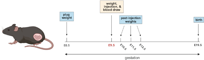
Figure 1: Timeline of MIA generation. Timed pregnant dams are administered poly(I:C) or saline at E9.5, followed by a blood draw 3 h later. Maternal weights are collected at E0.5, E9.5, E10.5, E11.5, and E12.5 to monitor the pregnancy progression post-injection. Please click here to view a larger version of this figure.
Access restricted. Please log in or start a trial to view this content.
Protocol
All animal experiments were conducted in accordance with ethical guidelines at the University of Oklahoma and were approved by the Institutional Animal Care and Use Committee (IACUC). Animals were housed in individually ventilated cages on 1/8-inch cellulose bedding with ad libitum access to food and water, and cage enrichments including a nestlet and a hut. The animal breeding facility is maintained on a 14-hour on, 10-hour off light cycle in accordance with Jackson Laboratory to reflect the photoperiod of late spring, a peak mouse breeding season.
Refer to Table 1 for specific data fields to be collected throughout the protocol. Refer to the supplemental statistics table (Supplementary Table 1) and GitHub (https://hayes-lab.github.io/2026_Camfield_JoVE/) for detailed statistical analyses.
Table 1: Example data collection sheet. Template of important data to collect while generating the MIA model. Please click here to download this Table.
1. Generation of timed pregnancies
- Set up two 8-10 week-old, C57BL/6, female mice with C57BL/6 breeder males in the late afternoon or evening. Using experienced males aged 2-10 months who are isolated prior to breeding results in faster, more reliable pregnancies.
- Assess females for vaginal plugs the following morning and continue checking once daily until plugging occurs. Importantly, the plugs are temporary and may fall out by the afternoon if not checked in the morning.
NOTE: For a detailed protocol for plug checking, refer to Behringer et al. (2016)33. - After confirming a vaginal plug, weigh the female and record the weight to monitor pregnancy status by weight gain across gestation.
- Separate plugged females into individual cages and maintain under standard housing conditions with ad libitum access to food and water. Designate plugged females as embryonic day 0.5 (E0.5). Record the date of plug to ensure accurate tracking of gestational age.
2. Preparation of polyinosinic:polycytidylic acid (poly(I:C))
- Reconstitute lyophilized poly(I:C) to a high-concentration stock solution (10-20 mg/mL) by dissolving in either sterile saline or sterile water according to the manufacturer's instructions, which results in a clear solution. Record vendor, catalog number, and lot number of poly(I:C).
NOTE: Poly(I:C) is a chemical hazard; handle in accordance with institutional biosafety guidelines and dispose of in appropriate biohazard waste receptacles using appropriate PPE to minimize exposure. - Vortex and further dilute to a low-concentration stock solution (4 mg/mL) in saline. To avoid repeated freeze-thaw cycles, aliquot single-use volumes of low-concentration stock (~125 µL) into 1.5 mL tubes and store at -20 °C or -80 °C. Keep the reconstituted poly(I:C) on ice during preparation and handling to minimize degradation.
NOTE: For a comprehensive review on the variability of poly(I:C), refer to Mueller et al. (2019)14. - Immediately before injection on E9.5, dilute the low-concentration poly(I:C) stock (4 mg/mL) to the desired working concentration (2 mg/mL) in sterile saline.
NOTE: Typical poly(I:C) injection doses for mice range between ~5-20 mg/kg, but the optimal dose should be empirically determined by each researcher. - Heat working stock (2 mg/mL) at 65 °C for 10 min to ensure proper re-annealing of the double-stranded RNA structure.
- Mix by vortexing or pipetting to ensure the solution is homogeneous prior to injection.
3. Injecting poly(I:C)
- On the morning of E9.5, weigh the dam to confirm pregnancy and use the weight to calculate the poly(I:C) injection volume for MIA and 0.9% saline injection volume for controls (CON). In the described results, a dose of 10 mg/kg is achieved using an injection volume equal to 5× the animal's body weight. Injections of more than 10x the weight of the animal should be avoided.
E9.5 body weight * 5 = µL of poly(I:C) (at 2 mg/mL) or 0.9% saline
NOTE: A typical weight gain of 2-3 grams is expected between E0.5 and E9.5 to indicate a viable pregnancy. - Draw the appropriate volume of prepared poly(I:C) working solution (2 mg/mL) or saline into a sterile, 27 or smaller gauge syringe.
- Properly restrain the dam to ensure minimal pain and distress. Inject the poly(I:C) or saline intraperitoneally (IP) on the lower left or right quadrant just above the hip level or at the second set of nipples. Properly dispose of syringes in a designated biosafety sharps container. Record the injection side to maintain consistency across the cohort.
- Immediately return the dam to its home cage and monitor for signs of distress. Supplement with hydrogel for at least 24 hours after injection to prevent dehydration due to sickness behavior.
- To ensure thorough documentation, record poly(I:C) dosage, µL of poly(I:C) injected, date/time of injection, gestational age, and lot number of poly(I:C) used (Table 1).
NOTE: Analgesics were not administered because they can alter inflammatory responses induced by poly(I:C).
4. Blood draw
- Three hours after injection, mice were observed individually for 1 min. The presence of any of the following behaviors, such as reduced activity, hunched posture, facial grimacing, or watery stool, was recorded on a three-point scale (no phenotype, mild, prominent), and mice exhibiting one or more behaviors were classified as showing acute sickness behaviors.
NOTE: No sickness behaviors should be observed in CON dams. - Additionally, carefully collect maternal blood from injected dams using a minimally invasive method, such as tail bleed. After blood collection, apply sterile gauze with moderate pressure to the tail until bleeding has stopped. Apply an anti-hemorrhagic agent such as styptic powder and return the dam to the home cage. Aim to collect approximately 50 µL of blood to quantify inflammatory cytokines.
NOTE: All materials that contact blood should be considered biohazardous and disposed of using appropriate PPE and in accordance with institutional biosafety guidelines. - Allow the blood to clot for 30 min at room temperature (20-25 °C). Centrifuge at 2,000 x g for 5 min at room temperature to separate the serum from the clot. The upper, clear layer represents the serum, while the dark pellet at the bottom contains the clot. Carefully collect the serum without disturbing or aspirating any portion of the clot. The serum can be stored at -20 °C until ready for downstream cytokine analysis.
5. Monitoring of injected dams
- Weigh the dams daily for three days following the injection (E10.5, E11.5, and E12.5) to monitor their response to poly(I:C). It is common to observe weight loss of up to 1 g on the day after poly(I:C) injection (E10.5), followed by a gradual recovery, with weight typically exceeding the E9.5 baseline by E12.5.
NOTE: Weight loss exceeding 1 g or failure to regain weight by E12.5 is typically a sign of a lost litter. - For the remainder of gestation, ensure the dams have continuous access to food, water, and nesting materials. Additionally, limit handling of animals or movement of cages, as it may result in additional stress and contribute to decreased litter viability.
- On the day of expected parturition (E19.5), record litter outcomes such as litter size, pup viability, or no litter, indicating an unsuccessful pregnancy. Promptly remove any deceased pups and dispose of them according to institutional biosafety and IACUC-approved animal carcass disposal guidelines.
NOTE: MIA can have smaller litter sizes and may experience delivery complications, leading to a late delivery on E20.5.
6. Validation of immune response
- Using the maternal serum collected after MIA and CON injections, quantify inflammatory cytokines, such as IL-6 or TNF-α, using enzyme-linked immunosorbent assays (ELISA) or multiplex assays according to the manufacturer's instructions. Typical serum dilutions are 1:100 for MIA serum and 1:25 for CON serum.
- After quantification, use the results to establish a representative threshold for the MIA response. The MIA threshold is defined as a maternal serum IL-6 response greater than 1,000 pg/mL (approximately 25% lower than the MIA group mean response), and any MIA dams with responses below this threshold are excluded from the dataset as low or non-responders.
NOTE: CON dams should have very low or undetectable cytokine levels; any CON dams with cytokine levels above 25% of the CON group mean (i.e., 250 pg/mL threshold) should be excluded.
7. Statistical analysis
- Perform all statistical analyses using the R statistical software. Consider dam as the biological unit in all analyses. Define statistical significance as p < 0.05. Present data as boxplots representing median and interquartile range (IQR; 25th-75th percentiles), whiskers extend to 1.5 × IQR, and points represent individual dam.
- Assess normality using the Shapiro-Wilk test to guide the selection of parametric or non-parametric analyses. Perform comparisons between two independent groups using unpaired, two-sided t-tests (normal) or Wilcoxon rank sum tests (non-normal) as appropriate. Analyze paired measurements within the same animals (i.e., weights) using paired t-tests (normal) or Wilcoxon signed-rank tests (non-normal). Conduct one-sample tests against a mean of zero when evaluating relative weight gain across embryonic days.
- Assess the relationship between maternal serum IL-6 and TNF-α levels using Spearman's rank correlation. Use Fisher's exact test to compare the proportion of successful litters across vendors and poly(I:C) formulations. Refer to the detailed statistical summary provided in Supplementary Table 1 and analysis code on GitHub (https://hayes-lab.github.io/2026_Camfield_JoVE/).
Access restricted. Please log in or start a trial to view this content.
Results
Following administration of poly(I:C) or saline at E9.5, maternal serum cytokines were assessed to evaluate the efficiency and robustness of MIA induction (Figure 2). MIA dams displayed significantly increased interleukin-6 (IL-6) levels relative to CON dams (Figure 2A). Additionally, assessment of IL-6 levels allowed for exclusion of MIA dams that did not meet the predefined threshold of 1,000 pg/mL. Thus, IL-6 quantification serves as a valuable biomarker for ...
Access restricted. Please log in or start a trial to view this content.
Discussion
In this protocol, we established checkpoints to increase the efficiency, robustness, and reproducibility of the early-gestational maternal immune activation (MIA) model, focusing on maternal cytokine responses, weight trajectories, and experimental variables that influence pregnancy outcomes. Our results demonstrate that MIA induces variable maternal immune responses, with IL-6 serving as a reliable biomarker for identifying dams with suboptimal immune activation. Approximately 31% of MIA-treated dams failed to reach the...
Access restricted. Please log in or start a trial to view this content.
Disclosures
The authors have no conflicts of interest to disclose.
Acknowledgements
The research was generously funded by a K01 from NIH (K01AG087810), a Burroughs Wellcome Fund Next Gen pregnancy initiative grant (1266845), and a COBRE pilot award (P20GM134973). Figure 1 was created with BioRender.com.
Access restricted. Please log in or start a trial to view this content.
Materials
| Name | Company | Catalog Number | Comments |
|---|---|---|---|
| Animal weighing scale | Kent Scientific | SCL-4000 | For weighing mcie |
| BD Lo-Dose U-100 Insulin syringes - 28G | Fisher Scientific | 14-826-79 | For PIC, intraperitoneal injection |
| C57BL/6J female mice | The Jackson Laboratory | 000664 | 10-12 week old, virgin females |
| C57BL/6J male mice | The Jackson Laboratory | 000664 | Breeding males |
| C57BL/6NCrl female mice | Charles River Laboratories | 027 | 10-12 week old, virgin females |
| Centrifuge (24-place lab standard) | Eppendorf | 5425 R | Spin down blood to collect serum |
| Data documentation platform | Airtable, Microsoft Excel, etc. | Keep records of important data, such as weights, time of injection, etc. (Table 1) | |
| Digital dry bath - One block | Medicus Health | 5277M1 | Use to heat PIC before injection |
| Eppendorf tubes - 1.5 mL | Eppendorf | 30123611 | Use when diluting PIC to stock nd working concentrations |
| Fisherbrand gauze sponges | Fisher Scientific | MSD-1400250 | Use to stop bleeding of tail after blood collection |
| Fisherbrand mall probe 6 in. | Fisher Scientific | 13-820-026 | Use to plug check |
| HydroGel | Clear H20 | 70-01-5022 | Supplement after injections |
| Mouse IL-6 uncoated ELISA kit | Thermo Fisher Scientific | 88-7064-88 | Quantify IL-6 levels in maternal serum |
| Nalgene polypropylene Griffin low-form plastic beakers | Thermo Fisher Scientific | 1201-1000PK | Put mice in beaker when weighing |
| Poly(I:C) (LMW) | Invivogen | tlrl-picw | PIC (LMW) for MIA injection |
| Polyinosinic–polycytidylic acid sodium salt | Sigma | P9582 | PIC (Sigma) for MIA injection |
| Scalpel blades, #22, 10 pack | Home Science Tools | DE-SC22BLD | Use to snip end of tail for blood collection |
| Sodium chloride 0.9% (Normal saline), USP, Sterile grade, Cytiva Hyclone | Cytiva | Z1376 | Inject for generation of control mice |
| Tail snipping platform | Braintree Scientific, Inc. | IL-200 | Properly restrains mice during tail bleeds |
| Vortex-Genie 2 | Scientific Industries, Inc. | SI-0236 | Vortex PIC after dilution |
References
- Taylor, M. J., et al. Association of Genetic Risk Factors for Psychiatric Disorders and Traits of These Disorders in a Swedish Population Twin Sample. JAMA Psychiatry. 76, 280-289 (2019).
- Azidane, S., et al. Identification of novel driver risk genes in CNV loci associated with neurodevelopmental disorders. Hum. Genet. Genomics Adv. 5, (2024).
- Price, K. M., et al. Hypothesis-driven genome-wide association studies provide novel insights into genetics of reading disabilities. Transl. Psychiatry. 12, 495(2022).
- Ripke, S., et al. Genome-wide association analysis identifies 13 new risk loci for schizophrenia. Nat. Genet. 45, 1150-1159 (2013).
- Talkowski, M. E., et al. Sequencing chromosomal abnormalities reveals neurodevelopmental loci that confer risk across diagnostic boundaries. Cell. 149, 525-537 (2012).
- Sanders, S. J., et al. Insights into Autism Spectrum Disorder Genomic Architecture and Biology from 71 Risk Loci. Neuron. 87, 1215-1233 (2015).
- Han, V. X., et al. Maternal acute and chronic inflammation in pregnancy is associated with common neurodevelopmental disorders: a systematic review. Transl. Psychiatry. 11, 71(2021).
- Barr, C. E., Mednick, S. A., Munk-Jorgensen, P. Exposure to influenza epidemics during gestation and adult schizophrenia. A 40-year study. Arch. Gen. Psychiatry. 47, 869-874 (1990).
- Brown, A. S. Epidemiologic studies of exposure to prenatal infection and risk of schizophrenia and autism. Dev. Neurobiol. 72, 1272-1276 (2012).
- Meyer, U. Sources and Translational Relevance of Heterogeneity in Maternal Immune Activation Models. Curr. Top. Behav. Neurosci. 61, 71-91 (2023).
- Mueller, F. S., Polesel, M., Richetto, J., Meyer, U., Weber-Stadlbauer, U. Mouse models of maternal immune activation: Mind your caging system. Brain. Behav. Immun. 73, 643-660 (2018).
- Kentner, A. C., et al. Maternal immune activation: reporting guidelines to improve the rigor, reproducibility, and transparency of the model. Neuropsychopharmacology. 44, 245-258 (2019).
- Schaer, R., Mueller, F. S., Notter, T., Weber-Stadlbauer, U., Meyer, U. Intrauterine position effects in a mouse model of maternal immune activation. Brain. Behav. Immun. 120, 391-402 (2024).
- Mueller, F. S., et al. Influence of poly(I:C) variability on thermoregulation, immune responses and pregnancy outcomes in mouse models of maternal immune activation. Brain. Behav. Immun. 80, 406-418 (2019).
- Carloni, E., Ramos, A., Hayes, L. N. Developmental Stressors Induce Innate Immune Memory in Microglia and Contribute to Disease Risk. Int. J. Mol. Sci. 22, 13035(2021).
- Smolders, S., Notter, T., Smolders, S. M. T., Rigo, J. -M., Brône, B. Controversies and prospects about microglia in maternal immune activation models for neurodevelopmental disorders. Brain. Behav. Immun. 73, 51-65 (2018).
- Mueller, F. S., et al. neuroanatomical, and molecular correlates of resilience and susceptibility to maternal immune activation. Mol. Psychiatry. 26, 396-410 (2021).
- Meyer, U. Prenatal poly(i:C) exposure and other developmental immune activation models in rodent systems. Biol. Psychiatry. 75, 307-315 (2014).
- Guma, E., et al. Differential effects of early or late exposure to prenatal maternal immune activation on mouse embryonic neurodevelopment. Proc. Natl. Acad. Sci. 119, e2114545119(2022).
- Guma, E., et al. Early or Late Gestational Exposure to Maternal Immune Activation Alters Neurodevelopmental Trajectories in Mice: An Integrated Neuroimaging, Behavioral, and Transcriptional Study. Biol. Psychiatry. 90, 328-341 (2021).
- Smith, S. E. P., Li, J., Garbett, K., Mirnics, K., Patterson, P. H. Maternal Immune Activation Alters Fetal Brain Development through Interleukin-6. J. Neurosci. 27, 10695-10702 (2007).
- Haddad, F. L., Patel, S. V., Schmid, S. Maternal Immune Activation by Poly I:C as a preclinical Model for Neurodevelopmental Disorders: A focus on Autism and Schizophrenia. Neurosci. Biobehav. Rev. 113, 546-567 (2020).
- Gzielo, K., et al. The Effect of Maternal Immune Activation on Social Play-Induced Ultrasonic Vocalization in Rats. Brain Sci. 11, 344(2021).
- Tillmann, K. E., et al. Differential effects of purified low molecular weight Poly(I:C) in the maternal immune activation model depend on the laboratory environment. Transl. Psychiatry. 14, 300(2024).
- Lipina, T. V., Zai, C., Hlousek, D., Roder, J. C., Wong, A. H. C. Maternal immune activation during gestation interacts with Disc1 point mutation to exacerbate schizophrenia-related behaviors in mice. J. Neurosci. Off. J. Soc. Neurosci. 33, 7654-7666 (2013).
- Haddad, F. L., De Oliveira, C., Schmid, S. Investigating behavioral phenotypes related to autism spectrum disorder in a gene-environment interaction model of Cntnap2 deficiency and Poly I:C maternal immune activation. Front. Neurosci. 17, 1160243(2023).
- Möhrle, D., et al. Characterizing maternal isolation-induced ultrasonic vocalizations in a gene-environment interaction rat model for autism. Genes Brain Behav. 22, e12841(2023).
- Atanasova, E., et al. Immune activation during pregnancy exacerbates ASD-related alterations in Shank3-deficient mice. Mol. Autism. 14, 1(2023).
- Abazyan, B., et al. Prenatal Interaction of Mutant DISC1 and Immune Activation Produces Adult Psychopathology. Biol. Psychiatry. 68, 1172-1181 (2010).
- Capellán, R., et al. Interaction between maternal immune activation and peripubertal stress in rats: impact on cocaine addiction-like behaviour, morphofunctional brain parameters and striatal transcriptome. Transl. Psychiatry. 13, 84(2023).
- Zhao, Q., et al. Microglia-mediated neurogenesis is linked to cognitive deficits in a two-hit model of maternal immune activation and juvenile stress. Brain. Behav. Immun. 129, 649-661 (2025).
- Guma, E., et al. Investigating the "two-hit hypothesis": Effects of prenatal maternal immune activation and adolescent cannabis use on neurodevelopment in mice. Prog. Neuropsychopharmacol. Biol. Psychiatry. 120, 110642(2023).
- Behringer, R., Gertsenstein, M., Nagy, K. V., Nagy, A. Selecting Female Mice in Estrus and Checking Plugs. 2016, Cold Spring Harb. Protoc. (2016).
- Urakubo, A., Jarskog, L. F., Lieberman, J. A., Gilmore, J. H. Prenatal exposure to maternal infection alters cytokine expression in the placenta, amniotic fluid, and fetal brain. Schizophr. Res. 47, 27-36 (2001).
- Solek, C. M., Farooqi, N., Verly, M., Lim, T. K., Ruthazer, E. S. Maternal immune activation in neurodevelopmental disorders. Dev. Dyn. Off. Publ. Am. Assoc. Anat. 247, 588-619 (2018).
- Ozaki, K., et al. Maternal immune activation induces sustained changes in fetal microglia motility. Sci. Rep. 10, 21378(2020).
- Kim, S., et al. Maternal gut bacteria promote neurodevelopmental abnormalities in mouse offspring. Nature. 549, 528-532 (2017).
- Sal-Sarria, S., Conejo, N. M., González-Pardo, H. Maternal immune activation and its multifaceted effects on learning and memory in rodent offspring: A systematic review. Neurosci. Biobehav. Rev. 164, 105844(2024).
- Martín-Guerrero, S. M., et al. Maternal immune activation imprints translational dysregulation and differential MAP2 phosphorylation in descendant neural stem cells. Mol. Psychiatry. 30, 2994-3007 (2025).
- Yu, D., et al. Microglial GPR56 is the molecular target of maternal immune activation-induced parvalbumin-positive interneuron deficits. Sci. Adv. 8, eabm2545(2022).
- Hayes, L. N., et al. Prenatal immune stress blunts microglia reactivity, impairing neurocircuitry. Nature. 610, 327-334 (2022).
Access restricted. Please log in or start a trial to view this content.
Reprints and Permissions
