Here, we describe a protocol for ex vivo high-density microelectrode array recordings from acute cerebellar brain slices of a dystonic hamster model that received in vivo. continuous pallidal deep brain stimulation for 11 days.
Method Article
Investigating The Network-Wide Mechanisms of Pallidal Deep Brain Stimulation Using High-Density Microelectrode Arrays
In This Article
Summary
Abstract
Dystonia is a neurological movement disorder characterized by abnormal muscle control affecting voluntary movements and sustained postures. Although dystonia is associated with basal ganglia dysfunction, increasing evidence suggests that the cerebellum is involved in its pathophysiology. This protocol presents in vivo pallidal deep brain stimulation (DBS) in a dystonic hamster model (dtsz hamster) combined with ex vivo high-density microelectrode array (HD-MEA) recordings to investigate cerebellar activity. The protocol includes (1) DBS surgery for electrode and stimulator implantation, (2) acute cerebellar slice preparation, (3) high-resolution electrophysiological recordings, and (4) data analysis. Representative results indicate that 11 days of pallidal DBS restored spike activity toward normal levels in the molecular, Purkinje, and granular layers, consistent with cerebellar network involvement in dystonia. This protocol enables detailed analysis of cerebellar activity and its modulation by DBS, providing a platform to investigate network-level mechanisms underlying neuromodulation therapies.
Introduction
Dystonia is the third most common movement disorder1 characterized by distinct clinical signs and symptoms2,3, although it may sometimes be mistaken for other motor disorders4,5,6. The most prominent clinical features include involuntary muscle contractions, repetitive movements, and abnormal and/ or twisted posture7. These features may be accompanied by symptoms such as pain, impaired motor performance, and fatigue, which may worsen during periods of stress, anxiety, or physical exhaustion8,9,10,11. The pathophysiology of dystonia remains not totally understood12,13. Although studies have traditionally focused on the basal ganglia, increasing evidence supports a network-level concept of the disorder, involving multiple brain regions, including the cerebellum14,15,16,17. This protocol aims to investigate extracellular network activity across distinct layers of the cerebellar cortex to elucidate their role in the pathophysiology of dystonia and to understand the network-wide mechanisms of deep brain stimulation (DBS).
Deep brain stimulation (DBS) targeting the globus pallidus internus (GPi) or the subthalamic nucleus (STN) is often used to treat certain forms of dystonia18,19, particularly those that are drug-resistant. Additionally, the treatment outcome is heterogeneous and unpredictable, as patients with similar clinical manifestations may respond differently to the same therapeutic approach20. A more comprehensive understanding of the neural network underlying dystonia could improve treatment outcomes21. Therefore, large-scale and experimental approaches are needed, which can only be achieved through preclinical studies in animal models. Combining in vivo DBS experiments with ex vivo high-density microelectrode array (HD-MEA) recordings enables high-resolution monitoring of the (almost) physiological neuronal network activity to better understand the pathophysiology of dystonia and the mechanisms of DBS.
The basal ganglia and cerebellum each influence motor control, at least in part, through their connections with the primary motor cortex (M1)22. An anatomical study with a striatal lesion and surgical removal of the cerebellum supports this idea23. In our study, we use the animal model of the dystonic dtsz hamster24,25, which exhibits cerebellar abnormalities associated with dystonia26. Using high-density microelectrode arrays (HD-MEAs), we investigated cerebellar cortical network activity in dystonic and healthy hamsters. However, conventional electrophysiological approaches lack the spatial resolution to capture layer-specific network dynamics, whereas this method addresses this limitation. On the other hand, we investigated the effect of deep brain stimulation in the globus pallidus internus on the cerebellar cortical networks.
The HD-MEA system offers the advantage of detailed electrophysiological recordings in parallel, enabling analysis across the layers of the cerebellar cortex (granular, Purkinje, and molecular layers), with each layer playing an essential role in the processing of movement sequences27. The granular layer, densely packed with granule cells, receives and relays sensory and motor information from mossy fibers, allowing initial signal integration before further processing27. The Purkinje layer, which contains Purkinje cells, transmits inhibitory signals to the deep cerebellar nuclei, where the granular and molecular layers modulate the outgoing signal. The molecular layer, rich in dendrites and interneurons, supports complex modulation through synaptic interactions with Purkinje cells, fine-tuning motor output28. HD-MEA, unlike other recording techniques, is not limited by spatial resolution or sampling area, allowing the unique contributions of each layer to be captured in the altered neural dynamics seen in dystonia15. This approach provides higher spatial resolution and parallel recording capabilities compared to single-electrode or low-density recording methods. There are HD-MEA systems based on the complementary metal-oxide-semiconductor (CMOS)29,30 technology, combined with the Active Pixel Sensor (APS) concept, that provide functional imaging for visualizing the electrical activity of large populations of neurons over time31,32. With this technique, the dynamics of each layer of the cerebellar cortex can be monitored, and specific synaptic transmission processes can be investigated by washing with various substances. This method is particularly suitable for studies investigating complex neuronal network activity and layer-specific alterations in neurological disease models.
This work addresses technical challenges regarding encapsulation of the stimulation system as well as tissue preparation, data acquisition, and the analysis of a large electrophysiological dataset. With this work, we present an option for in vitro investigation of highly complex neuronal processing, which, in our case, on the one hand, contributes to a more differentiated understanding of the pathophysiological dynamics in dystonic neuronal networks and, on the other hand, provides knowledge about the therapeutic mechanism of action of DBS.
Protocol
Use dystonic dtsz hamsters and control Syrian golden hamsters for experiments26. In total, four experimental groups were examined in the present study: (1) non-dystonic control hamsters (wild type, WT), (2) native untreated dtsz hamsters, (3) dtsz-Sham (underwent implantation, but without active stimulation), and (4) dtsz-DBS (with continuous DBS in vivo for 11 days before slice preparation). All animal experiments were conducted in accordance with the EU Directive 2010/63/EU and the federal laws for the protection of animals, under license 7221.3-1-029/20.
1. Animals
- House the animals under standard conditions of GV Solas33 at a 14 h light/ 10 h dark cycle, and with free access to water and food (standard diet hamster, Altromin).
- Screen the dtsz mutant hamsters for dystonic symptoms using the triple stimulation technique26, at the ages of 20 ± 1 and 27 ± 4 days
NOTE: This standardized mild stress procedure consists of removing the hamster from its home cage and transferring it to a new empty cage, followed by intraperitoneal or subcutaneous injection of saline, and short-term sleep deprivation. This sequence of mild anxiety-provoking stimuli reliably induces dystonic attacks with reproducible latency and is therefore used for phenotypic screening before inclusion in experimental groups. Non-dystonic control hamsters undergo the same procedure to ensure comparable experimental conditions between groups. - For analgesia, treat the animals of the dtsz-Sham and dtsz-DBS groups with 100 mg·kg-1 peroral (p.o.) metamizole. Implant both groups according to the surgical procedure described below.
2. Encapsulation of the STELLA stimulation system
- The STELLA stimulation systems are encapsulated using a biocompatible approach to protect the devices from liquid penetration during chronic implantation. The electronic is housed in a custom 3D-printed PETG capsule.
- Seal between the lid and the capsule using silicone.
- Additionally, seal the housing with silicone at the cable exit.
- If gaps in the silicone are visible after drying, repeat steps 1 and 2.
- After full solidification of the silicone, prepare the epoxy resin at a 100:35 mixing ratio (weight).
- Remove air bubbles from the resin under vacuum (Vevor RS-1.5).
- Prior to mould assembly, apply a thin layer of vaseline to the mating surface of the custom silicone mould to improve sealing and facilitate demoulding after epoxy curing. Place the stimulator capsule into the silicone mould and slowly fill the mould with epoxy resin until the stimulator and cable junctions are fully covered. Avoid air bubbles during filling.
- Once the resin has cured, remove the capsule from the mould and smooth the epoxy surface using a fine file or polishing tool to eliminate sharp edges.
- Apply a final thin silicone coating over the cured epoxy capsule by immersing the capsule in liquid silicone. Use a plastic pipette to apply additional silicone around the cable near the junction area. Excess silicone accumulation should be removed with a stick before curing to avoid coating thickness.
- Finally, perform a post-encapsulation functionality test to verify correct stimulation output before implantation or experimental use. Place the STELLA stimulation system in Ringer’s solution and activate the magnetic BIST function. Confirm the correct LED blink pattern (2 × 2), indicating proper stimulation output and the absence of short circuits, open circuits, or low battery conditions34.
3. Surgical procedure for implanting a stimulation system
- Prepare surgical instruments (Figure 1) and the STELLA (software-defined implantable modular platform) stimulation system34.
- Sterilize the metal instruments from Figure 1 for approximately 5 min at 200 °C, e.g., with the FST Sterilizer 250. Note that the instruments are hot when removing them.
- Disinfect the STELLA stimulation system with the two DBS electrodes with 70 % ethanol. Place the dried stimulation system in a Falcon (50 mL conical tube) filled with Ringer’s solution overnight.
- Prepare the animal (at the age of 25 –32 days)
- Place the anesthesia chamber (self-made) in the hamster cage. The hamster will walk into the tube voluntarily after a short time (Figure 2A).
- When the hamster is in the tube, close the tube at the end and connect the tube to the volatile isoflurane output for 1-2 min (induction of anesthesia with 4 % isoflurane) (Figure 2B).
- Remove the animal from the cylinder gently. Place the animal on the temperature-controlled
surface (approximately 38 °C) and fit with a breathing mask (GM-4; maintenance of anesthesia with 3 % isoflurane; test depth of anesthesia with the interdigital reflex). - Shave the surgical area on the head with a suitable hair clipper (Figure 2C). Clean the area with a wet swab and inject 500 µL of 0.25 % bupivacaine subcutaneously (s.c.) to the scalp for additional local anesthesia of the periosteum. Wait at least 5 min before opening the scalp.
- Transfer the animal carefully into the stereotaxic frame and fix it with the ear bars so that bregma and lambda are at the same height (reduce isoflurane to 1.5–2.5 % for maintaining the anesthesia).
- Protect the eyes with a tear substitute (TVM Ocry-Gel).
4. Implant the STELLA stimulation system
- Make a 2 cm long skin incision medially on the scalp. A retractor (Figure 1, No. 14 and Figure 2D) pulls the skin away. Prepare the skull by locally removing the periosteum with the blade by scratching. Stop possible bleeding with swabs.
- Apply 3 % H2O2 with a cotton-tip applicator for roughening.
- Take the stimulation system from Ringer’s solution and protect the DBS electrodes with parafilm.
- Insert a blunt pair of scissors through the skin incision just made on the head and form a 5 cm skin pocket subcutaneously towards the neck and flank.
- Insert the stimulator, which has been removed from the Falcon and soaked in dermal disinfection.
- Apply approximately 500 µL of antibiotic (10 mg·g-1 fusidic acid) to the skin pocket with a cotton tip applicator.
- Drill two 7 mm holes (with FST#19007-07) in the skull for DBS electrodes at AP: −0.6 mm and ML: ± 2.2 mm according to the bregma.
- Drill two more holes (9 mm; FST#19007-09) laterally on each electrode hole. Screw four screws (DIN 84 A2 M1x2) into the prepared holes (Figure 2D).
- Implant two DBS electrodes (SNEX-100) with DV: 6.0 mm according to the bregma. Please note that the electrode holder of your specific stereotactic frame determines which side the electrode is implanted first (the distance between the electrodes is 4.4 mm).
- Fix the electrode with UV-light-curing bonding (Heliobond) to the two screws. When both electrodes are implanted, a final layer with UV-light-curing bonding is applied between them. Finally, the electrodes are cemented with Compoglass Flow (Figure 2E).
- Activate the stimulator of the DBS group magnetically after at least three days of recovery for continuous 11 days of DBS. In this study, we used stimulation parameters similar to those used in clinics (130 Hz, 50 μA, and 60 μs).
- Check the light pattern of the stimulator by swiping over the hamster’s flank with a magnet daily (Figure 2F).
5. Preparation of acute cerebellar slices
- Prepare 1 L of artificial cerebrospinal fluid (ACSF) and 0.5 L of sucrose for each animal (see Table 1 and Table 2 for details).
- Use sucrose solution at approximately 4 °C during brain extraction and slicing, with continuous carbogenation. Additionally, prepare ice-cold slushy sucrose to maintain low tissue temperature during rapid dissection.
- Prepare the workplace with dissection tools, sucrose solution, Petri dishes, and vibratome setup (see Figure 3A).
- Animals' sacrifice must occur within approximately 15 min after off-DBS, and cerebellar extraction should begin immediately thereafter. Decapitate the animal in deep anesthesia induced with volatile isoflurane. Place the head in a higher Petri dish filled with ice-cold slushy sucrose.
- Dissect the brain by avoiding medial cuts through the spinal canal to protect the cerebellar tissue from damage.
- Transfer the brain into a new Petri dish filled with ice-cold slushy sucrose, with a filter paper at the bottom to prevent the cerebellum from slipping during sectioning.
- First, remove the cerebellum through coronal cutting through the pons with a blade (Figure 3B). Then, remove the cerebellar hemispheres through parasagittal cuts with a blade (Figure 3C).
- Prepare the vibratome plate with a drop of super glue. Carefully remove the cerebellar vermis from the Petri dish. Dab the parasagittal section plane onto a filter paper to dry before gluing this side (Figure 3D).
- Place the vibratome plate into the tray and fill it with enough sucrose (half slushy, half solution) to completely submerge the vermis. Do not forget to gas with carbogen (CB).
- Cut parasagittal cerebellar slices with a thickness of 200 μm at a constant speed of 0.08 mm·s-1 (Figure 3E). Store the slices in ACSF in a beaker constantly gassed with CB at room temperature (Figure 3F).
- Allow the slices to recover in carbonated ACSF at room temperature for approximately 40 min before transfer to the HD-MEA, which serves as the rewarming and stabilization period before recording. During this period, maintain ACSF at room temperature.
6. Preparing the CorePlateTM Single-Well of the BioCAM DupleX MEA system for recording
- Switch on the BioCAM DupleX recording system with the Brainwave 5 software (BW5) at least 30 min before starting the experiments.
- Before placing the tissue slice on the CorePlateTM Single-Well for recording, assess the background noise level by inserting the single-well into the recording setup. The optimal noise level for high-quality recording should be 9-20 µV.
- Clean the gold-plated pads with 100 % ethanol (Figure 4A-1), the electrode array with 70 % ethanol, and a soft brush (Figure 4A-2). Finally, rinse the electrode array with MilliQ water.
- Connect the HD-MEA/CorePlateTM Single-Well into the recording system (Figure 4D-1).
- Pipette approximately 2 mL ACSF into the well. Ensure no air bubbles are on the electrodes or the ground (Figure 4D-2).
- Start live acquisition and record for at least 30 seconds with BW5.
7. Analyze peak-to-peak amplitude
- To analyze the noise interface, open the recorded *.bxr file, which contains 30 seconds of intrinsic noise recording (generated by the resistive components of the electrodes and amplifier circuits).
- Select “Activity Map” and, under “Metric,” choose Peak-to-Peak Amplitude. The scale is set to 500 µV, allowing visualization of signal variability across the electrode array (Figure 5A).
- In the "Activity Map" window, position the mouse pointer over the recorded electrode to display the "Well Wide Mean" amplitude value.
- Alternatively, you can determine the average amplitude value by creating an Excel file (*.xlsx) under "Graph Export." Then open that Excel file and calculate the mean amplitude value [=Average(Number1; Number2;...)].
8. Analyze the baseline noise (signal standard deviation)
- Analyze the baseline noise by clicking "Playback" to generate the *.brw-file. Then, select "Activity Map" and under "Metric" select "Standard Deviation". The scale is automatically set to 30 µV, allowing visualization of signal variability across the electrode array (Figure 5B).
- In the "Activity Map" window, hover the mouse pointer over the recorded electrode to display the "Well Wide Mean" standard deviation.
- Alternatively, you can determine the average standard deviation using "Chart Export" and create the *.xlsx Excel file. Then, open the Excel file and calculate the mean standard deviation [=Average(Number1; Number2;...)].
9. For evaluating the signal quality:
- Calculate the signal-to-noise Ratio (SNR)

SNR ≥ 5: Good quality
SNR between 2 and 5: Moderate quality
SNR < 2: Poor quality
10. Slice positioning in CorePlate single-well for data acquisition
- With the help of a Pasteur pipette, position the slice at the electrode array so that the region of interest is covered by recording electrodes (Figure 4 B-1). Remove the solution from the well (Figure 4 B-2). Take a picture with an upright microscope.
- Fix the tissue with the slice anchor (Figure 4C).
- Using a 3 mL Pasteur pipette, add one drop of ACSF to the slice.
- Remove the liquid using a Pasteur pipette with a smaller volume. Remove the remaining liquid with a piece of twisted paper (repeat this 7-10 times) (Figure 4C-2).
- For perfusion during recordings, use ACSF at room temperature and set the rate to 2 mL·min-1. The inflow position is above the tissue slice, and the outflow is at the well's wall (Figure 4D).
- An additional grounding of the inflow and outflow is recommended to minimize perfusion-induced noise, which can be achieved by connecting them to the recording system via a wire (Figure 4D-1).
- To reduce disturbances, turn off the microscope and the room's lights.
11. Recordings of network activity with the CorePlateTM Single-Well
- Set the recording parameters to a sampling frequency of 20,000 Hz with a high-pass filter at 100 Hz.
- On the BrainWave 5 Software, go to File and open a new recording session.
- Click on “Source”.
- Open: chip pilot, and select for the following adjustments:
Recording spontaneous neural activity at a sampling rate: 20,000 Hz/electrode.
Hardware high-pass filter cut-off: 100 Hz.
Light compensation Rate: short (direct light stimulation).
- To select the chip calibration channel and the type of raw trace.
- Open: Control signal, and select: On Internal chip calibration, select output channel: 1.1.
- Click on “+” and “Charting”.
- Open: Chart setting.
- Select raw traces: raw and spikes.
12. Insert and align the microscope image on the activity map
- Click on “+”.
- “Overlays and Annotations” to insert the microscope picture into the activity map on BW5:
- Go to Well layers:
- Select manage layers and the cerebellar picture.
- Align selected image layer: click on start.
- Click on the four corners of the picture following the numeric sequence and apply.
13. Data compression setup before recording
- To compress the data, go to the first page of RECORDER:
- Cockpit mode: advanced
- Save raw compressed: wavelet compression + events: spikes.
- Play for 10 min before starting the recording.
- Clean the gold-plated pads with 100 % ethanol after finishing the recordings. Next, fill the well with pure water and brush it for 30 s.
- Next, fill the well with 70 % ethanol and gently brush the electrode array for approximately 30 seconds. Finally, fill the well with pure water and gently brush it for 30 s (repeat this step 2 or 3 times).
- Keep the CorePlateTM Single-Well dry in the appropriate container.
14. Offline analysis of recorded data
- Open the desired file with the BW5.
- Divide the cerebellar cortex into three groups by selecting electrodes according to anatomical layer assignment (molecular layer, Purkinje cell layer, granule cell layer).
NOTE: The number of electrodes within the regions of interest (ROIs) per layer depended on the spontaneous activity detected in each layer and therefore varied across recordings. Typically, 25-250 electrodes per ROI were analyzed from a single slice. Electrodes located in regions spanning two anatomical layers were excluded to avoid ambiguity in layer assignment. Only electrodes clearly attributable to one specific cerebellar layer were included for further evaluation.- Selection and grouping: Go to Unit groups.
- On Manage unit groups, click on “+” to form one group and name it.
- To include more electrodes in the group, select the electrodes and click “add or remove selected units”.
- Analyze raw traces using the spike detection method, known as precise timing spike detection (PTSD). It focuses on detecting relative maxima and minima with peak-to-peak amplitudes exceeding a predefined threshold, ensuring precise and accurate spike identification.
- Data processing: Go to Spike detection and set the mode to advanced.
- Use data from: Raw.
- Algorithm: PTSD (Precise timing spike detection).
- Standard deviation factor: 8.0.
- Peak lifetime period: 2.0 ms.
- Refractory period: 1.0 ms.
- Spike assignment: negative peak.
- Pre-peak wave duration: 1.0 ms.
- Post-peak wave duration: 1.5 ms.
- Go to Spike sorting and set the mode to advanced. Adjust the following parameter:
- Pre-peak wave taken duration: 0.5 ms.
- Post-peak wave taken duration: 1.0 ms.
- Feature extraction algorithm: Legendre moments.
- Number of features: 3.
- Clustering algorithm: K-Means & Silhouette.
- Minimum number of spikes per cluster: 20.
- Maximum number of clusters: 3.
- Outline threshold: 2.
- Exporting data to Excel.
- Click on “Charting”.
- Go to Chart Export.
- Select the spreadsheet and export.
Results
Pallidal DBS increased the number of spikes to normal levels in cortical layers between groups.
We analyzed the total number of spikes in three layers of the cerebellar cortex—molecular layer (ML), Purkinje cell layer (PL), and granular cell layer (GL) — across four experimental groups: wild-type (WT, N = 8), dtsz (N = 8), dtsz-Sham (N = 7), and dtsz-DBS (N = 7), with analyses performed on slices obtained from these animals.
While dystonic tissue (dtsz) generated significantly fewer spikes, pallidal DBS increased the number of spikes to levels comparable to those of healthy controls (Table 3). The Kruskal-Wallis H test further confirmed this, showing a statistically significant increase in the number of spikes in the ML for the dtsz DBS group compared to both the dtsz and dtsz-Sham groups (χ2(3) = 20.728, p = 0.001; Figure 6A). The dtsz-DBS group also showed a similar increase in spikes in PL (χ2(3) = 16.855, p = 0.001; Figure 6B) and GL (χ2(3) = 13.164, p. = 0.004; Figure 6C), reaching values comparable to those of the WT group (Table 3).
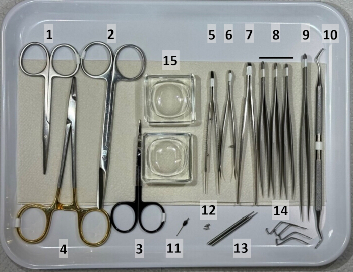
Figure 1. Overview of surgical instruments. The image shows an arranged tray containing a set of surgical and microsurgical instruments designed explicitly for procedures involving the implantation of deep electrodes and stimulators. The instruments are organized in sequential order of use, enhancing procedural efficiency. They are placed on tissue to maintain cleanliness and ensure easy accessibility during the procedure. This arrangement focuses on precision, sterility, and readiness for complex surgical workflows. (1) Scissors blunt/blunt, (2) Suture scissors, (3) Scissors sharp/sharp, (4) Needle holder, (5) Suture tying forceps, (6) Splinter forceps/curved, (7) Forceps serrated, (8) Forceps standard short, (9) Forceps standard, long, (10) Micro spatula, (11) Reused electrode, (12) Screws, (13) Drill accessories, (14) Retractor Tip/clamp, (15) Glass cavity block to be filled with 0.9 % NaCl and xylocaine. For more information, check the material list. Please click here to view a larger version of this figure.
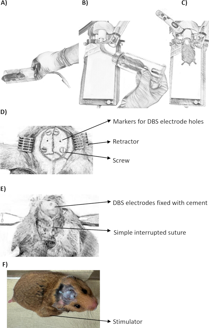
Figure 2. Experimental setup for anesthetizing and positioning the animal in the stereotaxic apparatus for implantation of DBS electrodes and the stimulator. (A) The animal is gently placed inside a custom-made induction chamber to initiate anesthesia. (B) The induction chamber is connected to a breathing mask to ensure proper administration of anesthesia (induction with isoflurane at 4%) while maintaining safety during handling. (C) Once anesthetized, the animal is transferred to a heating plate with a connected breathing mask (maintenance with isoflurane at 3%) to prevent hypothermia and ensure continuous anesthesia during shaving. (D) The animal is carefully positioned in the stereotaxic frame. A longitudinal incision is made, and clamps are used to retract the skin. Four dots indicate screw placement. Screws are inserted into the marked areas, and two additional dots are marked for electrode placement. (E) The electrode is inserted into the GPi and secured with glue before the stereotaxic holder is removed. For better fixation, cement is applied around the electrodes. Interrupted longitudinal sutures are used to close the surgical site, and (F) the stimulator is placed in the subcutaneous flank area of the animal. The animal remains alive post-surgery, and the arrow indicates the location of the stimulator. Please click here to view a larger version of this figure.
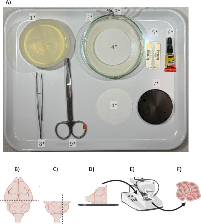
Figure 3. Illustration of brain sectioning and slicing. (A) surgical instruments for the dissection procedure. (1*) Homemade Agar Petri dish, (2*) Agar Petri dish, (3*) Clamp fixing the carbogen tube, (4*) Paper filter, (5*) Blade, (6*) Super glue, (7*) Vibratome plate/holder, (8*) Curved forceps, (9*) Scissor, Sharp/Blunt. (B) Brain sample preparation: after dissection, the brain is cut, and the dashed lines indicate the orientation of the coronal plane sectioning. (C) Parasagittal sectioning: the brain is cut along the specified parasagittal plane. (D) Tissue fixation: The resulting brain tissue is affixed to a plate/holder with glue to secure it for precise sectioning. (E) Vibratome sectioning: The plate with attached tissue is transferred to a vibratome, where ultra-thin slices are obtained (200 µm). (F) Final slices: Precise parasagittal cerebellar slices are produced, ready for electrophysiological recording35. For more information, check the material list. Please click here to view a larger version of this figure.
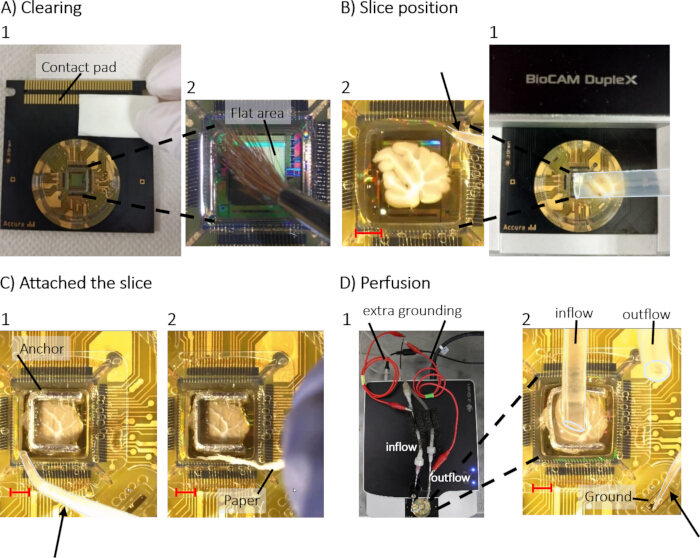
Figure 4. Illustration of the steps to get a good cell activity. (A) Cleaning: This step involves preparing the HD-MEA by cleaning its surface to ensure that no dirt interferes with the attachment (Contact pad, A-1) or recording process (flat area, A-2). (B) Slice position: The tissue slice is carefully positioned on the HD-MEA (B-1), ensuring proper alignment for effective electrode contact (B-2). A thin tip of the pipette (arrow) is used to remove the remaining ACSF, allowing the anchor to be inserted. (C) Attachment of the slice: the slice is securely positioned on the HD-MEA using the anchor. At this step, it is crucial to place a drop of ACSF and then carefully remove the excess liquid using a pipette (C-1; arrow) and a twisted piece of precision wipe paper (C-2). This ensures the slice remains stable and well-connected to the electrodes for accurate recording. (D) Perfusion: Finally, a perfusion system is connected and initiated to maintain the tissue slice's viability during the experiment, where the inflow has to be on top of the slice and the outflow close to the wall (D-2). It is also essential to remove all bubbles from the ground to minimize noise during recording (indicated by the arrow). Additional grounding is attached to both the inflow and outflow tubing (D-1). The red scale bars are 1.5 mm long. Please click here to view a larger version of this figure.
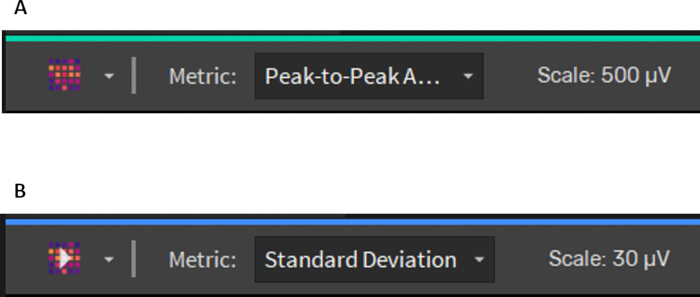
Figure 5. Metrics for amplitude and standard deviation in HD-MEA recordings using Brain Wave 5 software (BW5) to evaluate the signal quality. (A) Peak-to-Peak Amplitude metric selected to extract intrinsic electrode amplitude values (scale: 500 µV). (B) Standard Deviation metric selected to quantify baseline noise across electrodes (scale: 30 µV). Please click here to view a larger version of this figure.
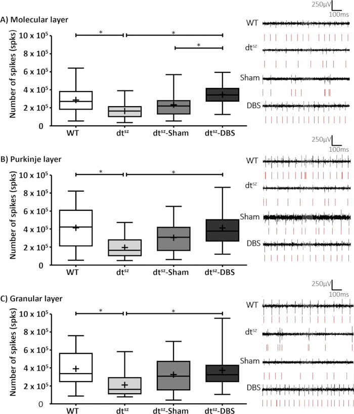
Figure 6: Effects of pallidal DBS on spike activity across cerebellar cortical layers: (A) molecular layer (ML), (B) Purkinje cell layer (PL), and (C) granular layer (GL). The Box-Whisker plots display the mean and median with a cross and horizontal line, respectively, along with the 25th and 75th percentiles. The respective groups are wild type (WT, n = 22 slices), dtsz without surgery (n = 20 slices), dtsz DBS inactive (Sham, n = 20 slices), and dtsz-DBS active (n = 20 slices). Statistical significance was determined using the Kruskal-Wallis test, followed by Dunn's pairwise comparisons (*p .< 0.05). Please click here to view a larger version of this figure.
| Name | Molecular weight [g/ mol] | Final concentration [mol/ l] |
| NaCl | 58.44 | 0.124 |
| KCl | 74.55 | 0.003 |
| Glucose | 180.16 | 0.01 |
| CaCl2 (water-free) | 110.98 | 0.0025 |
| NaHCO3 | 84,007 | 0.026 |
| NaH2PO4 | 119.98 | 0.00125 |
| pH | 7.35 to 7.40 (adjustable with HCl) | |
| Osmolality [mosmol/ kg] | 290 to 300 | |
Table 1: Artificial cerebrospinal fluid (ACSF) solution: Composition and final molar concentrations of components used to prepare ACSF for cerebellar slice experiments. pH was adjusted to 7.35–7.40 with HCl, and osmolality was maintained between 290–300 mOsm·kg-1.
| Name | Molecular weight [g/mol] | Final concentration [mol/L] |
| Saccharose | 342.30 | 0.075 |
| KCl | 74.55 | 0.0025 |
| NaCl | 58.44 | 0.087 |
| CaCl2 (water-free) | 110.98 | 0.0005 |
| MgCl2 | 95.21 | 0.007 |
| Glucose | 180.16 | 0.01 |
| NaHCO3 | 84.01 | 0.025 |
| NaH2PO4 | 119.98 | 0.00125 |
| pH | 7.35 to 7.40 (adjustable with HCl) | |
| Osmolality [mosmol/ kg] | 320 to 330 | |
Table 2: Sucrose solution. Composition and final molar concentrations of the sucrose solution used during cerebellar slice preparation. pH was adjusted to 7.35–7.40, and osmolality was maintained between 320–330 mOsm·kg-1.
| Groups: | WT | dtsz | dtsz-Sham | dtsz-DBS |
| n | 22 | 20 | 20 | 20 |
| Coefficient of variance | ||||
| ML | 0.53 | 0.55 | 0.6 | 0.35 |
| PL | 0.57 | 0.57 | 0.52 | 0.46 |
| GL | 0.48 | 0.66 | 0.61 | 0.51 |
| Number of spikes | ||||
| ML | 287240±32605 | 163434±19998 | 234209±31284 | 345915±27113 |
| PL | 413223±50171 | 194277±24786 | 304568±35692 | 412707±42509 |
| GL | 390817±40139 | 211274±31193 | 325533±44318 | 373208±42220 |
Table 3: Summary of spike metrics across cerebellar layers and experimental groups. WT – wild type, non-dystonic control hamsters; dtsz - native dtsz hamsters; dtsz-DBS - dtsz hamsters continuously stimulated for 11 days; dtsz-Sham - dtsz hamsters undergoing the surgery, however, with no active DBS; ML – molecular layer; PL – Purkinje cell layer; GL. – granular cell layer.
Discussion
We described a methodological approach to investigate the mechanisms of deep brain stimulation (DBS) in the pathophysiological dystonic neuronal network in the dtsz hamster. Critical steps include precise electrode implantation, rapid cerebellar slice preparation, and stable HD-MEA recording conditions to ensure reliable data acquisition. We demonstrate the surgery procedure, preparation of cerebellar slices, recording, and analysis techniques to explore the ex vivo cerebellar activity and its modulation by DBS. The electrophysiological measurements using HD-MEA provided real-time recordings of cerebellar spontaneous activity with high temporal and spatial resolution36, which we consider necessary for investigating the mechanisms of pallidal deep brain stimulation in the treatment of dystonia.
Dystonia is currently known as a network disorder characterized by abnormal motor output arising from dysfunctional interactions among basal ganglia, cerebellar, and cortical circuits37,38. Cerebellar abnormalities have been shown to play a significant role in dystonia, as supported by both clinical and experimental studies15,22,39. Furthermore, an anatomical study involving the injection of retrograde rabies virus (RV) into the cerebellar cortex of Cebus monkeys shows that the subthalamic nucleus of the basal ganglia has a substantial projection to the cerebellar cortex40. These findings underscore the importance of exploring cerebellar circuits alongside the basal ganglia networks when studying the therapeutic mechanisms of DBS40,41,42. This method is particularly suitable for studies investigating neuronal interactions and neuromodulation effects in movement disorders.
Regarding the network-wide mechanisms of pallidal DBS, studies have demonstrated that DBS of the internal segment of the globus pallidus (GPi) induces widespread changes in brain activity, not only within the basal ganglia but also across interconnected regions such as the cortico-striatal network43, the thalamus44, and the cerebellum15. These observations support the hypothesis that pallidal DBS can modulate activity throughout the basal ganglia-cerebellar network, contributing to its therapeutic efficacy in dystonia. This demonstrates the need for preclinical studies to investigate DBS mechanisms and the advantage of HD-MEA recordings for capturing network-wide electrophysiological changes. Furthermore, the study by Kotyra et al. (2025) supports the presence of long-lasting neuroplastic remodeling induced by longer-term GPi-DBS within the dystonic motor network45. In vivo findings showed that pallidal stimulation progressively reduced dystonia severity in dtsz hamsters and that the beneficial effect persisted after stopping stimulation, suggesting that DBS-induced effects are not limited to acute stimulation alone45. In addition, recent electrophysiological studies demonstrated network-wide modulation of synaptic plasticity and spike patterns after longer-term DBS, including increased cerebellar activity15, enhanced excitatory input to motor thalamic neurons, and more clustered excitatory input to cortical M146.
Representative results indicate a network-wide impact of deep brain stimulation (DBS), as evidenced by the normalization of spike activity in the molecular, Purkinje cell, and granular cell layers of the cerebellar cortex in dystonic hamsters (Figure 6). The increased spike activity observed in the dtsz DBS group reached levels comparable to the wild-type controls. This normalization of spike activity supports the therapeutic potential of DBS for treating dystonia. Notably, the dtsz-Sham group showed intermediate spike values compared to the WT group across all cerebellar layers, suggesting that electrode implantation alone may influence cerebellar network activity. This interpretation is consistent with the clinically described microlesion effect in dystonia, where electrode implantation itself may transiently improve symptoms before stimulation onset47. Although the dtsz hamster reproduces key features of paroxysmal generalized dystonia, it does not fully reflect the clinical heterogeneity observed across human dystonia syndromes. Therefore, caution should be taken when translating these findings to human dystonia.
This work addresses technical challenges related to stimulator encapsulation, DBS surgery, slice preparation, and large-scale electrophysiological data analysis. The combination of CMOS-based HD-MEA technology provides a robust platform for in vitro investigation of highly complex neuronal interactions48. Our results contribute to a more nuanced understanding of the pathophysiological dynamics underlying dystonia, offering valuable insights into the network-level mechanisms of DBS therapy. Facilitating a detailed functional analysis of specific brain regions and their interactions contributes to a more differentiated understanding of the pathophysiological dynamics in dystonic neuronal networks, and, on the other hand, provides insight into the therapeutic mechanism of action of DBS.
In this study, technical problems led to increased background noise and disrupted stable signal acquisition during HD-MEA recordings. Common troubleshooting steps include addressing noise sources, ensuring proper grounding, and maintaining stable tissue-electrode contact. In cases of high noise levels or unstable measurements, grounding, cable connections, and nearby electrical devices should be checked first. Air bubbles on the electrode array can also impair tissue-electrode contact and recording stability and must be carefully removed (Figure 4). Furthermore, stable data transmission from the amplifier to the computer via a high-quality USB-C-to-USB-C cable must be ensured. Electrodes with persistent noise should be excluded from further analysis. While recording extracellular signals with HD-MEA allows for high spatial resolution of the network activity and, together with the superimposed microscopic image, assignment to the individual layers of the cerebellar cortex, it cannot be ruled out that the detected extracellular activity was generated by crossing axons from a layer other than the assigned one. Furthermore, statistical analyses were performed at the slice level, as each slice represented an independent electrophysiological recording under standardized experimental conditions, although some slices originated from the same animal.
During DBS, further problems can occur due to electrode loosening or a technical defect in the stimulator. To minimize these issues, ensure proper electrode fixation and maintain sterile handling conditions throughout the procedure. This happened in 5% of all trials in the present study. To minimize electrode loosening, the skull surface must be dried before fixing the electrode with adhesive or dental cement to improve adhesion and prevent slippage. To ensure sterility and avoid mechanical contamination, the stimulator, electrode tip, and monofilament thread must only be handled with forceps or hemostasis forceps. Additionally, gloves should be disinfected with ethanol and dried before handling the animal to minimize the risk of infection. Rapid preparation of cerebellar brain slices is essential for high-quality electrophysiological recordings. However, this meant that the position of the implanted DBS electrodes could not be verified post-hoc. In another study investigating the effect of different stimulation parameters on dystonia severity in the dtsz-Hamster, the same stimulation electrodes and surgical techniques were used. Here, we were able to confirm the correct electrode position in 94 % of trials37.
In conclusion, this protocol provides a powerful platform for studying the electrophysiological mechanisms of DBS and contributes valuable insights into its mechanism of action. The dtsz hamster uniquely enables preclinical studies of the pallidal DBS effects on the dystonic phenotype, together with ex vivo high-resolution recording of neuronal network activity. Our results support the concept of cerebellar involvement in dystonia and highlight the importance of network-level analysis in understanding and optimizing neuromodulation therapies.
Disclosures
The authors have nothing to disclose.
Acknowledgements
This study is supported by the German Research Foundation (DFG) within the Collaborative Research Center (SFB 1270/1,2 ELAINE 299150580).
Materials
| Name | Company | Catalog Number | Comments |
|---|---|---|---|
| 3% H2O2 | Caelo | 15301730 | |
| 50 ml conical tube | Greiner Bio-One | Falcon-type conical tube | |
| Agar Petri dish | Handmade | ||
| Anaesthesia Unit | BioMedical Instruments | Univentor 1200 | |
| BioCam DupleX | 3Brain | BioCAM DupleX | CMOS-HD-MEA platform |
| Blade (5*) | Apollo Herkenrath | 09-025-056 | |
| Brainwave Software 5 | 3Brain | Version 5 | CMOS-HD-MEA platform |
| Breathing mask | Narishige | GM-4 | |
| Bulk Fill Flowable: Compula Refill | Dentsply Sirona | 267-0195 | For cement |
| Capsule Dispenser Gun | Dentsply Sirona | 296-0003 | For cement |
| Carbogen | Air Liquide | Air Liqiuide (House system) | |
| Cavity Block Glass | AIM | AIM | 40 mm x 40 mm |
| Clamp (3*) | DIEFFENBACH | 12-1003-04 | stainless steel 4cm |
| Computer | Dell | Precission 3431 | |
| CorePlateTM 1W 38/60 | 3Brain | Accura HD-MEA | CMOS-HD-MEA chip |
| Drill accessory (13) | FST | 19007-07 | Tip: 7mm |
| Drill accessory (13) | FST | 19007-09 | Tip: 9mm |
| Eco wipes - wipe dispenser | Dr Schumacher | 00-915SE001 (1x1) | |
| Eco wipes - wipe rolls | Dr Schumacher | 00-915SE001 (6x1) | |
| ELASTOSIL | Wacker | RT 601 A/B | |
| Electrode | Microprobes | SNEX-100 | |
| Ethanol | Carl Roth GmbH + Co. KG | Art-Nr. T913.3 | |
| Excavator (10) | HLW Dental-Instruments Germany | S-10.28-6 | |
| Forceps (5) | FST | 18025-10 | Stainless Steel 10cm |
| Forceps (6) | Aesculap | BD311R | 9cm |
| Forceps (7) | FST | 91100-12 | Stainless Steel 12cm |
| Forceps (8) | FST | 11231-20 | Stainless Steel 11cm |
| Forceps (8*) | Karl Hammacher | 9.160 160 | Wironit 10,5cm |
| Forceps (9) | FST | 11200-33 | Dumoxel 13,5cm |
| Forceps (9) | FST | 11200-33 | Dumoxel 13,5cm |
| Heated surgical table+integrated gas exhaust | BioMedical Instruments | BioMedical Instruments | |
| heliobond | Ivoclar Vivadent | 42040 | |
| Ice Machine | Scotsman | AF 80 AS / WS | |
| Inflow and outflow needles | BD Microlance | 1,25 x70mm | |
| Isathal 10mg/g | Dechra | Zul.-Nr. 400216.00.00 | Fucidin |
| Isoflurane | Sedana medical | N001254 | |
| Mili-Q Water Filter | Elga | PURELAB flex 3 | |
| Needle Holder (4) | FST | 12501-13 | Tungsten Carbide 13cm |
| Octenisept | Schülke & Mayr | Antiseptic solution | |
| Paper Filter (4*) | Carl Roth GmbH + Co. KG | AP75.1 | |
| Parafilm | Sigma Aldrich | HS234526C | |
| Platinum anchor | 3Brain | 3Brain | |
| Retractor Tip (14) | FST | 18200-10 | 2.5mm |
| Reused Electrode (11) | Microprobes | SNEX-100 | For Coordinates |
| Ringer | B. Braun | 3570030 | |
| Scissor (1) | FST | 14078-10 | Stainless Steel 10cm |
| Scissor (2) | Aesculap | BC144R | |
| Scissor (3) | FST | 14058-09 | Stainless Steel 9cm |
| Scissor (9*) | FST | 91401-12 | Stainless Steel 12cm |
| Screw (12) | Online-Schrauben.de | DIN 84 A2 M 1x2 | |
| Shaver | Exacta | GT415 | |
| STELLA (software defined implantable modular platform) stimulation system | non-commercial; developement of the University of Rostock | open source (https://github.com/SFB-ELAINE/STELLA) | battery-driven DBS stimulator |
| Sterelizer 250 | FST | 18000-45 | |
| Stereotaxic | Narishige | Model SR-AR | |
| Super glue | UHU | 64212 | |
| Tear cream (Panthenol Nose Cream) | Jenapharm | 5541249 | |
| Vibratome | Laica | VT12005 | |
| Xylocain Gel 2% | Aspen Pharma | 1138060 | |
| Weigh Boat | Carl Roth GmbH + Co. KG | HYT9.1 | |
| Weigher | Voltcraft | TS-5000/1 | |
| Compounds used for solutions preparation | |||
| Calcium chloride (CaCl2) | Sigma-Aldrich | 10043-52-4 | |
| D-Glucose(C6H12O6) | Sigma-Aldrich | 50-99-7 | |
| Magnesium Chloride (MgCl2) | Sigma-Aldrich | 7786-30-3 | |
| Potassium Chloride(KCl) | Sigma-Aldrich | 7447-40-7 | |
| Sodium Bicarbonate (NaHCO3) | Sigma-Aldrich | 144-55-8 | |
| Sodium Chloride (NaCl) | Sigma-Aldrich | 7647-14-5 | |
| Sodium phosphate monobasic (NaH2PO4) | Sigma-Aldrich | 7558-80-7 | |
| Sucrose | Sigma-Aldrich | 57-50-1 |
References
- Beylergil, S. B., et al. Tremor in cervical dystonia. Dystonia. , (2024).
- Stephen, C. D., et al. Dystonias: Clinical recognition and the role of additional diagnostic testing. Semin Neurol. , (2023).
- Fung, V. S. C., Jinnah, H. A., Bhatia, K., Vidailhet, M. Assessment of the patient with isolated or combined dystonia: An update on dystonia syndromes. Mov Disord. , (2013).
- Klepitskaya, O., Neuwelt, A. J., Nguyen, T., Leehey, M. Primary dystonia misinterpreted as Parkinson disease. Neurol Clin Pract. , (2013).
- Drivenes, B., et al. A child with myoclonus-dystonia (DYT11) misdiagnosed as atypical opsoclonus myoclonus syndrome. Eur J Paediatr Neurol. , (2015).
- Valeriani, D., Simonyan, K. A. Microstructural neural network biomarker for dystonia diagnosis identified by a DystoniaNet deep learning platform. Proc Natl Acad Sci U S A, , (2020).
- Albanese, A., et al. Definition and classification of dystonia. Mov Disord. , (2025).
- Balint, B., et al. Dystonia. Nat Rev Dis Primers. , (2018).
- Bohne, P., et al. Cerebellar α1D-adrenergic receptors mediate stress-induced dystonia in totteringtg/tg mice. Cell Mol Life Sci. , (2025).
- Gwinn-Hardy, K. A., et al. Effect of hormone variations and other factors on symptom severity in women with dystonia. Mayo Clin Proc, , (2000).
- Kim, J. E., et al. Cerebellar 5HT-2A receptor mediates stress-induced onset of dystonia. Sci Adv. , (2021).
- Vitek, J. L. Pathophysiology of dystonia: A neuronal model. Mov Disord. , (2002).
- Rissardo, J. P., et al. Neurobiology of dystonia: Review of genetics, animal models, and neuroimaging. Brain Sci. , (2025).
- Bologna, M., Berardelli, A. The cerebellum and dystonia. Handb Clin Neurol. , (2018).
- Kragelund, F. S., et al. Network-wide effects of pallidal deep brain stimulation normalised abnormal cerebellar cortical activity in the dystonic animal model. Neurobiol Dis. , (2025).
- Löscher, W., Hörstermann, D. Abnormalities in amino acid neurotransmitters in discrete brain regions of genetically dystonic hamsters. J Neurochem. , (1992).
- Lüttig, A., et al. Short-term stimulations of the entopeduncular nucleus induce cerebellar changes of c-Fos expression in an animal model of paroxysmal dystonia. Brain Res. , (2024).
- Yousef, O., et al. Subthalamic nucleus versus globus pallidus internus deep brain stimulation in the treatment of dystonia: A systematic review and meta-analysis of safety and efficacy. J Clin Neurosci. , (2025).
- Lin, S., et al. Globus pallidus internus versus subthalamic nucleus deep brain stimulation for isolated dystonia: A 3-year follow-up. Eur J Neurol. , (2023).
- Liker, M. A., et al. Stereotactic awake basal ganglia electrophysiological recording and stimulation (SABERS): A novel staged procedure for personalized targeting of deep brain stimulation in pediatric movement and neuropsychiatric disorders. J Child Neurol. , (2024).
- Hock, A. N., et al. A randomised double-blind controlled study of deep brain stimulation for dystonia in STN or GPi: A long-term follow-up after up to 15 years. Parkinsonism Relat Disord. , (2022).
- Shakkottai, V. G., et al. Current opinions and areas of consensus on the role of the cerebellum in dystonia. Cerebellum. , (2017).
- Neychev, V. K., et al. The basal ganglia and cerebellum interact in the expression of dystonic movement. Brain. , (2008).
- Fredow, G., Löscher, W. Effects of pharmacological manipulation of GABAergic neurotransmission in a new mutant hamster model of paroxysmal dystonia. Eur J Pharmacol. , (1991).
- Löscher, W., et al. The sz mutant hamster: A genetic model of epilepsy or of paroxysmal dystonia? Mov Disord. , (1989).
- Richter, A., Löscher, W. Pathophysiology of idiopathic dystonia: Findings from genetic animal models. Prog Neurobiol. , (1998).
- Hull, C., Regehr, W. G. The cerebellar cortex. Annu Rev Neurosci. , (2022).
- van der Heijden, M. E., Sillitoe, R. V. Interactions between Purkinje cells and granule cells coordinate the development of functional cerebellar circuits. Neuroscience. , (2021).
- Emery, B. A., et al. High-resolution CMOS-based biosensor for assessing hippocampal circuit dynamics in experience-dependent plasticity. Biosens Bioelectron. , (2023).
- Viswam, V., et al. Impedance spectroscopy and electrophysiological imaging of cells with a high-density CMOS microelectrode array system. IEEE Trans Biomed Circuits Syst. , (2018).
- Imfeld, K., et al. Large-scale, high-resolution data acquisition system for extracellular recording of electrophysiological activity. IEEE Trans Biomed Eng. , (2008).
- Berdondini, L., et al. Extracellular recordings from locally dense microelectrode arrays coupled to dissociated cortical cultures. J Neurosci Methods. , (2009).
- Busch, M., et al. Species-appropriate housing of laboratory rats. , GV-SOLAS Committee for Laboratory Animal Husbandry. (2019).
- Plocksties, F., et al. The software defined implantable modular platform (STELLA) for preclinical deep brain stimulation research in rodents. J Neural Eng. , (2021).
- Kragelund, F. S. Illustration of brain sectioning and slicing. , BioRender. (2026).
- Schröter, M., et al. Advances in large-scale electrophysiology with high-density microelectrode arrays. Lab Chip. , (2025).
- Paap, M., et al. Deep brain stimulation by optimized stimulators in a phenotypic model of dystonia: Effects of different frequencies. Neurobiol Dis. , (2021).
- Takada, M., et al. Two separate neuronal populations of the rat subthalamic nucleus project to the basal ganglia and pedunculopontine tegmental region. Brain Res. , (1988).
- Filip, P., Lungu, O. V., Bareš, M. Dystonia and the cerebellum: A new field of interest in movement disorders? Clin Neurophysiol. , (2013).
- Bostan, A. C., Strick, P. L. The basal ganglia and the cerebellum: Nodes in an integrated network. Nat Rev Neurosci. , (2018).
- Bostan, A. C., Dum, R. P., Strick, P. L. The basal ganglia communicate with the cerebellum. Proc Natl Acad Sci U S A, , (2010).
- Middleton, F. A., Strick, P. L. Basal ganglia and cerebellar loops: Motor and cognitive circuits. Brain Res Rev. , (2000).
- Heerdegen, M., et al. Mechanisms of pallidal deep brain stimulation: Alteration of cortico-striatal synaptic communication in a dystonia animal model. Neurobiol Dis. , (2021).
- Pralong, E., et al. Effect of deep brain stimulation of GPI on neuronal activity of the thalamic nucleus ventralis oralis in a dystonic patient. Neurophysiol Clin. , (2003).
- Kotyra, M., et al. Effects of longer-term pallidal stimulation on the severity of dystonia in a phenotypic animal model. Exp Neurol. , (2026).
- Franz, D., et al. Network-wide modulation of synaptic plasticity and spike patterns in motor circuits after pallidal deep brain stimulation in a dystonia model. Neurobiol Dis. , (2025).
- Cersosimo, M. G., et al. Micro lesion effect of the globus pallidus internus and outcome with deep brain stimulation in patients with Parkinson disease and dystonia. Mov Disord. , (2009).
- Blotter, M. L., et al. High-quality seizure-like activity from acute brain slices using a complementary metal-oxide-semiconductor high-density microelectrode array system. J Vis Exp. (211), e67065(2024).
Reprints and Permissions
