This protocol describes the fabrication of 3D bone constructs using human osteocytes embedded in methacrylated gelatin (GelMA) hydrogels supplemented with decellularized extracellular matrix. The method is low-cost, scalable, and compatible with basic laboratory equipment, providing a translational platform for studying musculoskeletal disease processes.
A subscription to JoVE is required to view this content. Sign in or start your free trial.
Method Article
A 3D Bone Culture Platform Using Human Osteocytes and Decellularized Extracellular Matrix for Modeling Musculoskeletal Diseases
In This Article
Summary
Abstract
Modeling musculoskeletal diseases such as osteoporosis requires in vitro platforms that accurately reproduce key features of human bone biology. Conventional 3D culture systems provide valuable insight into cell-cell and cell-matrix interactions, but their dependence on costly materials, bioprinters, or specialized infrastructure limits their broader adoption. Here, we describe a robust and cost-effective protocol for generating 3D bone constructs using human-derived osteocytes embedded in GelMA hydrogels, supplemented with decellularized extracellular matrix (dECM) obtained from donated femoral heads. This pipetting-based method enables the fabrication of reproducible, bone-like constructs at physiological temperature (37 °C), using standard laboratory equipment.
The resulting constructs maintain high viability and structural integrity, supporting downstream applications such as immunofluorescence imaging and molecular assays. The incorporation of patient-derived dECM enhances translational relevance by better reflecting human bone remodeling and disease progression. This approach provides an accessible, scalable platform for drug testing, regenerative medicine, and mechanistic studies of bone biology. Overall, the protocol offers a practical alternative to high-cost bioprinting techniques and can be readily adapted to other tissue-specific dECM sources.
Introduction
This method aims to establish a cost-effective and reproducible 3D cell culture platform for modelling musculoskeletal diseases, including osteoporosis, by combining GelMA with dECM derived from human bone tissue. This strategy enables the generation of bone-like constructs that more closely replicate the physiological microenvironment than conventional two-dimensional cultures, supporting the study of cell-cell and cell-matrix interactions under under biologically relevant conditions1. The use of patient-derived osteocytes and clinically sourced dECM from femoral heads enhances translational value and provides a practical link between in vitro experimentation and clinical research needs2.
This method was developed to address key limitations of existing 3D culture and biofabrication systems, which often require bioprinters, proprietary biomaterials, or costly consumables (e.g., Matrigel), limiting accessibility accessibility in many3,4. Moreover, synthetic hydrogels frequently fail to recapitulate the biochemical complexity of mineralized bone extracellular matrix or the behavior of resident osteocytes5. In contrast, this protocol incorporates human bone-derived dECM, preserving mineralized content, native proteins, and signaling molecules while avoiding the infrastructure demands associated with additive manufacturing technologies6.
The combination of GelMA and dECM provides several advantages. GelMA supports cellular adhesion, viability, and differentiation, and can be processed at 37 °C, enabling cell encapsulation without specialized equipment. Crosslinking using low-cost ultraviolet (UV) sources (365 nm) further increases accessibility7. dECM supplementation enriches the construct with tissue-specific cues that are absent in synthetic substitutes, improving osteogenic compatibility and facilitating downstream assays such as immunofluorescence and molecular profiling8.
This method aligns with current efforts to develop human-relevant bone models that reduce dependence on animal studies, whose translational accuracy is often limited by interspecies differences1,9,10. The protocol is consistent with the principles of the 3Rs11, and can be adapted to generate constructs using other tissue-specific dECMs, expanding potential applications to cartilage, skin, and cardiovascular tissue engineering12.
Overall, this technique provides an accessible and scalable platform for studying bone biology in physiologically relevant conditions. Its compatibility with routine laboratory infrastructure and its capacity to incorporate human clinical samples make it particularly suitable for academic and hospital-based research groups. The method supports not only a platform for further bone studies of bone processes but also could be a starting point for more translational applications in regenerative medicine and personalized therapeutic development.
Access restricted. Please log in or start a trial to view this content.
Protocol
This study was approved by the Scientific Ethics Committee of the Concepción Health Service. Human bone tissue was obtained from femoral heads donated by patients undergoing hip replacement surgery at the Traumatology Hospital of Concepción, Chile, following written informed consent. Samples were stored at −80 °C until use. All procedures complied with institutional guidelines for the ethical use of human tissue in research. All human bone tissue was obtained from femoral heads donated by patients at the Traumatology Hospital of Concepción, Chile, following informed consent and with approval from the Scientific Ethics Committee of the Concepción Health Service. All procedures comply with institutional guidelines for human tissue use.
1. GelMA synthesis
- Dissolve 10 g of type A gelatin A in 100 mL of phosphate-buffered saline (PBS, pH 7.5) at 60 °C under constant stirring.
- Reduce the temperature to 50 °C and add 5 mL of methacrylic anhydride at a rate of 0.5 mL/min with stirring gently for 1–3 h.
- Dilute the reaction mixture 5× with PBS prewarmed at 40 °C to stop the reaction.
- Dialyze the solution using a 12–14 kDa membrane against 3–5 L of distilled water at 40 °C for 5–7 days, replacing the water daily.
NOTE: (Critical step) Ensure complete daily water replacement to remove unreacted methacrylic anhydride. - Lyophilize the dialyzed solution for 24 h and store the lyophilized GelMA at 4 °C until use.
2. GelMA hydrogel preparation
- Prepare 10 mL batches of GelMA hydrogels at 5, 10, or 15 w/v%.
- Dissolve 0.5, 1.0, or 1.5 g of lyophilized GelMA in 9.5 mL of deionized water at 50 °C.
- Cool the solution to 37 °C.
- Add 0.5 mL of Irgacure 2959 photoinitiator solution (0.5 w/v% in absolute ethanol).
- Mix gently and maintain the solution at 37 °C until sterile filtration through a 0.22 µm cellulose acetate membrane filter.
3. Bone decellularization
- Thaw femoral heads gradually to 25 °C. Cut each femoral head in half and then into smaller pieces.
- Immerse pieces in sterile distilled water at 60 °C and sonicate for 15 min (40 kHz, continuous).
- Wash samples in 300 mL of distilled water preheated at 60 °C while shaking at 200 rpm for 5 min.
- Centrifuge at 1850 × g for 15 min at 25 °C. Repeat the wash and centrifuge steps three times.
- Sterilize the tissue by sonication (40 kHz, continuous) for 10 min at 60 °C in a solution containing 3% hydrogen peroxide and 0.02% peroxyacetic acid.
- Transfer the tissue to 300 mL of 70% ethanol and sonicate (40 kHz, continuous) for 10 min at 21 °C.
- Wash the samples twice with 300 mL distilled water, shake at 200 rpm for 10 min at 60 °C, and centrifuge at 1850 × g for 15 min at 25 °C.
- First, pre-freeze the bone at -80 °C for 4 h, then lyophilize the decellularized bone for 24 h in a wide-base to ensure even drying and to avoid excessive sample height at 0.05 mbar at -40 to -60 °C.
NOTE: (Critical step) Ensure the sample does not exceed 2–3 cm in height inside the container to prevent incomplete lyophilization.
4. Bone microparticle preparation
- Place the decellularized bone fragments in 250 mL Erlenmeyer flasks in the sterilizing solution and homogenize using a homogenizer until a uniform white solution is obtained (Supplementary Video 1).
- Centrifuge at 1850 × g for 15 min at 25 °C, discard the supernatant, and wash the pellet twice with 300 mL distilled water at 25°C under agitation at 200 rpm for 10 min. Perform a final centrifugation at 1850 × g for 15 min at 25 °C.
- Lyophilize (0.05 mbar at -40 to -60 °C) the resulting material for 48 h and store at -80 °C until use.
- Mechanically mill the lyophilized cortical or medullary bone using a sterilized ceramic mortar until fine powder is obtained (Figure 1).
- Pass the powder through a 200 µm mesh or sieve, weigh the final material, and store at -80 °C.
NOTE: (Critical step) Ensure the powder is completely dry prior to sieving to prevent aggregation and inaccurate particle size selection.
Supplementary Video 1: Bone homogenization procedure. Homogenization of decellularized bone fragments using a homogenizer until a uniform white suspension is produced.Please click here to download this file.
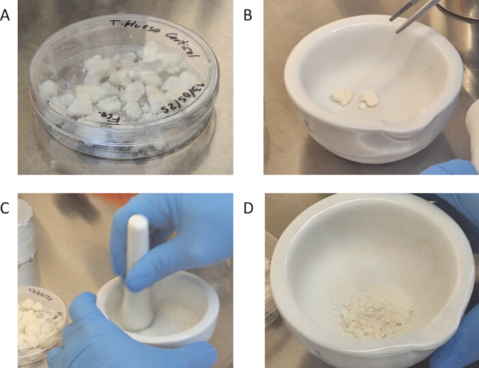
Figure 1: Bone microparticle preparation. (A) Decellularized bone fragments after homogenization with OV5 homogenizer and storage at -80 °C. (B,C) Sterilized ceramic mortar used to mill decellularized bone into a fine powder. (D) Final decellularized bone microparticles after sieving. Please click here to view a larger version of this figure.
5. Bone gel fabrication
- Prepare 10 w/v% GelMA by dissolving 1 g of GelMA in 9.5 mL deionized water at 50 °C.
- Cool the solution to 37 °C and add Irgacure 2959 photoinitiator solution.
- Maintain the solution at 37 °C and perform sterile filtration using a 0.22 µm filter.
- Add decellularized bone microparticles (DBMP) at 1% w/v.
- Mix and pipette up and down carefully, avoiding creating bubbles, at 40 °C until a homogeneous suspension is obtained to form bone gel (BG).
- Store BG at 4 °C for up to 2 weeks.
NOTE: (Critical step) Maintain the hydrogel above its gelling temperature (≥37 °C) before cell incorporation to avoid premature solidification.
6. Osteocyte primary culture
- Bone marrow extraction and initial washing
- Extract and mince bone marrow tissue from the interior of the femoral head using sterile surgical instruments (Figure 2).
- Transfer the minced tissue to a sterile Petri dish.
- Wash the tissue three times with α-MEM culture medium supplemented with antibiotic-antimycotic solution (100 µg/mL penicillin, 100 µg/mL streptomycin, 0.25 µg/mL amphotericin).
- Rinse once with Hank’s Balanced Salt Solution (HBSS).
NOTE: (Critical step) Minimize time between tissue retrieval and processing to preserve cell viability.
- Enzymatic digestion
- Incubate the tissue in collagenase Type I solution for 25 min at 37 °C with gentle agitation.
- Wash with HBSS.
- Repeat steps 6.2.1–6.2.2 two additional times (three total collagenase digestions).
- Incubate the tissue in 0.5 mM ethylenediaminetetraacetic acid (EDTA) for 25 min at 37 °C.
- Wash with HBSS and repeat the EDTA step two additional times.
- Perform one final collagenase Type I digestion (25 min at 37 °C), followed by an HBSS wash.
NOTE: (Troubleshooting) Incomplete digestion may result in low cell yield; extend enzymatic incubation by 5–10 min if needed.
- Cell recovery and culture
- Collect the collagenase solution and transfer the final HBSS wash into a 50 mL centrifuge tube.
- Centrifuge at 1,000 × g for 5 min at room temperature.
- Discard the supernatant and resuspend the pellet in 2 mL of complete α-MEM.
- Seed the cell suspension into two T75 culture flasks and incubate at 37 °C with 5% CO₂.
NOTE: Replace culture medium every 2–3 days until adequate confluency (Figure 3) is achieved.
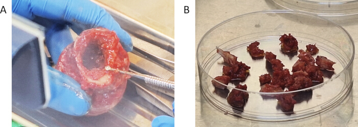
Figure 2: Bone marrow tissue retrieval. (A) Femoral head obtained from hip replacement surgery, showing the medullary region used for cell isolation. (B) Minced medullary bone pieces placed in a sterile Petri dish prior to enzymatic digestion. Please click here to view a larger version of this figure.
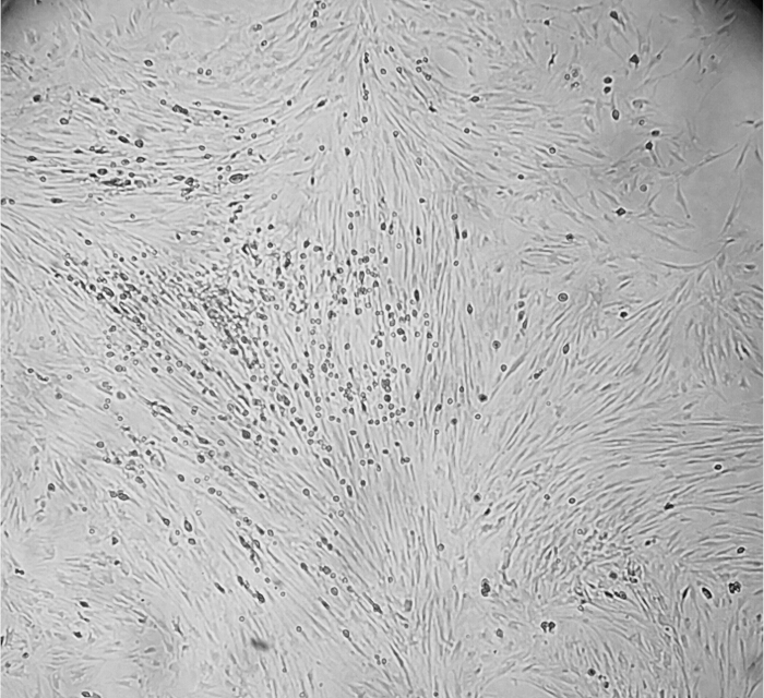
Figure 3: Primary osteocyte culture. Osteocytes attached and proliferating in a monolayer within a T75 following isolation from human medullary bone. Please click here to view a larger version of this figure.
7. 3D cell culture
- Prewarm the amount of BG hydrogel at 37 °C.
NOTE: (Critical step) Ensure the working area is free of drafts; cooling accelerates premature gelling. - Detach cultured osteocytes and centrifuge at 500 × g for 5 min in a 15 mL tube.
- Resuspend the pellet in 200 µL of culture medium and mix with the BG hydrogel to obtain a final cell density of at least 1000 cells/µL (from now on referred to as bioink).
- Aspirate 50 µL of the bioink using a 1000 µL pipette and maintain at 37 °C until deposition.
- Pipette 50 µL of the bioink per construct into each well of the culture plate (Supplementary Video 2); allow 5 min approximately for physical gelation at room temperature (Figure 4).
- Expose each well to 365 nm UV light for 1 min to achieve irreversible crosslinking (0.30 J/cm2).
- Add culture medium and incubate the constructs at 37 °C, 5% CO2 for the required time points. Use a separate plate for each time point.
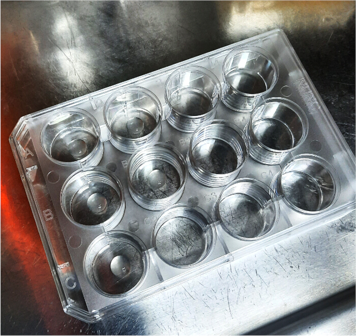
Figure 4: 3D cell culture construct preparation. Well plate containing cell-laden GelMA constructs immediately after pipetting and following UV (365 nm) crosslinking. Please click here to view a larger version of this figure.
Supplementary Video 2: Preparation of 3D cell-laden constructs. Deposition of hydrogel-cell mixtures into a 12-well plate using a simple pipetting technique prior to UV crosslinking.Please click here to download this file.
8. Biocompatibility testing
- Live/dead staining
- Culture the cell-laden scaffolds for the desired time intervals.
- Wash gently with sterile PBS.
- Incubate the constructs for 30 min in the dark with live/dead solution containing 0.2% ethidium homodimer-1 (EthD-1) and 0.05% calcein AM in PBS.
- Image immediately using a fluorescence microscope with filters for calcein AM (~495/515 nm) and EthD-1 ( ~495/635 nm). Standard fluorescein isothiocyanate (FITC) and Texas Red filter sets are compatible with calcein AM and EthD-1 detection, respectively.
- Identify live cells by green cytoplasmic fluorescence and dead cells by red nuclear fluorescence.
NOTE: Planar images can be processed using ImageJ (Fiji). The objective lens, exposure time, gain, and illumination intensity were kept constant for all compared samples and adjusted only to avoid signal saturation. To minimize misinterpretation caused by autofluorescence, the same acquisition settings were maintained for all samples within each experiment, and control observations of scaffold/bone microparticle constructs without cells were used to recognize the background fluorescence pattern associated with the biomaterial. These controls allowed us to distinguish particle-derived signal from true rounded cell staining during image interpretation.
- Alamarblue quantification
- Prepare a 5% alamarblue working solution in complete medium.
- Incubate scaffolds in alamarBlue solution for 2.5 h at 37 °C and 5% CO₂, protected from light.
- Transfer 100 µL of supernatant to a 96-well plate in triplicate, including controls.
- Measure fluorescence at 360/40 nm excitation and 460/40 nm emission.
- Normalize fluorescence values to controls to determine relative metabolic activity.
Access restricted. Please log in or start a trial to view this content.
Results
Primary osteocytes were successfully isolated from human femoral heads and were detectable in culture approximately 5 days after extraction. The cells displayed a stellate morphology and formed interconnected networks characteristic of osteocyte morphology, confirming successful isolation, attachment, and viability (Figure 5).
Access restricted. Please log in or start a trial to view this content.
Discussion
The presented protocol offers an accessible and translational method for generating 3D bone-like constructs, addressing key limitations of current approaches such as reliance on expensive bioprinters or proprietary matrices13. By combining GelMA with human decellularized bone extracellular matrix8, physiologically relevant constructs can be fabricated using only standard laboratory equipment. This simplicity enables adoption in resource-limited environments while preserving...
Access restricted. Please log in or start a trial to view this content.
Disclosures
The authors have nothing to disclose.
Acknowledgements
The authors are grateful to Dr. Carolina Delgado from the Pathological Anatomy Unit of the University of Concepción for her support and for providing access to microscopy resources.
The authors would like to acknowledge the funding support from the Agencia Nacional de Investigación y Desarrollo ANID codes VIU24P0041 and SUC260026.
Access restricted. Please log in or start a trial to view this content.
Materials
| Name | Company | Catalog Number | Comments |
|---|---|---|---|
| 2-Hydroxy-4′-(2-hydroxyethoxy)-2-methylpropiophenone (Irgacure 2959) | Sigma-Aldrich | 410896-10G | Hydrogel polymerization photoinitiator |
| AlamarBlue cell viability reagent | Invitrogen | DAL1100 | For quantitative viability assay |
| Antibiotic-Antymycotic (100´) | Gibco | 15240-062 | Culture medium component |
| Cell culture flask (T75) | SPL Life Sciences | 70075 | For osteocyte culture |
| Collagenase Type 1 | Gibco | 17100-017 | For enzymatic digestion of bone samples |
| Conical tube, 15 mL | SPL Life Sciences | 50015 | For Hydrogel preparation |
| DarkDawn D15-365 nm | DarkDawn | - | For UV Crosslinking of hydrogel |
| DPBS, powder, no calcium, no magnesium | gibco | 21600069 | Hydrogel solvent, LIVE/DEAD kit solvent |
| EDTA Disodium Salt 2-HID | Winkler | ED-0760 | For enzymatic digestion of bone samples |
| Eppendorf centrifuge 5702 | Eppendorf | 5702000010 | For bone microparticle preparation |
| Ethanol absolute for analysis | Supelco | 1,00,98,32,511 | For bone descellularization (diluted to 70%), photoinitiator solvent |
| Fetal bovine serum | Gibco | A52567-01 | Culture medium component |
| Gelatin from porcine skin | Sigma-Aldrich | G1890-500G | Hydrogel component |
| HBSS (10´) | Gibco | 14185-052 | For washing of bone marrow samples |
| L-Ascorbic acid 2-phospate | Sigma-Aldrich | A8960-5G | Culture medium component |
| LIVE/DEAD viability/cytotoxicity Kit | Invitrogen | L3224 | For qualitative viability/cytotoxicity assay |
| MEM Alpha (1´) | Gibco | 12571-063 | Culture medium component |
| Methacrylic anhydride | Sigma-Aldrich | 276685-500G | Hydrogel component |
| Micro centrifuge tube | Citotest | 4610-1844 | For preparation of alamarBlue and LIVE/DEAD dyes |
| NuncIon delta surface | Thermoscientific | 150628 | 12 well plate for seeding of samples |
| OLYMPUS BX43 fluorescence light source Olympus U- LH100HG | Evident | U- LH100HG | Fluorescence analysis light source |
| RC-6 Plus centrifuge | Thermo Scientific Sorvall | 46910 | For bone descellularization and bone microparticle preparation |
| Synergy HTX multi mode reader | BioTek Instruments | - | Spectrophotometer |
| Syringe with needle | NIPRO | SY3-WN-R-03 | For Hydrogel filtering |
| Tissue culture plate 96 well | Jet Biofil | TCP001096 | 96 well for alamarBlue viability assay. |
| Ultrasonic cleaner | VWE | 97043-958 | For sonication of bone samples |
References
- Horvath, P., et al. Screening out irrelevant cell-based models of disease. Nat Rev Drug Discov. 15 (11), 751-769 (2016).
- Hatt, L. P., et al. Standard in vitro evaluations of engineered bone substitutes are not sufficient to predict in vivo preclinical model outcomes. Acta Biomater. 156, 177-189 (2023).
- Kozlowski, M. T., Crook, C. J., Ku, H. T. Towards organoid culture without Matrigel. Commun Biol. 4 (1), 1387 (2021).
- Matai, I., Kaur, G., Seyedsalehi, A., McClinton, A., Laurencin, C. T. Progress in 3D bioprinting technology for tissue/organ regenerative engineering. Biomaterials. 226, 119536 (2020).
- Sun, Q., Choudhary, S., Mannion, C., Kissin, Y., Zilberberg, J., et al. Ex vivo replication of phenotypic functions of osteocytes through biomimetic 3D bone tissue construction. Bone. 106, 148-155 (2018).
- Metzger, W., et al. Expansion and differentiation of human primary osteoblasts in two- and three-dimensional culture. Biotech Histochem. 88 (2), 86-102 (2013).
- Walsh, C. M., Wychowaniec, J. K., Costello, L., Brougham, D. F., Dooley, D. An In vitro and ex vivo analysis of the potential of GelMA hydrogels as a therapeutic platform for preclinical spinal cord injury. Adv Healthc Mater. 12 (26), e2300951 (2023).
- Smith, C. A., Board, T. N., Rooney, P., Eagle, M. J., Richardson, S. M., et al. Human decellularized bone scaffolds from aged donors show improved osteoinductive capacity compared to young donor bone. PLoS One. 12 (5), e0177416 (2017).
- Ryan, S. L., et al. Drug discovery approaches utilizing three-dimensional cell culture. Assay Drug Dev Technol. 14 (1), 19-28 (2016).
- Sittampalam, S., et al. Three-dimensional cell culture assays: Are they more predictive of in vivo efficacy than 2D monolayer cell-based assays?. Assay Drug Dev Technol. 13 (5), 254-261 (2015).
- Knight, A. Systematic reviews of animal experiments demonstrate poor human clinical and toxicological utility. Altern Lab Anim. 35 (6), 641-659 (2007).
- Crapo, P. M., Gilbert, T. W., Badylak, S. F. An overview of tissue and whole organ decellularization processes. Biomaterials. 32 (12), 3233-3243 (2011).
- Schlander, M., Hernandez-Villafuerte, K., Cheng, C. Y., Mestre-Ferrandiz, J., Baumann, M. How Much Does It Cost to Research and Develop a New Drug? A Systematic Review and Assessment. Pharmacoeconomics. 39 (11), 1243-1269 (2021).
- Shah, K. M., Stern, M. M., Stern, A. R., Pathak, J. L., Bravenboer, N., et al. Osteocyte isolation and culture methods. Bonekey Rep. 5, 838 (2016).
- Zhang, W., Lee, W. Y., Siegel, D. S., Tolias, P., Zilberberg, J. Patient-specific 3D microfluidic tissue model for multiple myeloma. Tissue Eng Part C Methods. 20 (8), 663-670 (2014).
- Jiang, Q., Nagano, K., Moriishi, T., Komori, H., Sakane, C., et al. Roles of Sp7 in osteoblasts for the proliferation, differentiation, and osteocyte process formation. J Orthop Translat. 47, 161-175 (2024).
Access restricted. Please log in or start a trial to view this content.
Reprints and Permissions
