This protocol describes a screening platform using freshly isolated porcine buccal epithelial tissue mucosae to evaluate peptide permeation. The method integrates permeation of a GLP-1RA in the presence of a permeation enhancer from solutions, Hilltop chambers, and films in Franz Diffusion Cells, and 3D-printed inserts, accompanied by histological mucosal assessment.
Method Article
A Standardized Ex Vivo Porcine Oromucosal Model for Evaluating Peptide Fluxes
In This Article
Summary
Abstract
Oromucosal administration of drugs in films, tablets, lozenges, and other dosage forms provides a convenient systemic administration route for pediatric and elderly patients, and for patients who cannot swallow capsules or tablets easily. Developing buccal and sub-lingual peptide formulations requires preclinical models that align with the non-keratinized oromucosal epithelial structures of humans, of which pigs and dogs are the most suitable. We sourced discarded fresh porcine oromucosal tissues from an abattoir. The mucosae were dissected from the underlying smooth muscle using controlled heating and then mounted in either Franz Diffusion Cells or in novel low-volume 3D-printed resin-cured inserts. Peptide-permeation enhancer combinations were added to the donor side, and permeated molecules were sampled from the receiver side and quantified by ultra-high performance liquid chromatography (uHPLC). For the inserts, transepithelial electrical resistance (TEER) values of mounted mucosae were measured using EVOM chopstick electrodes. Tissue histology was examined following incubation in both Franz diffusion cells and 3D inserts. The optimal concentration of permeation enhancer was selected when aqueous mixtures of peptide and enhancer produced an increase in permeation. Peptide permeation reached 4.3% over 3 h in three-dimensional (3D) inserts and 1.7% in Hilltop chamber models, the latter simulating direct exposure from a buccal film. Layer-by-layer polymeric mucoadhesive films containing peptide-enhancer combinations were developed and applied to mucosal tissue for permeation studies using diffusion chambers. Ex vivo peptide permeation across porcine buccal mucosae reached 2% when mucosae were exposed to the optimal combination of glycodeoxycholate (GDC) incorporated into the buccal films. These ex vivo screening studies provide a pathway to subsequent pharmacokinetic evaluation of peptide-loaded film formulations administered via the oromucosal route in pigs and people.
Introduction
Buccal and sub-lingual oromucosae are attractive routes of oral drug administration because they offer directly accessible sites and avoidance of hepatic first-pass metabolism, potentially improving bioavailability and patient acceptability compared to other routes, including oral1,2. Peptides are generally given by injection in preference to oral administration due to poor permeation through intestinal epithelia, due to their higher molecular weight, hydrophilicity, and susceptibility to enzymatic degradation3. The FDA has approved 102 peptides for therapeutic applications, with ~450 therapeutic peptides under evaluation in preclinical or clinical trials4. These include oral tablets formulated with permeation enhancers (e.g., semaglutide, Rybelsus)5 and oromucosal platforms (Minirin Melt and Nocdurna sublingual desmopressin acetate) developed using Catalent's Zydis orally disintegrating tablet (ODT) technology6. Despite extensive investigation into buccal drug permeability studies, permeability values of molecules show poor reproducibility and limited comparability across studies7,8. These inconsistencies largely stem from variations in the experimental animal models used and from differences in the handling and preparation of isolated buccal mucosae9.
Ex vivo permeability assays using isolated buccal and sub-lingual tissue mucosae are a valuable tool for predicting in vivo oromucosal small molecule permeability and for preclinical screening of formulation components relevant to dosage forms10,11,12; however, their use in peptide screening has been limited because basal permeability is negligible. In recent years, the porcine buccal mucosa has become the preferred model because its non-keratinized structure and physiological and biochemical characteristics closely resemble those of humans13. Buccal samples from dogs are even less accessible compared to porcine tissue from abattoirs, though both sources are non-keratinized14. Considerable variability in epithelial thickness and processing methods persists. Standardization of buccal and sub-lingual oromucosal preparation is essential. Variables such as short- and long-term storage conditions, tissue thickness, and maintenance of viability during experiments all influence ex vivo permeability results8. Tissue preservation is critical, as both the structural integrity and metabolic activity of the mucosa directly affect diffusion profiles12,15. Loss of barrier function can result in artificially elevated drug permeation rates16,17. Beyond storage, the method used to dissect porcine buccal mucosae for mounting in permeation chambers introduces further variability. Differences in removing submucosal connective tissue, whether by mechanical, chemical, or thermal means, can alter permeability18. Although some reports suggest that heat separation to isolate the epithelium from smooth muscle preserves permeability19, consensus is lacking. Currently, no universally accepted method exists to dissect and assess the integrity of isolated buccal mucosae20. The labeled hydrophilic sugar fluorescein isothiocyanate-dextran (FITC dextran 4000, FD-4) is commonly used as an integrity marker for porcine buccal mucosa because only small molecules below ~500 Da can passively permeate the tissue. By contrast, an intact mucosa restricts FD-4 transport, and a donor-to-receiver transfer below 0.6% is typically considered acceptable over 2-4 h21,22. Additionally, haematoxylin and eosin tissue staining provides a straightforward assessment of epithelial morphology23.
In this work, we established a standardized protocol for mucosal handling of isolated porcine buccal mucosae designed to optimize mucosal integrity and permeability over a set period. Our objectives were to provide an experimental framework for dissecting porcine buccal epithelial tissue and to enhance the reliability of ex vivo permeability assessments of peptides across oromucosae mounted in diffusion chambers and inserts. We first conducted visual analyses to identify the damage-free buccal mucosa from the abattoir. We optimized tissue processing techniques, mucosal thickness, and methods to maintain tissue integrity during experiments. Using a stable low-potency Glucagon-like peptide-1 receptor agonist (GLP-1 RA) analog as a tool molecule, we carried out peptide permeation studies in the presence and absence of a bile salt permeation enhancer, sodium glycodeoxycholate (GDC), on buccal mucosae mounted in Franz diffusion cells and customized home-made 3D printed transwell-type inserts. GDC and the GLP-1 RA were presented to buccal mucosae from solutions, Hilltop chambers, and mucoadhesive polymeric bilayer films. We used permeability markers, tissue histology, and Trans-epithelial/endothelial electrical resistance (TEER) values as our primary evaluation endpoints.
Protocol
The pig cheeks used in this study were collected from a local abattoir; therefore, no ethical approval was required.
1. Porcine buccal epithelium isolation, preparation, and storage
- Collect pig cheek tissues from a local abattoir and transport within 90 min to the laboratory in ice-cold phosphate buffer saline (PBS, pH 7.4). Within the next 60 min, prepare the tissues according to the methodology previously described19,24, with minor modifications for different oromucosae sites as shown in Figure 1.
- Using a scalpel, separate the buccal mucosae from the underlining tissue and rinse the tissue thrice with phosphate-buffered saline (PBS). Discard mucosae with visual surface damage.
- Immerse intact mucosae in PBS at 65 °C for 3 min and separate the epithelium and remaining underlying connective tissue from smooth muscle using a size 5 watchmaker's forceps. This method is also applicable for isolating sublingual and tongue epithelial mucosae.
- Gently dry the dissected oromucosae on cellulose filter paper and proceed to permeation studies (the preferable option) or freeze for a maximum of 4 weeks.
- For the freezing process after separation, immerse oromucosae for 2 h in a cryoprotectant, 85% glycerol25, with gentle shaking. High viscosity glycerol replaces intercellular water to prevent the formation of water crystals, which could damage the tissue structure.
- After this period, replace glycerol with fresh PBS and 10% glycerol. Freeze the mucosal samples at -20 °C or -70 °C, depending on the storage duration required. Perform the freezing procedure at a controlled rate of ~-1 °C·min-1.
NOTE: All experiments are conducted using at least three specimens of tissues from the cheeks of different animals.
2. Ex vivo permeation study in mucosae mounted in Franz diffusion cells and use of Hilltop chambers to present with the GLP-1 RA and GDC
NOTE: Ex vivo permeability studies are performed initially on fresh isolated porcine buccal mucosae using Franz diffusion cells (FDC).
- Mount the dissected tissue mucosae in chambers (window area 1.2 cm2) shown in Figure 2 and equilibrate for 30 min with PBS, pH 6.8 (donor side, 1 mL volume) and PBS, pH 7.4 (receiver side, 10 mL volume).
- After 30 min of equilibration, replace the donor solution PBS (pH 6.8) with 1 mL of PBS containing a 1:1 ratio of the GLP-1RA (25 mg/mL) and GDC (25 mg/mL).
- Keep stirring the receiver chamber with 10 mL of PBS pH 7.4, maintained at 37 °C at 300 rpm. Sample 1 mL from the receiver compartment every 60 min for 180 min. Take triplicate samples from donor compartments at the start and end of the experiment. After each sampling, replenish the chambers with 1 mL of PBS.
- At the end of the experiment, store the tissue in 10% formalin for histology or homogenize it to quantify the GLP-1RA peptide remaining in the tissue.
- Similarly, carry out the permeability of the GLP-1RA using Hilltop chambers (HTC).
NOTE: The purpose of using HTC is to confirm the oromucosal permeation-enhancing effect of GDC on the GLP-1 RA in solution before formulating a bilayer buccal film. The HTC is a chamber constructed from Kraton G-2705, a biocompatible styrene-ethylene-butylene-styrene (SEBS) block copolymer, with an outer diameter of 19 mm, and contains a Webril pad (12.7 mm diameter) composed of 100% nonwoven cotton fibers, which serves as an absorbent non-stick material for solution application26. - Likewise, assess ex vivo permeability of GLP-1RA and GDC (1:1 ratio) loaded in mucoadhesive bilayer films by placing the films in donor chambers of FDCs or in 3D inserts, as described for HTC.
- To confirm the integrity of porcine buccal epithelial tissue, use FD-4 (0.05 mM) as a fluorescent flux marker and add it to the donor side to assess leakage across the tissue.
NOTE: Tissues showing leakage are excluded from permeability calculations. - After the 3 h experiment, quantify the cumulative GLP-1 RA flux by uHPLC analysis of the 200 µL samples withdrawn from the basolateral compartment in triplicate. Repeat all the permeation experiments three times. Quantify the cumulative flux of GLP-1RA from a calibration curve generated from set concentrations of GLP-1RA.
3. Preparation of buccal films entrapped with the GLP-1RA and GDC
NOTE: Buccal bilayer films containing a mucoadhesive and backing layer are manufactured using solvent casting with a semi-automated coater27.
- Prepare a bilayer film for entrapping GLP-1RA using HPMC and PVP-K90 as the film-forming polymers, and a mucoadhesive component, Na-CMC.
NOTE: Refer to Table 1 for the composition of the mucoadhesive and backing layer of the buccal film. GDC is incorporated with a GLP-1 RA peptide in model films in low mg quantities to see its effect on peptide permeation across porcine buccal mucosae mounted in chambers. - Prepare the mucoadhesive layer by dissolving the film-forming polymers, 45 mg of HPMC, 10 mg of Na-CMC, and 25 mg of PVP-K90, in deionized water containing 3% glycerol at 400 rpm for 10 min using a magnetic hot plate stirrer.
NOTE: HPMC is soluble at room temperature, whereas PVP-K90 requires heating at 40 °C for 30 min to dissolve. - Add 2 mg of Aerosil 200 F under stirring, followed by the addition of the GLP-1 RA. Then, add GDC and disperse it at 600 rpm for 30 min.
- Ultrasonicate the final polymer solution for 90 min using an ultrasonication bath to remove bubbles.
- Cast films onto a polyethylene terephthalate (PET) liner and coat with a doctor blade set to 2600 µm at 2 mm/s. Dry the films in a hot air oven at 40 °C for ~1 h.
- For the backing layer, dissolve 5% ethyl cellulose (EC) and 5% propylene glycerol (PG) in hydroalcoholic water (ethanol:water), then cast the dissolved slurry onto a PET liner. Finally, cut the dried films into 1 cm × 1 cm squares, pack in aluminum sachets, and store at 4 °C.
4. Printing of a custom 3D transwell-mimicking insert
NOTE: The 3D-printed custom inserts are fabricated following a previously described method with minor modifications28.
- Briefly, design the 3D model using computer-aided design (CAD) software.
- Keep the dimensions of the 3D insert the same as the main cylindrical body (donor chamber), with a maximum surface area of 1659.06 mm2, and overall external dimensions of 25.20 mm along the x-axis, 14.50 mm along the y-axis, and 21.63 mm along the z-axis.
- In addition, design a detachable circular retaining ring to support the epithelial tissue. The ring has an outer diameter of approximately 19.90 mm and an inner diameter of 17.93 mm, corresponding to the dimensions of the main cylindrical body, and can fit in Transwell 12-well plates (Figure 3).
- Fabricate all the components using a desktop stereolithography (SLA) 3D printer with a UV-curable clear resin. All dimensions are approximate and may be adjusted as required to ensure proper fitting in Transwells and experimental compatibility.
NOTE: To ensure the removal of unreacted monomers and potential leachables, all parts underwent a standardized post-processing protocol, including a 10-min wash in pristine 99% isopropyl alcohol (IPA) followed by UV curing according to manufacturer specifications, i.e., 1 min at ambient temperature in a Form Cure 2. Stereolithography file (STL) for 3D model design can be found at https://doi.org/10.5281/zenodo.19368577 or supplementary files (Supplementary File 1 and Supplementary File 2).
5. Permeation across mucosae mounted in 3D inserts
NOTE: Ex vivo permeability of GLP-1RA and GDC incorporated into mucoadhesive bilayer buccal films can also be assessed using low-volume, custom-designed Transwell-mimicking 3D inserts (Figure 2) as an alternative to FDCs. This is because the 10 mL receiver side of FDCs dilutes the peptide, challenging the detection sensitivity of uHPLC. The FDC design is therefore not cost-effective or practical for peptide fluxes, as it would require enzyme-linked immunosorbent assay (ELISA) or radioimmunoassay (RIA) detection on an ongoing basis.
- Manufacture/print the inserts in a UV-curable resin using a desktop SLA 3D printer28, and mount them in 12-well tissue culture plates.
- Position the porcine buccal mucosae across a window area of 1.4 cm2 between the apical and basolateral chambers, measure the baseline transepithelial electrical resistance (TEER) using EVOM 2 voltohmmeter with STX2 chopstick electrodes placed in the donor and receiver sides of the inserts. TEER is calculated by Ohm's Law.
- After placing the film (1.0 × 1.0 cm2) on the top of the tissue, pipette 0.5 mL of PBS (pH 6.8) into the apical chamber and 1.5 mL of PBS (pH 7.4) into the basolateral chamber.
- Collect samples (0.2 mL) from the receiver sides at 1 h intervals over 3 h and replace with equal volumes of fresh buffer.
- Quantify the fluxed GLP-1 RA peptide by uHPLC.
NOTE: No adsorption of GLP-1RA with 3D inserts is observed after 3 h, as shown in Supplementary Figure 1 and Supplementary File 3. - Measure the transepithelial electrical resistance (TEER) using EVOM 2 voltohmmeter with STX2 chopstick electrodes at 1 h intervals over 3 h. TEER is calculated by Ohm's Law.
- To confirm porcine buccal and sublingual epithelial tissue integrity, use FD-4 (0.05 mM) as a fluorescent marker.
NOTE: All experiments are performed in triplicate, and reported results are the average ± SD of three sets of data.
6. uHPLC detection of the GLP-1 RA
- Quantify the quantity of the GLP-1 RA permeated across oromucosae mounted in FDCs and inserts, from each of donor side solutions, HTCs, and films by uHPLC using a CSH C18 column (130 Å, 1.7 µm, 2.1 mm × 50 mm) with a mobile phase of 0.1% TFA in acetonitrile (Eluent A) and 0.1% TFA in water (Eluent B), gradient elution over 8 min at 30 °C, with 20 µL injection volume and 0.7 mL/min flow rate, and UV detection at 214 nm (retention time: ~4.2 min).
- Perform a linearity study.
- Design a linearity study to estimate the analytical response of the optimized uHPLC conditions using GLP-1 calibration standards in the range of 2.5–20.0 µg·mL⁻1.
- Prepare calibration standards (n = 6–8 levels) independently and inject samples in triplicate. Perform a least-squares linear regression of peak area (mAU) versus nominal concentration, and assess linearity using the coefficient of determination (r2), the % y-intercept, and the distribution of residuals (observed–predicted response).
NOTE: Visual inspection of the calibration plot and the residuals plot indicated an insignificant deviation from linearity at the lowest sensitivity level (2.5 µg·mL⁻1), with residuals within the predefined acceptance limit (±3%). The calibration range is subsequently restricted to a range of 2.5–20.0 µg·mL⁻1, at which the regression showed acceptable linearity (r2 ≥ 0.999, % y-intercept < 2%, and all residuals within ±3%).
7. Estimation of GLP-1RA retained in buccal tissue
- Remove the porcine buccal epithelial mucosae exposed to the GLP-1 RA and GDC from FDCs/inserts.
- Rinse the tissue thrice with PBS to remove adsorbed drug and film excipients.
- Then, homogenize the tissue in 5 mL of PBS using a tabletop homogenizer until completely fragmented.
- Store the sample overnight at 4 °C to allow equilibration of peptide partitioning between homogenized tissue and PBS.
- Subsequently, centrifuge the mixture at 5000 × g for 5 min to precipitate the fragments and collect the supernatant for quantitation.
- Sterilize the supernatant through 0.22 µm centrifuge filters to remove residual particles before uHPLC analysis.
8. Oromucosal tissue histology
NOTE: After 3 h of permeation of the GLP-1 RA from mixtures, HTCs, or films, mucosae are processed for assessment of damage.
- Fix the tissues in 10% formalin for 48 h, and then embed the fixed tissues in paraffin.
- Section the tissues at a size of 5 µm using a microtome.
- Then stain tissues with hematoxylin and eosin (HE) to examine the histological integrity using light microscopy at 4× magnification.
- Slides are imaged with an upright microscope and analyzed using image analysis software.
NOTE: The overall workflow is summarized in Figure 4.
Results
Isolation and dissection of porcine buccal epithelial tissues
Porcine buccal mucosa is widely used as a surrogate tissue for human oromucosae because they share comparable anatomical features and physiological barrier properties29. Porcine buccal epithelial tissues are isolated with both surgical and heating methods, as shown in Figure 4. Initially, a scalpel is used to remove muscles and deeper connective tissues from the cheeks. The isolated pieces of tissue with combined and intact mucosal layers and connective tissue are then placed into warm PBS heated to 65 °C for 3 min. The tissue is removed from the buffer, and the epithelium and underlying lamina propria are peeled off with the size 5 forceps. Heating allows for a consistent, uniform thickness in the resulting dissected epithelium.
Ex vivo permeation of a GLP-1RA across porcine buccal oromucosae mounted in FDCs and 3D-printed inserts
Ex vivo permeation of GLP-1RA was assessed using porcine buccal epithelial tissue mucosae mounted in FDCs and inserts. To assess the optimum concentration ratio of the GLP-1 RA: GDC, HTCs containing cotton pads with an area of 12.7 mm were affixed to the donor side of mucosae mounted in FDCs, and the peptide-enhancer mixture was pipetted onto the cotton pad. The cumulative permeation of the GLP-1 RA reached approximately 4.3% over 3 h when GDC was included at a 1:1 ratio (GLP-1RA 25 mg: GDC 25 mg). In contrast, in the absence of GDC, no detectable GLP‑1RA flux was observed27. Quantitative permeability parameters, including the Papp are calculated using the following formula:
Papp = 
Where:
Papp: apparent permeability coefficient (cm/s).
dQ/dt: transport rate of the GLP-1RA at steady state, calculated as the amount of peptide transported per unit time (µg/s).
A: area of 3D insert (cm2).
Co: the initial concentration of the peptide in the donor (apical) compartment (µg/mL).
Apparent permeability coefficient (Papp) of GLP-1RA across porcine buccal mucosae is shown in Supplementary Figure 2. When the GLP‑1RA:GDC ratio was 1:1, the same concentrations as for HTCs were incorporated into bilayer films; however, GLP‑1RA permeation across isolated porcine buccal tissues was not quantifiable using the conventional FDC setup, as the presentation of the GLP-1RA from the films to mucosae is slower than from either mixtures in solution or from HTCs. To overcome this limitation, customized low-volume 3D‑printed inserts were developed to reduce the receiver volume to 1.5 mL, thereby improving detection sensitivity. Using 3D inserts, the cumulative permeation of GLP‑1RA from buccal films over 3 h was measurable and reached approximately 1.8 %, as shown in Figure 5. TEER values and FD-4 fluxes were used to evaluate epithelial barrier integrity and paracellular permeability. A time-dependent decline in TEER was observed (Supplementary Figure 3) in the treatment groups and corresponded with increased GLP‑1RA permeation. PBS and blank film–treated tissues showed no changes in TEER, indicating that the bilayer film matrix did not disrupt the barrier. These data implicate GDC as the primary contributor to the permeation-enhancing effect.
Porcine tissue histological assessment
Histological evaluation of ex vivo porcine buccal mucosa was performed to assess the impact of tissue isolation methods and to examine epithelial integrity following 3 h exposure to GLP-1RA and GDC. As shown in Figure 6, freshly isolated tissues obtained by the heating method preserved normal epithelial physiological architecture, with intact stratified layers and continuous surface mucus, and showed no evidence of structural damage. In contrast, tissues isolated by mechanical separation or subjected to storage/freezing exhibited minor alterations in epithelial cell morphology and a reduction in surface mucus thickness. Buccal mucosae exposed to 25 mg of GLP-1RA in the presence of 25 mg of GDC maintained epithelial integrity and closely resembled untreated controls. Only mild edema and superficial epithelial erosion were observed. Exposure to GLP-1RA alone or to blank and GLP-1RA: GDC-loaded bilayer films did not produce visible tissue damage, and histological features were comparable to those of PBS-treated control tissues.
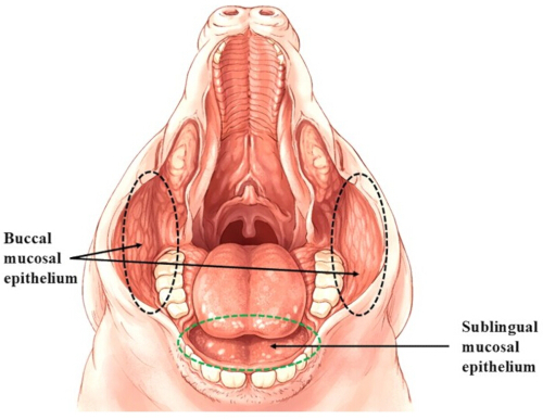
Figure 1: Porcine oral cavity showing buccal and sublingual mucosal areas. Please click here to view a larger version of this figure.
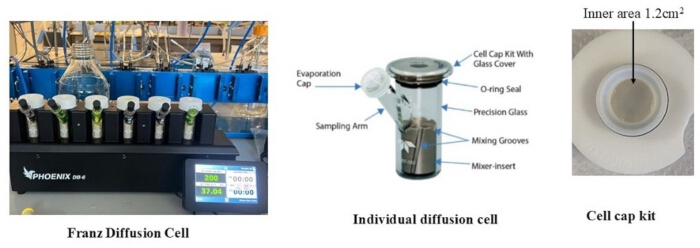
Figure 2: FDCs and components. Six chambers are mounted in an integrated system (left). An individual, Franz Cell, is shown (middle). The mounting system for buccal tissue in an individual chamber (right). Please click here to view a larger version of this figure.
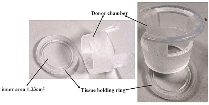
Figure 3: Components of 3D-printed 1 cm2 resin inserts. Please click here to view a larger version of this figure.
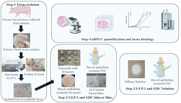
Figure 4: Workflow for porcine buccal epithelium isolation, mounting, permeation, and histological characterization processes. Created in BioRender.com. Please click here to view a larger version of this figure.
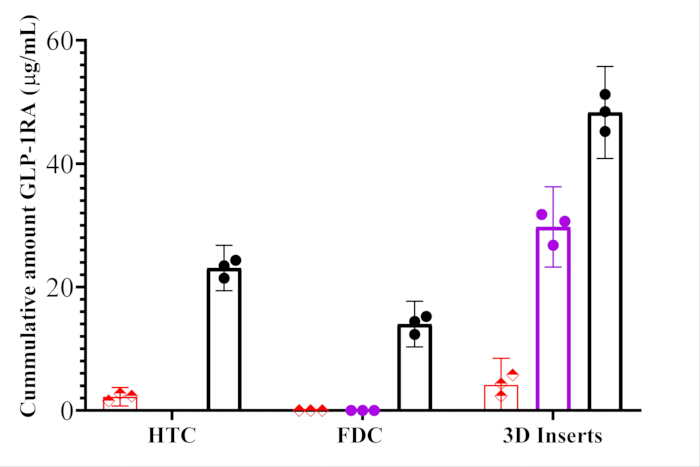
Figure 5: Cumulative amount of the GLP-1 RA. Ex vivo cumulative amount of the GLP-1 RA permeated on the receiver side of porcine buccal epithelial mucosa after 180 min application of 25 mg/mL GLP-1RA (Red), 25 mg/mL GLP-1RA: 25 mg/mL GDC solution (Black), and 25 mg/mL GLP-1RA: 25 mg/mL GDC loaded in films (purple) applied through HTC, FDC, and 3D inserts. Please click here to view a larger version of this figure.
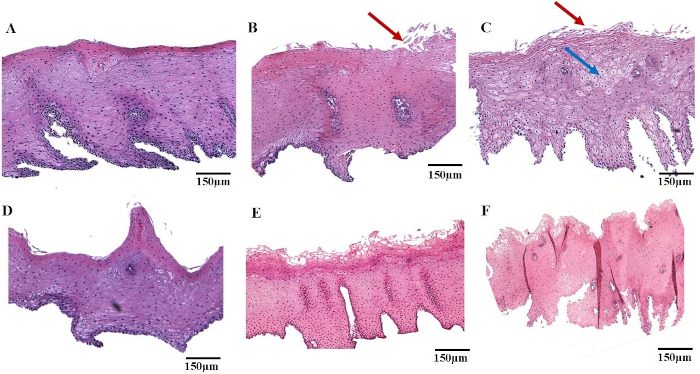
Figure 6: HE-stained microscopic images of porcine buccal epithelial mucosae under different exposure conditions. (A) Blank film, (B) aqueous mixture of GLP-1RA: GDC, (C) films entrapped with GLP-1RA:GDC. The red arrows show superficial epithelial erosion, and the blue arrow shows mild edema. Please click here to view a larger version of this figure.
| Composition of Mucoadhesive layer | |
| Components | Concentration (mg) |
| GLP-1RA | 12.5 mg |
| GDC | 12.5 mg |
| HPMC | 45 mg |
| Na-CMC | 10 mg |
| PVP-K90 | 25 mg |
| Glycerol | 3 mg |
| Aerosil 200F | 2 mg |
| Backing layer | |
| Ethyl cellulose | 5 mg |
| Propylene glycol | 5 mg |
Table 1: Composition of mucoadhesive and backing layers of buccal film.
Supplementary Figure 1: Adsorption of GLP-1RA after incubating 0.5 mg of GLP-1RA for 6 h with 3D inserts. Data is mean ± SD of three experiments (n = 3).Please click here to download this file.
Supplementary Figure 2: Apparent permeability coefficient (Papp) of a GLP-1RA after 3-h exposure of porcine buccal mucosal tissue mounted in 3D inserts. 25 mg/mL GLP solution (red), 25 mg/mL GLP + 25 mg/mL GDC aqueous solution (black), and 25 mg/mL GLP + 25 mg/mL GDC incorporated in buccal films (purple). Data represented is mean ± SD (n = 3). Statistical significance between groups was observed: p < 0.05.Please click here to download this file.
Supplementary Figure 3: Transepithelial resistance (TEER). Gradual reduction of transepithelial resistance (TEER) from isolated porcine buccal epithelial tissue fixed in 3D inserts, after exposure to 25 mg/mL GLP-1 + 25 mg/mL GDC solution (red), 25 mg/mL GLP-1 + 25 mg/mL GDC in buccal films (green), and 25 mg/mL GLP-1RA in PBS (blue).Please click here to download this file.
Supplementary Figure 4: Tissue integrity and leakage. Tissue integrity and leakage were evaluated by exposing porcine buccal epithelial tissues in 3D inserts to 0.05 mM FD4 on the donor side. Samples were withdrawn at fixed intervals of 60 min over 6 h. Mean +SD of three experiments.Please click here to download this file.
Supplementary File 1: transwell-Oring-v7 (JOVE).stl.Please click here to download this file.
Supplementary File 2: transwell-v9 (JOVE).stl.Please click here to download this file.
Supplementary File 3: Additional experimental details.Please click here to download this file.
Discussion
This methodology describes experimental protocols for the isolation of porcine buccal and other oromucosal epithelia, along with permeability assays for peptide-based therapeutics using a low-potency stable GLP-1RA analog as an exemplar30. Peptides are hydrophilic macromolecules with negligible permeability across mucosal membranes17. The successful execution of the above protocol relies on precise isolation and mounting of the buccal and sublingual mucosa. Isolation requires specialized skill and rigorous temperature control; even minor thermal fluctuations during the heating process can induce deleterious physiological and morphological changes in the epithelial tissue, ultimately compromising the integrity of permeation data. Furthermore, the fixing of the isolated tissue within the customized 3D-printed inserts represents a critical step. Given the delicate nature of the epithelial layers, there is a higher risk of tissue rupture or leakage during mounting. To mitigate this, we employed an FD4 leakage test, which serves as a quality control measure to ensure membrane integrity before commencing flux studies. FD4 leakage was initially assessed visually and further quantified using UV-visible spectroscopy. The corresponding data is provided in the supplementary data (Supplementary Figure 4 and Supplementary File 3).
Conventional FDC, considered as standard for ex vivo studies, often presents challenges when quantifying peptide therapeutics due to the high volumes in the receiver chamber, typically 10 mL. The high volume leads to excessive sample dilution, pushing permeated peptide concentrations below the lower LOD for standard analytical methods. To avoid this, we developed a novel 3D-printed insert that mimics the scale and volumes of transwells system used for epithelial cell monolayers, including Caco-231. The inserts can be directly placed in standard 12-well tissue culture plates and securely accommodate isolated porcine buccal epithelial mucosae. This modification significantly reduces the donor compartment volume nearly 7-fold to 1.5 mL, thereby concentrating on the analyte and overcoming sensitivity limitations of uHPLC. This adaptation is particularly important for the quantification of costly low-permeability peptide drugs.
An increase in GLP-1 RA permeation in the presence of GDC was observed using the 3D insert system. The cumulative permeated amount of GLP-1RA reached ~55.3 µg at 3 h, corresponding to ~4.3 % of the initially applied concentration to the donor side. In contrast, no measurable GLP-1 RA flux was detected when similar concentrations of GLP-1RA and GDC entrapped in buccal films were applied to buccal mucosae mounted in FDCs, suggesting that peptide permeation may have occurred but remained analytically undetectable due to excessive dilution in the large receiver volume. However, cumulative permeation of GLP-1 RA from the bilayer buccal film formulation reached approximately 1.8 % at 3 h in 3D inserts over 3 h, demonstrating the feasibility of detecting low-level peptide transport across buccal tissue from dosage forms using the miniaturized receiver system. The ex vivo permeation of GLP-1 RA was also evaluated using HTCs containing a cotton pad loaded with the peptide and GDC. This is a very useful initial screening device to prove the concept that presentation of a peptide from films with a permeation enhancer can be possible. Barrier integrity of porcine buccal epithelium can be monitored during the experiment using TEER and FD-4 leakage. A slow, time-dependent decrease in TEER was observed during exposure to GLP-1 receptor agonist (GLP-1 RA), which is consistent with increased paracellular transport induced in buccal mucosae by the bile salt32.
A limitation of this technique is a reliance on fresh porcine tissue, which is not always available on demand from abattoirs, and tissue quality can be variable. To address the constraints of obtaining fresh tissue at a reasonable distance from the lab, we evaluated cryoprotection for storing high-quality samples that we could use later. While using fresh tissue is optimal, the use of isolated stored mucosal tissues requires careful use of cryoprotectants and freezing rates to prevent ice crystal formation and subsequent barrier damage. The protocol has the capacity to bridge the gap between standard macro-scale permeation studies and high-sensitivity peptide analysis. By optimizing both the tissue isolation process and permeation protocol using 3D inserts, we have achieved a higher degree of reproducibility and sensitivity than is typically reported with FDCs. Furthermore, the use of 3D inserts provides a framework for researchers to provide detection limits for costly peptide molecules, representing a significant improvement in the flexibility and throughput of ex vivo mucosal research.
These protocols provide a foundation for reliable measurement of mucosal tissue permeation using drug classes beyond peptides. The capacity to generate high-resolution permeation profiles from ex vivo models will assist predictions for preclinical absorption data derived from animal models. The GLP-1 RA ex vivo permeation was also evaluated using HTCs containing a cotton pad loaded with the peptide and GDC, a permeation enhancer solution. This delivery approach ensured intimate, reproducible contact between the admixture and the mucosal surface. Significant increase in the permeation of GLP-1RA was observed with 1:1 ratio of GDC in transwells-mimicking 3D inserts. This is a very useful screening device to prove the concept that presentation of a peptide from films with a permeation enhancer can be possible in a constructed design adapted from cultured monolayers of epithelial cells to tissue mucosae. Barrier integrity of porcine buccal epithelium can be monitored during the experiment using TEER measurement and FD4 leakage.
Overall, the ex vivo permeation and histological assessments demonstrate that reliable assessment of buccal and sub-lingual peptide transport in the presence of a permeation enhancer depends on tissue preparation, flux protocols, and analytical sensitivity. Among the isolation approaches examined, separation of freshly sourced porcine buccal and sublingual epithelium using the heat method provided a good balance between maintaining epithelial integrity and tissue dissection reproducibility, as evidenced by stable baseline TEER, minimal FD4 leakage, and preserved tissue cell structures. Importantly, the 3D‑printed insert markedly improved experimental reliability by reducing receiver compartment dilution, enabling quantification of low peptide fluxes that were undetectable in FDCs. Taken together, these findings support the use of isolated porcine buccal epithelial mucosae in a tissue insert chamber as an ex vivo platform for evaluating buccal administration of therapeutic peptides for systemic delivery.
Disclosures
The authors have no financial involvements or conflicts of interest to disclose.
Acknowledgements
The authors thank Ben Shovlin (UCD School of Veterinary Medicine) for technical assistance. We also thank Dr. Hans Eckhardt (UCD School of Chemistry) for support with uHPLC analysis. This project has received funding from the European Union's Horizon Europe research and innovation program under grant number 101071054. We also acknowledge additional funding support from Grant Number 13/RC/2073_P2, the Research Ireland CURAM Center for Medical Devices, and the European Development Fund. The authors would like to thank Kenneth McNamara of Rosderra Irish Meats for the provision of porcine cheeks.
Materials
| Name | Company | Catalog Number | Comments |
|---|---|---|---|
| Computer / Laptop | Generic | Any standard system | ≥8 GB RAM recommended |
| Data acquisition script | Custom | collect.py | Python-based acquisition script |
| Disposable gel electrodes | Generic | Pre-gelled Ag/AgCl electrodes | Single-use, adhesive |
| EEG acquisition module | Upside Down Labs | BioAmp EXG Pill | Analog front-end for biopotential signals |
| Electrode gel (if reusable electrodes used) | Generic | Conductive gel | Improves signal conductivity |
| Interactive application (game) | Generic | Any simple game (e.g., racing) | Keyboard-controlled interface |
| Jumper wires | Generic | Male–Female connectors | Standard breadboard wires |
| Matplotlib / Seaborn | Open-source | Latest versions | Visualization libraries |
| Microcontroller board | Arduino | Uno / Maker Uno | 10-bit ADC, USB interface |
| Python programming environment | Python Software Foundation | Python ≥3.8 | Open-source programming language |
| PyTorch library | Meta AI | Version ≥1.12 | Deep learning framework |
| Scikit-learn | Open-source | Latest stable version | ML utilities |
| Signal processing libraries | SciPy | Latest version | Filtering, STFT |
| USB cable | Generic | USB Type-A to B | Data transfer cable |
References
- Karki, S., et al. Oromucosal films for peptide delivery: formulation strategies using permeation enhancers and polymers. Drug Deliv Transl Res. , (2025).
- Karavasili, C., Eleftheriadis, G. K., Gioumouxouzis, C., Andriotis, E. G., Fatouros, D. G. Mucosal drug delivery and 3d printing technologies: a focus on special patient populations. Adv Drug Deliv Rev. 176, 113858(2021).
- Brayden, D. J., Hill, T., Fairlie, D., Maher, S., Mrsny, R. Systemic delivery of peptides by the oral route: formulation and medicinal chemistry approaches. Adv Drug Deliv Rev. 157, 2-36 (2020).
- Al Musaimi, O. FDA's stamp of approval: unveiling peptide breakthroughs in cardiovascular diseases, ACE, HIV, CNS, and beyond. J Pept Sci. 30 (11), e3627(2024).
- Lewis, A. L., McEntee, N., Holland, J., Patel, A. Development and approval of Rybelsus (oral semaglutide): ushering in a new era in peptide delivery. Drug Deliv Transl Res. 12 (1), 1-6 (2022).
- Zhang, Y., Zhang, H., Ghosh, D., Williams, R. O. Just how prevalent are peptide therapeutic products? a critical review. Int J Pharm. 587, 119491(2020).
- Wanasathop, A., Patel, P. B., Choi, H. A., Li, S. K. Permeability of buccal mucosa. Pharmaceutics. 13 (11), 1814(2021).
- Mazzinelli, E., et al. Oral mucosa models to evaluate drug permeability. Pharmaceutics. 15 (5), 1559(2023).
- Djekic, L., Martinovic, M. In vitro, ex vivo and in vivo methods for characterization of bioadhesiveness of drug delivery systems. Bioadhesives Drug Deliv. , (2020).
- Mura, P., et al. A preliminary study for the development and optimization by experimental design of an in vitro method for prediction of drug buccal absorption. Int J Pharm. 547 (1-2), 530-536 (2018).
- Majid, H., Bartel, A., Burckhardt, B. B. Development, validation and standardization of oromucosal ex-vivo permeation studies for implementation in quality-controlled environments. J Pharm Biomed Anal. 194, 113769(2021).
- Wakchaure, R. S., Dhial, K., Pathak, A. Ethical and Safety Considerations in Drug Delivery Systems. Next-Generation Drug Delivery Systems. , Springer, Humana. New York, NY. (2025).
- Wang, S., Zuo, A., Guo, J. Types and evaluation of in vitro penetration models for buccal mucosal delivery. J Drug Deliv Sci Technol. 61, 102122(2021).
- Sa, G., et al. Histological features of oral epithelium in seven animal species: as a reference for selecting animal models. Eur J Pharm Sci. 81, 10-17 (2016).
- Patel, V. F., Liu, F., Brown, M. B. Modeling the oral cavity: in vitro and in vivo evaluations of buccal drug delivery systems. J Control Release. 161 (3), 746-756 (2012).
- Tian, Y., et al. Oromucosal films: from patient centricity to production by printing techniques. Expert Opin Drug Deliv. 16 (9), 981-993 (2019).
- Malhotra, S., Lijnse, T., O'cearbhaill, E., Brayden, D. J. Devices to overcome the buccal mucosal barrier to administer therapeutic peptides. Adv Drug Deliv Rev. 220, 115572(2025).
- De Vries, M. E., et al. Localization of the permeability barrier inside porcine buccal mucosa: a combined in vitro study of drug permeability, electrical resistance and tissue morphology. Int J Pharm. 76 (1-2), 25-35 (1991).
- Diaz Del Consuelo, I., Pizzolato, G. P., Falson, F., Guy, R. H., Jacques, Y. Evaluation of pig esophageal mucosa as a permeability barrier model for buccal tissue. J Pharm Sci. 94 (12), 2777-2788 (2005).
- Pather, S. I., Rathbone, M. J., Şenel, S. Current status and the future of buccal drug delivery systems. Expert Opin Drug Deliv. 5 (5), 531-542 (2008).
- Junginger, H. E., Hoogstraate, J. A., Verhoef, J. C. Recent advances in buccal drug delivery and absorption—in vitro and in vivo studies. J Control Release. 62 (1-2), 149-159 (1999).
- Goswami, T., Jasti, B. R., Li, X. Estimation of the theoretical pore sizes of the porcine oral mucosa for permeation of hydrophilic permeants. Arch Oral Biol. 54 (6), 577-582 (2009).
- Otali, D., Fredenburgh, J., Oelschlager, D., Grizzle, W. A. Standard tissue as a control for histochemical and immunohistochemical staining. Biotech Histochem. 91 (5), 309-326 (2016).
- Franz-Montan, M., et al. Evaluation of different pig oral mucosa sites as permeability barrier models for drug permeation studies. Eur J Pharm Sci. 81, 52-59 (2016).
- Pegg, D. E. Cryopreservation and Freeze-Drying Protocols. , Springer, Humana. New York, NY. (2014).
- Hoogstraate, A., et al. In vivo buccal delivery of fluorescein isothiocyanate-dextran 4400 with glycodeoxycholate as an absorption enhancer in pigs. J Pharm Sci. 85 (5), 457-460 (1996).
- Karki, S., et al. A pullulan-based bilayer film for buccal delivery of a GLP-1 peptide analogue. Carbohydr Polym. 380, 125064(2026).
- Shu, W., et al. Computational and experimental analysis of drug-coated microneedle skin insertion: the mode of administration matters. Adv Mater Interfaces. 12 (17), 2500202(2025).
- Marxen, E., Axelsen, M. C., Pedersen, A. M. L., Jacobsen, J. Effect of cryoprotectants for maintaining drug permeability barriers in porcine buccal mucosa. Int J Pharm. 511 (1), 599-605 (2016).
- Rebollo, R., et al. Salcaprozate-based ionic liquids for GLP-1 gastric delivery: a mechanistic understanding of in vivo performance. J Control Release. 377, 267-276 (2025).
- Hubatsch, I., Ragnarsson, E. G., Artursson, P. Determination of drug permeability and prediction of drug absorption in Caco-2 monolayers. Nat Protoc. 2 (9), 2111-2119 (2007).
- Brayden, D. J., Stuettgen, V. Sodium glycodeoxycholate and sodium deoxycholate as epithelial permeation enhancers: in vitro and ex vivo intestinal and buccal bioassays. Eur J Pharm Sci. 159, 105737(2021).
Reprints and Permissions
