This protocol describes a standardized multi-voxel proton magnetic resonance spectroscopy approach used in a retrospective cohort study to evaluate intracranial gliomas and tumefactive demyelinating lesions by quantifying the N-acetylaspartate-to-choline ratio. The method supports metabolic assessment and their differential diagnosis.
Research Article
The N-Acetylaspartate to Choline Ratio Differentiates Intracranial Gliomas from Tumefactive Demyelinating Lesions in a Retrospective Cohort Study
* These authors contributed equally
In This Article
Summary
Abstract
Intracranial gliomas and tumefactive demyelinating lesions (TDLs) often exhibit overlapping features on conventional structural magnetic resonance imaging (MRI), making their differential diagnosis challenging. This retrospective cohort study evaluated the diagnostic performance of quantitative multi-voxel proton magnetic resonance spectroscopy (1H-MRS) for differentiating these lesions. A total of 63 patients with indeterminate intracranial masses, including gliomas, TDLs, and primary central nervous system lymphomas, underwent 1H-MRS. Absolute and relative metabolite ratios were measured within lesional solid cores and contralateral normal-appearing brain tissue. Regions of interest were independently delineated by two blinded neuroradiologists. Although both gliomas and TDLs demonstrated significantly increased choline-to-creatine (Cho/Cr) ratios relative to normal tissue, the Cho/Cr ratio showed limited diagnostic specificity. In contrast, the N-acetylaspartate-to-choline (NAA/Cho) ratio differed significantly between the two groups. The median NAA/Cho ratio was 1.22 (interquartile range [IQR], 0.75–1.60) in TDLs and 0.32 (IQR, 0.20–0.69) in high-grade gliomas. Receiver operating characteristic analysis demonstrated that an NAA/Cho threshold of ≤0.649 yielded a sensitivity of 72.7% (95% confidence interval [CI], 55.8%–84.9%), a specificity of 93.3% (95% CI, 70.2%–98.8%), and an area under the curve (AUC) of 0.848 (95% CI, 0.74–0.96). Inter-reader reproducibility was high, with intraclass correlation coefficients (ICCs) greater than 0.99. These findings suggest that quantitative 1H-MRS, particularly the NAA/Cho ratio, may provide a useful noninvasive metabolic biomarker to support the differentiation of intracranial gliomas from TDLs, although further validation in larger, multicenter cohorts is warranted.
Introduction
Primary brain and other central nervous system (CNS) tumors represent a major challenge in neuro-oncology because of their substantial morbidity and mortality. According to the Central Brain Tumor Registry of the United States (CBTRUS), the average annual incidence rate of primary brain tumors is 24.83 per 100,000 population1. Gliomas constitute approximately 26.3% of these tumors, with glioblastoma being the most common malignant primary brain tumor and accounting for more than half of all malignant cases1. The management of gliomas typically involves a multimodal approach comprising maximal safe surgical resection, followed by concurrent chemoradiotherapy and adjuvant chemotherapy. Accurate and timely diagnosis is therefore essential for surgical planning and prognostic assessment.
Conversely, tumefactive demyelinating lesions (TDLs) are relatively rare, atypical manifestations of inflammatory demyelinating diseases that can closely mimic primary brain tumors both clinically and radiologically2. TDLs are generally defined as solitary or multiple demyelinating lesions measuring more than 2.0 cm in diameter and are frequently accompanied by mass effect, perilesional edema, and contrast enhancement3. Unlike gliomas, TDLs are managed medically. Acute-phase treatment primarily relies on high-dose intravenous corticosteroids or plasma exchange, followed by appropriate disease-modifying therapies for underlying conditions such as multiple sclerosis (MS) or neuromyelitis optica spectrum disorders4,5. Because these conditions require fundamentally different therapeutic approaches, misdiagnosing a TDL as a high-grade glioma (HGG) may subject patients to unnecessary stereotactic biopsy or craniotomy, with the associated risks of permanent neurological deficits and other procedure-related complications6.
Differentiating these two entities using conventional magnetic resonance imaging (MRI) remains a persistent clinical challenge. Both HGGs and TDLs typically present as large supratentorial space-occupying lesions7. Although certain conventional MRI findings, such as the “open-ring” enhancement pattern, in which the non-enhancing portion of the ring faces the gray matter, are highly suggestive of atypical demyelination8, this feature is absent in a substantial proportion of cases. Furthermore, atypical TDLs may exhibit irregular, closed-ring, or garland-like peripheral enhancement with central hypointensity that is difficult to distinguish from the central necrosis commonly observed in glioblastomas9. Consequently, conventional structural MRI alone is often insufficient for confident differential diagnosis.
Advanced physiological imaging techniques, particularly proton magnetic resonance spectroscopy (1H-MRS), have emerged as valuable tools for noninvasively evaluating the biochemical microenvironment of brain lesions10. Magnetic resonance spectroscopy (MRS) provides a quantitative assessment of cerebral metabolites, including N-acetylaspartate (NAA), a marker of neuronal and axonal integrity; choline-containing compounds (Cho), which reflect cell membrane turnover and proliferation; and creatine (Cr), which serves as a reference for cellular energy metabolism11. In neuro-oncology, HGGs are typically characterized by increased Cho resulting from rapid tumor cell proliferation together with marked depletion or loss of NAA, reflecting destruction of normal neural tissue by infiltrating neoplastic cells12.
However, interpretation of MRS findings in TDLs remains challenging. During the acute inflammatory phase of demyelination, myelin breakdown and infiltration of lipid-laden macrophages increase the release of mobile membrane phospholipids, resulting in elevated Cho peaks13. Consequently, reliance on the absolute Cho peak or the Cho/Cr ratio alone may lead to considerable metabolic overlap between TDLs and gliomas, thereby contributing to diagnostic uncertainty14,15. Although previous studies have reported the utility of short echo-time MRS and magnetization transfer ratios in demyelinating diseases16,17, robust quantitative metabolic thresholds specifically designed to differentiate indeterminate gliomas from TDLs in routine clinical practice remain limited.
From a clinical perspective, multi-voxel 1H-MRS provides spatially resolved metabolic information across heterogeneous intracranial lesions. This metabolic characterization may support clinical decision-making and assist in identifying metabolically active targets for stereotactic biopsy, potentially reducing sampling error. However, several practical limitations may affect its routine implementation in neuroradiological practice. These include susceptibility to magnetic field inhomogeneity near the skull base or paranasal sinuses, voxel volume-averaging at the lesion-tissue interface, and degradation of metabolite quantification caused by baseline distortion. Therefore, the primary objective of this retrospective cohort study was to evaluate the diagnostic performance of quantitative MRS parameters. Specifically, this study investigated whether the absolute NAA/Cho ratio, which reflects the physiological balance between axonal preservation and membrane proliferation, could serve as a quantitative metabolic biomarker to support the differentiation of intracranial gliomas from TDLs. Although the findings may have potential clinical utility, additional validation in larger, multicenter cohorts is warranted before broader clinical implementation.
Protocol
All methods described in this protocol were carried out in accordance with relevant guidelines and regulations. This retrospective study analyzed de-identified clinical data collected during routine patient care and was approved by the Hebei General Hospital Ethics Committee (Approval No. 2026-LW-112; approved on April 10, 2026). Informed written consent was obtained from all subjects and/or their legal guardian(s) before participation.
Patient Selection and Study Design
Patients presenting with indeterminate intracranial space-occupying lesions on initial conventional MRI between January 2018 and December 2024 were retrospectively identified from routine clinical records according to the study eligibility criteria. An indeterminate intracranial space-occupying lesion was operationally defined as a brain mass demonstrating atypical, overlapping morphological characteristics on structural imaging that confounded the primary diagnosis and required formal diagnostic consensus by a multidisciplinary neuro-oncology board. The study included patients who had an untreated intracranial mass, were naïve to high-dose corticosteroid treatment before neuroimaging, completed a two-dimensional (2D) multi-voxel 1H-MRS examination before any clinical intervention, and had a definitive final diagnosis confirmed by either histopathology (via surgical resection or stereotactic biopsy) or strict clinico-radiological follow-up.
For non-biopsied cases, a follow-up diagnosis was deemed sufficiently reliable only if patients completed a documented longitudinal clinical and radiological follow-up period of at least 12 months. Standardized criteria for a confirmed TDL follow-up diagnosis required verified clinical neurological stabilization or improvement accompanied by a ≥50% volumetric reduction of the contrast-enhancing lesion component on consecutive follow-up scans without the administration of cytostatic or oncological therapies. Follow-up imaging examinations were performed at 3- to 6-month intervals to assess lesion regression. Patients were excluded if they lacked a definitive final diagnosis, were diagnosed with alternative pathologies (e.g., brain abscess, metastasis, or subacute infarction), or had non-diagnostic MRS data due to severe motion artifacts, poor shimming, or significant baseline distortion. All non-diagnostic spectra were quantitatively screened and excluded before the blinded independent evaluation to eliminate selection bias. A schematic representation of the study design and patient selection process is provided in Figure 1.
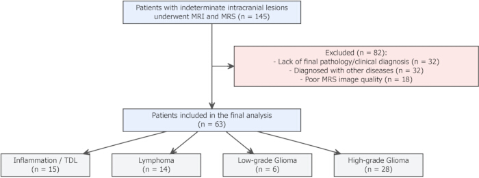
Figure 1. Patient selection and study design. Flowchart illustrating patient enrollment and study selection. A total of 145 patients with indeterminate intracranial lesions who underwent conventional magnetic resonance imaging (MRI) and proton magnetic resonance spectroscopy (1H-MRS) were screened. After application of the exclusion criteria, 63 patients were included in the final analysis. The final cohort comprised patients with high-grade glioma (n = 28), low-grade glioma (n = 6), primary central nervous system lymphoma (n = 14), and tumefactive demyelinating lesion (TDL; n = 15). Exclusion criteria included lack of a definitive diagnosis (n = 32), alternative diagnoses (n = 32), and poor-quality MRS examinations (n = 18). Please click here to view a larger version of this figure.
Conventional MRI Acquisition
All neuroimaging examinations were performed using a 3.0-T whole-body magnetic resonance scanner equipped with an 8-channel phased-array head coil. Before examination, patient safety screening was performed, establishing a mandatory exclusion threshold of an estimated glomerular filtration rate (eGFR) <30 mL/min/1.73 m2 to prevent nephrogenic systemic fibrosis. The conventional MRI protocol included axial T1-fluid-attenuated inversion recovery (FLAIR) images (repetition time [TR] = 1750 ms, echo time [TE] = 25.0 ms, field of view [FOV] = 240 mm × 192 mm, slice thickness = 6 mm, interslice gap = 1 mm, matrix size = 320 × 224), T2-weighted images (T2WI; TR = 4841 ms, TE = 102.8 ms, FOV = 240 mm × 240 mm, matrix = 416 × 416), and T2-FLAIR images (TR = 7000 ms, TE = 138.1 ms, FOV = 240 mm × 240 mm, matrix = 256 × 256).
Following intravenous administration of a macrocyclic gadolinium-based contrast agent at a standard dose of 0.2 mmol/kg and a flow rate of 2 mL/s, contrast-enhanced spoiled gradient recalled echo (SPGR) T1-weighted images were acquired (TR = 7.3 ms, TE = 3.1 ms, FOV = 240 × 240 mm, slice thickness = 1 mm with a 0.5 mm slice overlap, matrix = 256 × 256). Patients were continuously monitored within the facility for a minimum of 30 min after contrast administration to identify and manage any acute adverse or allergic reactions.
1H-MRS Acquisition and Prescribed Voxel Geometry
2D multi-voxel 1H-MRS was performed using a point-resolved spectroscopy (PRESS) sequence. The volume of interest (VOI) was prescribed on multiplanar contrast-enhanced T1-weighted and T2-FLAIR images to encompass the solid portion of the lesion, the peritumoral edema, and the adjacent normal-appearing brain tissue. To ensure institutional consistency across the retrospective cohort, the multi-voxel grid was uniformly aligned parallel to the anterior commissure-posterior commissure (AC-PC) line and centered over the maximum diameter of the target lesion.
The scanning parameters for the MRS acquisition included repetition time (TR) = 1000 ms, echo time (TE) = 144 ms, field of view (FOV) = 240 mm × 240 mm, slice thickness = 15.0 mm, interslice gap = 20.0 mm, and number of excitations (NEX) = 1.0. The nominal individual spectroscopic voxel dimensions within the acquisition matrix were 7.5 mm × 7.5 mm × 15.0 mm, acquired using a 32 × 32 spectroscopic acquisition matrix, corresponding to an individual voxel volume of approximately 0.84 mL. Standard automated procedures were consistently applied for water suppression and localized shimming. Strict quality-control checkpoints during acquisition required a localized water peak full-width at half-maximum (FWHM) linewidth of ≤15 Hz and automated water suppression attenuation exceeding 98%. A relatively long echo time (TE = 144 ms) was selected to provide a flat baseline by suppressing short-TE macromolecule signals and broad background lipid contamination commonly observed in acute inflammatory lesions, thereby improving phase clarity for the principal diagnostic metabolites.
Spectral Post-processing, Software Workflow, and Quantitative Analysis
The raw spectral data were transferred to a dedicated diagnostic post-processing workstation running specialized multi-modality evaluation software. The automated post-processing workflow consisted of consecutive steps: raw data apodization using a 2 Hz Gaussian filter, fast Fourier transformation, automated zero- and first-order phase correction, and automated polynomial baseline adjustment. Spectral quality degradation from residual rolling baselines was quantitatively assessed, and voxels with baseline distortion exceeding 10% of the maximum metabolite peak height were excluded.
Regions of interest (ROIs) were manually placed within the most representative solid portions of the lesions while avoiding macroscopic necrosis, cystic components, hemorrhage, calcifications, and areas adjacent to the skull base or ventricles to minimize spectral contamination and voxel volume-averaging artifacts. Specifically, three optimal target voxels were sampled within the active, contrast-enhancing, non-necrotic rim of the lesion. For internal standardization, a paired mirror ROI of identical size and symmetrical grid coordinates was placed in the contralateral normal-appearing white matter. The contralateral reference region was verified as metabolically unaffected by confirming normal, symmetric signal intensity across T1-weighted, T2-weighted, and T2-FLAIR images. A localized coil-intensity normalization filter was applied to minimize distance-related surface coil sensitivity bias.
The principal cerebral metabolites were identified according to their resonance frequencies: NAA at 2.0 ppm, Cr at 3.0 ppm, Cho at 3.2 ppm, lipids (Lip) at 0.9–1.3 ppm, and lactate (Lac) at 1.3 ppm. The detection threshold for metabolite peaks was defined as a signal-to-noise ratio (SNR) of ≥5. Owing to the phase modulation characteristics of the TE = 144 ms acquisition, lipid and lactate peaks were analyzed separately: lactate was identified by its characteristic inverted doublet at 1.33 ppm, whereas mobile lipids remained upright at 0.9–1.3 ppm, minimizing peak overlap. Absolute peak integrals were used to calculate the intra-lesional metabolite ratios (Cho/Cr, NAA/Cr, and NAA/Cho) by averaging measurements from the three target ROIs. Relative metabolite ratios (rCho/Cr, rNAA/Cr, and rNAA/Cho) were calculated by normalizing lesional values to those of the contralateral normal tissue. To assess inter-reader reliability, ROI selection and spectral quantification were performed independently by two experienced neuroradiologists who were blinded to the final clinical and histopathological diagnoses. Any procedural or positional discrepancies in voxel selection were resolved by consensus with a third senior neuroradiologist.
Statistical Analysis
All statistical modeling and graphical visualizations were performed using a verified statistical computing environment. The normality of continuous variables was assessed using the Shapiro–Wilk test. Continuous metabolic variables were compared across multiple disease groups using the Kruskal–Wallis H test, followed by the Wilcoxon rank-sum test for post hoc comparisons. Paired differences between the lesional core and contralateral normal tissue were evaluated using the Wilcoxon signed-rank test. Receiver operating characteristic (ROC) curves were constructed to evaluate diagnostic performance by calculating the optimal cut-off values, area under the curve (AUC), sensitivity, specificity, positive predictive value, and negative predictive value, with corresponding 95% confidence intervals (95% CIs) derived using Wilson’s score method for all point estimates. Inter-reader agreement was evaluated using the intraclass correlation coefficient (ICC) and Bland-Altman plots. A two-sided p-value <0.05 was considered statistically significant.
Results
Study Population and Baseline Characteristics
During the study period, a total of 145 patients with indeterminate intracranial lesions who underwent both conventional MRI and 1H-MRS were initially screened. Following application of the predefined exclusion criteria, primarily because of the absence of a definitive pathological or long-term clinical diagnosis (n = 32), confirmation of alternative pathologies (n = 32), or inadequate MRS spectral quality (n = 18), 63 patients were ultimately included in the quantitative analysis (Figure 1). Blinded inter-reader agreement for manual ROI placement and spectral quantification was excellent, with intraclass correlation coefficients (ICCs) of 0.996 for the Cho/Cr ratio and 0.990 for the NAA/Cho ratio (Supplementary Figure 1). Specifically, the 18 cases excluded because of inadequate spectral quality demonstrated visual and quantitative quality-control failures at the workstation level, including an inability to distinguish the principal metabolite peaks from background noise (SNR < 5), severe peak broadening caused by poor local shimming (FWHM > 15 Hz), or rolling baseline distortion exceeding 10% of the maximum metabolite peak height, which rendered automated curve fitting and quantitative peak integration unreliable (Supplementary Figure 2).
The final cohort comprised 28 patients with HGG, 6 with low-grade glioma (LGG), 14 with primary central nervous system lymphoma, and 15 with TDLs (Figure 1). Baseline demographic and qualitative imaging characteristics are summarized in Table 1. Median age differed significantly among the diagnostic groups (P = 0.004), with patients with HGG and lymphoma generally being older than those with TDLs. Lipid peaks were observed more frequently in malignant lesions, including lymphomas (43.0%) and HGGs (36.0%), than in TDLs (6.7%); however, the overall distribution did not differ significantly among the groups (P = 0.084).
| Characteristic | Overall (N = 63) | High-grade glioma (N = 28) | Low-grade glioma (N = 6) | Lymphoma (N = 14) | Tumefactive demyelinating lesion (TDL) (N = 15) | P value |
| Age | 60 (47, 69) | 67 (56, 69) | 39 (26, 52) | 63 (59, 72) | 47 (36, 65) | 0.004 |
| Male sex, n (%) | 32 (51%) | 17 (61%) | 4 (67%) | 5 (36%) | 6 (40%) | 0.3 |
| Lipid peak present, n (%) | 18 (29%) | 10 (36%) | 1 (17%) | 6 (43%) | 1 (6.7%) | 0.084 |
| Lactate peak present, n (%) | 7 (11%) | 1 (3.6%) | 0 (0%) | 3 (21%) | 3 (20%) | 0.15 |
Table 1: Baseline demographic and qualitative imaging characteristics of the study cohort. Baseline demographic characteristics and qualitative imaging findings of patients with high-grade glioma, low-grade glioma, primary central nervous system lymphoma, and tumefactive demyelinating lesion (TDL). Continuous variables are presented as median (interquartile range [IQR], Q1–Q3), and categorical variables are presented as number (%). P values indicate overall comparisons among the four diagnostic groups.
Inter-reader Agreement and Methodological Reproducibility
To evaluate the reproducibility of manual ROI placement and spectral quantification, blinded inter-reader agreement was assessed. ICCs demonstrated high agreement for the principal metabolite ratios, with an ICC of 0.996 for the Cho/Cr ratio and 0.990 for the NAA/Cho ratio (Supplementary Figure 1). Bland–Altman analysis demonstrated minimal mean differences between the two independent readers (mean difference, 0 for Cho/Cr and 0.01 for NAA/Cho), with nearly all paired measurements falling within the 95% limits of agreement (−0.55 to 0.55 for Cho/Cr and −0.16 to 0.18 for NAA/Cho).
Quantitative Metabolite Profiles and Paired Internal Comparisons
Quantitative metabolite ratios for the four diagnostic groups are summarized in Table 2. To account for individual baseline metabolic variation, a paired comparison was performed between the lesional core and the contralateral normal-appearing brain parenchyma (Figure 2). Patients with HGG demonstrated a significantly higher Cho/Cr ratio in lesion tissue than in the corresponding contralateral normal tissue (P < 0.001). Similarly, patients with TDLs exhibited a modest but statistically significant increase in the Cho/Cr ratio relative to the contralateral hemisphere (P = 0.030).
| Metabolite ratio | High-grade glioma (N = 28) | Low-grade glioma (N = 6) | Lymphoma (N = 14) | Tumefactive demyelinating lesion (TDL) (N = 15) | P value |
| Choline-to-creatine ratio (Cho/Cr), lesion | 2.88 (1.72, 5.40) | 1.19 (0.94, 1.60) | 2.48 (1.87, 4.33) | 1.34 (1.11, 1.68) | <0.001 |
| N-acetylaspartate-to-creatine ratio (NAA/Cr), lesion | 0.92 (0.61, 1.73) | 0.74 (0.35, 1.73) | 1.27 (0.64, 1.58) | 1.72 (1.28, 2.02) | 0.053 |
| N-acetylaspartate-to-choline ratio (NAA/Cho), lesion | 0.32 (0.20, 0.69) | 0.63 (0.25, 1.78) | 0.38 (0.25, 0.67) | 1.22 (0.75, 1.60) | <0.001 |
Table 2: Quantitative proton magnetic resonance spectroscopy metabolite ratios across the diagnostic groups. Absolute proton magnetic resonance spectroscopy (1H-MRS) metabolite ratios measured within the lesion in patients with high-grade glioma, low-grade glioma, primary central nervous system lymphoma, and tumefactive demyelinating lesion (TDL). Data are presented as median (interquartile range [IQR], Q1–Q3). P values indicate overall comparisons among the four diagnostic groups.
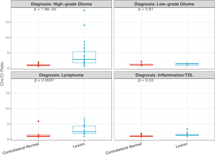
Figure 2. Comparison of choline-to-creatine ratios between contralateral normal tissue and lesional tissue. Paired box-and-whisker plots comparing the choline-to-creatine (Cho/Cr) ratio between contralateral normal-appearing brain tissue and the corresponding lesion in patients with high-grade glioma, low-grade glioma, lymphoma, and tumefactive demyelinating lesion (TDL). Individual data points represent individual patients. Horizontal lines indicate the median, boxes represent the interquartile range (IQR), and whiskers indicate the data range. P-values were calculated using the Wilcoxon signed-rank test. Please click here to view a larger version of this figure.
Discriminative Value of the N-Acetylaspartate-to-Choline Ratio Across the Disease Spectrum
Because increased Cho/Cr ratios were observed in both HGGs and TDLs, the NAA/Cho ratio was further evaluated (Figure 3). Kruskal–Wallis analysis demonstrated significant differences among the diagnostic groups (P = 0.00031). The median NAA/Cho ratio was 1.22 (IQR, 0.75–1.60) in the TDL group (Table 2). In comparison, the median NAA/Cho ratio was 0.32 (IQR, 0.20–0.69) in HGGs and 0.38 (IQR, 0.25–0.67) in primary central nervous system lymphomas (P < 0.001 and P < 0.01 versus TDL, respectively; Table 2 and Figure 3).
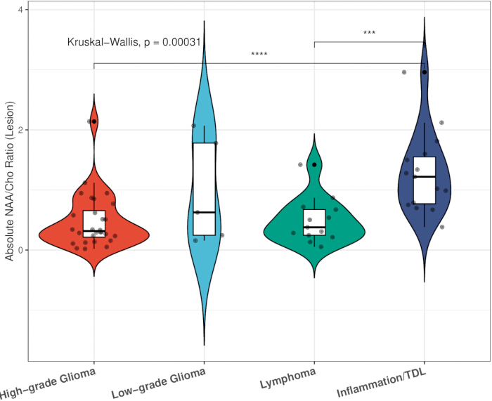
Figure 3. Distribution of the absolute N-acetylaspartate-to-choline ratio across disease groups. Violin plots with embedded box plots showing the distribution of the absolute N-acetylaspartate-to-choline (NAA/Cho) ratio in lesion tissue from patients with high-grade glioma, low-grade glioma, lymphoma, and tumefactive demyelinating lesion (TDL). Individual points represent individual patients. Box plots indicate the median (horizontal line) and interquartile range (IQR). Overall group differences were assessed using the Kruskal–Wallis test followed by post hoc comparisons. ***P < 0.001; ****P < 0.0001. Please click here to view a larger version of this figure.
Representative cases illustrating the imaging and spectroscopic findings are shown in Figures 4 and 5. In the representative TDL case, the conventional MRI findings (Figure 4A,B) demonstrated a peripheral enhancing lesion, and the corresponding multi-voxel 1H-MRS examination (Figures 4C,D) demonstrated the associated metabolite profile. Similarly, in the representative HGG case, the conventional MRI findings (Figure 5A,B) demonstrated an enhancing lesion, whereas the corresponding multi-voxel 1H-MRS examination (Figure 5C,D) demonstrated the measured metabolite profile.
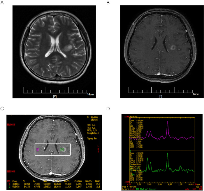
Figure 4. Representative magnetic resonance imaging and proton magnetic resonance spectroscopy findings in a tumefactive demyelinating lesion. Representative images from a 36-year-old female patient with a tumefactive demyelinating lesion (TDL). (A) Axial T2-weighted fluid-attenuated inversion recovery (T2-FLAIR) image demonstrating a hyperintense lesion with surrounding edema. (B) Contrast-enhanced T1-weighted image showing an open-ring enhancement pattern. (C) Placement of the multi-voxel proton magnetic resonance spectroscopy (1H-MRS) volume of interest over the enhancing lesion. (D) Representative spectra acquired from the lesion and contralateral normal-appearing brain tissue demonstrating the measured metabolite peaks, including choline (Cho), creatine (Cr), and N-acetylaspartate (NAA). Please click here to view a larger version of this figure.
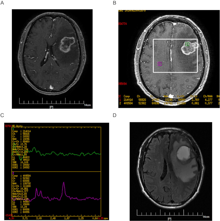
Figure 5. Representative magnetic resonance imaging and proton magnetic resonance spectroscopy findings in a high-grade glioma. Representative images from a 66-year-old male patient with an isocitrate dehydrogenase (IDH)-wildtype glioblastoma. (A) Contrast-enhanced T1-weighted image demonstrating a left frontal enhancing mass. (B) Placement of the multi-voxel proton magnetic resonance spectroscopy (1H-MRS) volume of interest over the enhancing lesion. (C) Representative spectra acquired from the lesion and contralateral normal-appearing brain tissue demonstrating the measured metabolite peaks, including choline (Cho), creatine (Cr), and N-acetylaspartate (NAA). (D) Axial T2-fluid-attenuated inversion recovery (T2-FLAIR) image demonstrating extensive perilesional edema. Please click here to view a larger version of this figure.
Diagnostic Performance and Clinical Thresholds
ROC curve analysis was performed to evaluate the diagnostic performance of the metabolite ratios for differentiating gliomas (high-grade and low-grade combined) from TDLs (Figure 6). The NAA/Cho ratio demonstrated an area under the curve (AUC) of 0.848 (95% CI, 0.740–0.957), whereas the Cho/Cr ratio demonstrated an AUC of 0.766 (95% CI, 0.630–0.902) (Table 3 and Figure 6).
| Biomarker | Optimal cut-off | Area under the curve (AUC) (95% CI) | Sensitivity (95% CI) | Specificity (95% CI) | Positive predictive value (PPV) (95% CI) | Negative predictive value (NPV) (95% CI) |
| Choline-to-creatine ratio (Cho/Cr) | >1.900 | 0.766 (0.630–0.902) | 60.6% (43.7%–75.3%) | 86.7% (62.1%–96.3%) | 90.9% (72.2%–97.5%) | 50.0% (32.1%–67.9%) |
| N-acetylaspartate-to-choline ratio (NAA/Cho) | ≤0.649 | 0.848 (0.740–0.957) | 72.7% (55.8%–84.9%) | 93.3% (70.2%–98.8%) | 96.0% (80.5%–99.3%) | 60.9% (40.8%–77.8%) |
Table 3: Diagnostic performance of metabolite ratios for differentiating intracranial gliomas from tumefactive demyelinating lesions. Diagnostic performance of the choline-to-creatine (Cho/Cr) ratio and the N-acetylaspartate-to-choline (NAA/Cho) ratio for differentiating intracranial gliomas from tumefactive demyelinating lesions (TDLs). The table summarizes the optimal cut-off values, area under the curve (AUC), sensitivity, specificity, positive predictive value (PPV), and negative predictive value (NPV). All point estimates are presented with corresponding 95% confidence intervals (CIs). The analysis included patients with high-grade and low-grade gliomas (n = 34) and TDLs (n = 15); patients with primary central nervous system lymphoma were excluded from the diagnostic performance analysis.
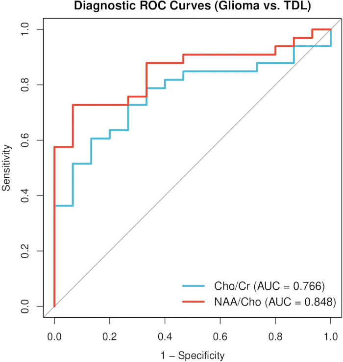
Figure 6. Receiver operating characteristic curves for differentiating gliomas from tumefactive demyelinating lesions. Receiver operating characteristic (ROC) curves comparing the diagnostic performance of the choline-to-creatine (Cho/Cr) ratio and the N-acetylaspartate-to-choline (NAA/Cho) ratio for differentiating gliomas from tumefactive demyelinating lesions (TDLs). The area under the curve (AUC) was 0.766 for the Cho/Cr ratio and 0.848 for the NAA/Cho ratio. The analysis included patients with high-grade and low-grade gliomas (n = 34) and TDLs (n = 15); patients with primary central nervous system lymphoma were excluded from the ROC analysis. Please click here to view a larger version of this figure.
Diagnostic performance analysis identified an optimal NAA/Cho cut-off value of ≤0.649 for differentiating gliomas from TDLs. At this threshold, the sensitivity was 72.7% (95% CI, 55.8%–84.9%), the specificity was 93.3% (95% CI, 70.2%–98.8%), the positive predictive value (PPV) was 96.0% (95% CI, 80.5%–99.3%), and the negative predictive value (NPV) was 60.9% (95% CI, 40.8%–77.8%) (Table 3). These findings indicate that, within this study cohort, the NAA/Cho ratio demonstrated greater diagnostic discrimination than the Cho/Cr ratio for differentiating gliomas from TDLs.
Data Availability:
The supplementary materials supporting this study are provided with the manuscript and include the Bland–Altman analysis of inter-reader agreement (Supplementary Figure 1), a representative example of a non-diagnostic proton magnetic resonance spectroscopy (1H-MRS) examination excluded during quality control (Supplementary Figure 2), and the anonymized patient-level dataset used for the quantitative analyses (Supplementary Table 1). The patient-level dataset includes anonymized demographic information, final diagnoses, quantitative metabolite ratios, relative metabolite ratios, lipid and lactate peak status, and independent measurements from the second reader used for inter-reader reliability analyses. All data have been anonymized to protect participant confidentiality.
Supplementary Figure 1. Bland–Altman analysis of inter-reader agreement for metabolite ratio measurements. Bland–Altman plots comparing measurements obtained independently by two blinded neuroradiologists. (A) Choline-to-creatine (Cho/Cr) ratio. (B) N-acetylaspartate-to-choline (NAA/Cho) ratio. The solid red line indicates the mean difference between the two readers, and the dashed black lines indicate the 95% limits of agreement (LoA). The corresponding intraclass correlation coefficients (ICCs) were 0.996 for the Cho/Cr ratio and 0.990 for the NAA/Cho ratio.Please click here to download this file.
Supplementary Figure 2. Example of a non-diagnostic proton magnetic resonance spectroscopy examination excluded during quality control. (A) Multi-voxel proton magnetic resonance spectroscopy (1H-MRS) positioning image showing the prescribed volume of interest and selected regions of interest (ROIs). (B) Corresponding spectra acquired from the indicated ROIs. The displayed spectra exhibit poor spectral quality with signal-to-noise ratios below the predefined acceptance threshold, resulting in exclusion from quantitative analysis according to the study quality-control criteria.Please click here to download this file.
Supplementary Table 1. Patient-level anonymized dataset used for quantitative magnetic resonance spectroscopy (1H-MRS) analyses. The table contains anonymized demographic information, final diagnosis, lesion and contralateral metabolite ratios (Cho/Cr, NAA/Cr, and NAA/Cho), relative metabolite ratios (rCho/Cr, rNAA/Cr, and rNAA/Cho), lipid and lactate peak status, and independent measurements from the second reader used for inter-reader reliability analyses. Patient identifiers have been anonymized to protect participant confidentiality.Please click here to download this file.
Discussion
Accurate differentiation between intracranial gliomas and TDLs before therapeutic intervention is important for appropriate clinical management. In this retrospective study, the diagnostic utility of quantitative 1H-MRS parameters was evaluated in patients with indeterminate intracranial lesions. The findings indicate that the NAA/Cho ratio provided greater diagnostic discrimination than the conventional Cho/Cr ratio for differentiating gliomas from TDLs. Specifically, an NAA/Cho ratio threshold of ≤0.649 yielded a specificity of 93.3% (95% CI: 70.2%–98.8%) and a PPV of 96.0% (95% CI: 80.5%–99.3%) for identifying neoplastic disease. The NAA/Cho ratio demonstrated an AUC of 0.848 (95% CI: 0.74–0.96), compared with 0.766 (95% CI: 0.63–0.90) for the Cho/Cr ratio. However, the observed sensitivity of 72.7% (95% CI: 55.8%–84.9%) and NPV of 60.9% (95% CI: 40.8%–77.8%) indicate that this threshold should be interpreted in conjunction with clinical and imaging findings rather than as a standalone diagnostic criterion. These findings support the proposed use of the NAA/Cho ratio as a complementary metabolic biomarker for differentiating gliomas from TDLs.
The metabolic profiles obtained by 1H-MRS reflect the distinct pathological processes underlying gliomas and TDLs. Choline is an essential component of phosphoglycerides in cell membranes. Paired analysis demonstrated that the Cho/Cr ratio was significantly elevated relative to the contralateral normal-appearing brain tissue, which served as an internal reference after verification that it was metabolically unaffected and free of structural or T2-FLAIR signal abnormalities according to the predefined imaging criteria, in both HGGs and TDLs. In gliomas, elevated choline is associated with increased cellular proliferation, cellular density, and membrane turnover characteristic of malignant neoplasia11,12. In contrast, elevated choline-containing compounds in TDLs are considered to result from active demyelination, during which myelin degradation and subsequent phagocytosis by reactive macrophages release mobile membrane phospholipids into the extracellular space13,18. In addition, transient reductions in NAA may occur during acute demyelination because of reversible mitochondrial dysfunction, even in the absence of irreversible axonal loss, which may further contribute to the observed metabolic profile in TDLs. Because both malignant proliferation and inflammatory demyelination increase regional choline concentrations, considerable overlap in Cho/Cr ratios was observed between the two groups. Within the present cohort, a Cho/Cr threshold of 1.900 yielded a specificity of 86.7% (95% CI: 62.1%–96.3%) and an NPV of 50.0% (95% CI: 32.1%–67.9%), indicating that the Cho/Cr ratio alone has limited discriminatory performance for differentiating these entities.
To overcome the diagnostic ambiguity associated with overlapping choline elevations, the NAA/Cho ratio was evaluated. NAA is synthesized exclusively in neuronal mitochondria and transported along axons, making it a well-established surrogate marker of neuronal integrity and axonal viability10,14. The results demonstrated significant differences in the NAA/Cho ratio among the diagnostic groups. In the TDL cohort, the median NAA/Cho ratio was 1.22 (IQR, 0.75–1.60). This metabolic profile is consistent with the characteristic pathological feature of demyelinating disease, namely the relative preservation of axons despite extensive myelin loss and inflammatory infiltration4,5. Even in the presence of perivascular lymphocytic cuffing and macrophage activity, preservation of the underlying axonal architecture allows the NAA signal to remain detectable15. In addition, a portion of the reduction in NAA observed during the acute phase of TDLs may reflect transient, reversible mitochondrial dysfunction rather than irreversible axonal loss, which may contribute to the relatively higher NAA/Cho ratio observed in inflammatory lesions compared with malignant tumors. In contrast, the median NAA/Cho ratio was 0.32 (IQR, 0.20–0.69) in the HGG group and 0.38 (IQR, 0.25–0.67) in patients with primary CNS lymphoma. Malignant gliomas exhibit infiltrative growth patterns that replace normal neuronal tissue with neoplastic cells, resulting in substantial reductions in the NAA signal within the enhancing tumor12. By combining decreased NAA with increased Cho, the NAA/Cho ratio provided greater diagnostic discrimination than the Cho/Cr ratio in the present cohort, yielding an AUC of 0.848 (95% CI: 0.74–0.96). However, several considerations should be taken into account when interpreting these findings. The diagnostic threshold was derived by combining a relatively small subgroup of LGGs (n = 6) with HGGs (n = 28), which may have introduced metabolic heterogeneity because LGGs generally demonstrate greater neuronal preservation than HGGs. Furthermore, although primary CNS lymphomas were analyzed to characterize their metabolic profiles, they represent a biologically distinct disease entity and were excluded from the ROC analysis used to derive the glioma-versus-TDL diagnostic threshold. These factors should be considered when interpreting the applicability of the proposed threshold and underscore the need for further validation in larger, independent cohorts.
These findings should be interpreted in conjunction with conventional MRI findings. Routine structural imaging frequently presents diagnostic challenges when evaluating TDLs. Although characteristic features such as open-ring enhancement can be helpful when present8,9, many TDLs, particularly those larger than 3.0 cm, may exhibit closed-ring, irregular, or nodular enhancement patterns accompanied by extensive vasogenic edema, resembling glioblastomas or primary CNS lymphomas16,17. As illustrated by the representative cases, both patients presented with large, irregular, peripherally enhancing frontal lobe lesions. Multi-voxel 1H-MRS demonstrated different metabolite profiles between these representative cases. In this study, spectroscopy was performed before the initiation of corticosteroid therapy because corticosteroids may alter inflammatory changes and metabolic profiles, potentially affecting spectroscopic measurements. The representative patient with relative preservation of the NAA signal subsequently underwent corticosteroid pulse therapy with lesion resolution, whereas the representative patient with marked NAA reduction underwent neurosurgical resection with a diagnosis of glioblastoma. These observations illustrate the potential complementary role of metabolic imaging within the diagnostic evaluation of atypical intracranial lesions19. To support the diagnostic validity of the non-biopsied TDL cohort (n = 9; 60.0% of the TDL group), while the remaining six patients (40.0%) had histopathological confirmation, all non-biopsied cases underwent a minimum of 12 months of clinical and radiological follow-up (median, 14 months), requiring documented lesion regression or long-term stability without evidence of neoplastic progression. Furthermore, the methodological design of this study supports the reproducibility of the proposed metabolic measurements. Manual ROI placement has been recognized as a potential source of variability in clinical 1H-MRS studies10. To address this issue, the post-processing protocol incorporated paired internal standardization and blinded inter-reader assessment. A total of 18 patients were excluded because of poor spectral quality, and 32 were excluded because a definitive reference diagnosis was unavailable. Although this level of attrition may introduce selection bias, all non-diagnostic spectra were excluded according to predefined quantitative quality-control criteria before blinded image review. Inter-reader agreement was excellent, with ICCs greater than 0.99 for both the Cho/Cr and NAA/Cho ratios. Bland–Altman analysis demonstrated minimal inter-reader bias (mean bias ≤0.01), with 95% limits of agreement of −0.55 to 0.55 for the Cho/Cr ratio and −0.16 to 0.18 for the NAA/Cho ratio. These findings support the reproducibility of the proposed quantitative approach when standardized acquisition, quality-control procedures, and ROI placement methods are used.
The reproducibility of the proposed protocol depends on several critical steps during data acquisition and post-processing. First, achieving optimal local B0 field homogeneity is a key determinant of spectral resolution. Automated phase-mapped shimming should be performed to achieve a localized water peak FWHM of ≤15 Hz. When local magnetic field distortions are present because of proximity to bone-air interfaces, the skull base, or the paranasal sinuses, additional localized shimming may be required to optimize spectral quality. Failure to achieve adequate field homogeneity may result in peak broadening and reduced spectral resolution. Second, during post-processing, representative lesional ROIs should be placed within the viable contrast-enhancing portions of the lesion while avoiding voxels containing extensive necrosis, cystic change, or gross hemorrhage. ROI selection should be performed using a standardized voxel placement strategy by readers who are blinded to the final diagnosis. Inclusion of predominantly necrotic tissue within the selected voxel may reduce metabolite signal intensities and affect calculation of the diagnostic metabolite ratios. Finally, spectral acceptance should be based on predefined quantitative quality-control criteria before diagnostic interpretation. Spectra with an SNR of <5, inadequate water suppression, significant phase instability, or excessive baseline distortion should be excluded from quantitative analysis. At the selected echo time (TE = 144 ms), the characteristic inverted lactate peak at 1.33 ppm should be distinguished from lipid resonances. The relatively long TE facilitates suppression of short-T2 macromolecular signals, improving baseline definition for quantification of NAA, Cho, and Cr.
Several limitations of this study should be acknowledged. First, the study cohort, particularly the low-grade glioma subgroup, was relatively small, limiting the statistical power to evaluate subtle metabolic differences between low-grade neoplasms and subacute inflammatory lesions. Second, this was a single-center study performed using a single 3.0-T MRI platform and post-processing workflow. Consequently, external validation in larger, multicenter cohorts using different scanner platforms, field strengths, and post-processing methods is needed to evaluate the generalizability of the proposed diagnostic threshold and reduce the potential for statistical overfitting. Third, the protocol employed a relatively long echo time (TE = 144 ms). Although this acquisition optimized the measurement of the principal metabolites by improving spectral baseline definition, it limited the evaluation of short-T2 metabolites, such as myo-inositol and glutamate/glutamine, which may provide additional information in demyelinating diseases. Finally, although 1H-MRS provides quantitative metabolic information, it represents one component of a comprehensive neuroimaging assessment20. Future studies should evaluate the proposed NAA/Cho threshold in larger, independent, and multicenter cohorts and investigate its integration with complementary advanced imaging techniques, including diffusion-weighted imaging and dynamic susceptibility contrast perfusion imaging. These multimodal approaches may facilitate the development of more comprehensive diagnostic models for indeterminate intracranial lesions. Automated image analysis and machine learning may further support this objective in future investigations; however, the present protocol relies exclusively on expert-guided manual voxel selection, standardized quality-control procedures, and quantitative threshold-based interpretation.
Disclosures
Conflict of Interest:
The authors declare that they have no competing interests.
Acknowledgements
This study was supported by the Medical Science Research Project of Hebei, China (Grant Nos. 20230402 and 20180161). The funding source had no role in the study design; data collection, analysis, or interpretation; the decision to publish; or the preparation of the manuscript.
Materials
| Name | Company | Catalog Number | Comments |
|---|---|---|---|
| 3.0-T MRI Scanner (Discovery MR750w) | GE Healthcare | N/A | MRI and 1H-MRS acquisition |
| 8-channel phased-array head coil | GE Healthcare | N/A | Head radiofrequency coil for MRI and 1H-MRS acquisition |
| Functional Tool post-processing software | GE Healthcare | Functool 9.4 | Spectral post-processing and metabolite quantification |
| Gadolinium-based contrast agent | Bayer Healthcare | N/A | Intravenous contrast agent for contrast-enhanced MRI |
| Multi-modality post-processing workstation | GE Healthcare | Advantage Workstation (AW4.6) | Dedicated workstation for spectral post-processing |
| R statistical computing software | R Foundation | RRID: SCR_001905 | Statistical analysis and graphical visualization |
References
- Ostrom QT, et al. CBTRUS statistical report: Primary brain and other central nervous system tumors diagnosed in the United States in 2016-2020. Neuro Oncol. 2023;25(Suppl 2):iv1-99.
- Lucchinetti CF, et al. Clinical and radiographic spectrum of pathologically confirmed tumefactive multiple sclerosis. Brain. 2008;131(Pt 7):1759-75.
- Haupts MR, et al. Fulminant tumefactive multiple sclerosis: Therapeutic implications of histopathology. J Neurol. 2008;255(8):1272-3.
- Algahtani H, Shirah B, Alassiri A. Tumefactive demyelinating lesions: A comprehensive review. Mult Scler Relat Disord. 2017;14:72-9.
- Altintas A, et al. Clinical and radiological characteristics of tumefactive demyelinating lesions: Follow-up study. Mult Scler. 2012;18(10):1448-53.
- Hu W, Lucchinetti CF. The pathological spectrum of CNS inflammatory demyelinating diseases. Semin Immunopathol. 2009;31(4):439-53.
- Jameson J, et al. Harrison's Principles of Internal Medicine. 20th ed. McGraw-Hill; New York; 2018.
- Masdeu JC, et al. Open ring imaging sign: Highly specific for atypical brain demyelination. Neurology. 2000;54(7):1427-33.
- Given CA, Stevens BS, Lee C. The MRI appearance of tumefactive demyelinating lesions. AJR Am J Roentgenol. 2004;182(1):195-9.
- Atlas SW. Magnetic Resonance Imaging of the Brain and Spine. 3rd ed. Lippincott Williams & Wilkins; Philadelphia; 2008.
- Hu X, et al. Combined application of MRS and DWI can effectively predict cell proliferation and assess the grade of glioma: A prospective study. J Clin Neurosci. 2021;83:56-63.
- Cianfoni A, Niku S, Imbesi SG. Metabolite findings in tumefactive demyelinating lesions utilizing short echo time proton magnetic resonance spectroscopy. AJNR Am J Neuroradiol. 2007;28(2):272-7.
- Saindane AM, et al. Proton MR spectroscopy of tumefactive demyelinating lesions. AJNR Am J Neuroradiol. 2002;23(8):1378-86.
- Cucurella MG, et al. Serial proton spectroscopy, magnetization transfer ratio and T2 relaxation in pseudotumoral demyelinating lesions. NMR Biomed. 2002;15(4):284-92.
- De Stefano N, et al. Chemical pathology of acute demyelinating lesions and its correlation with disability. Ann Neurol. 1995;38(6):901-9.
- Kiriyama T, et al. Characteristic neuroimaging in patients with tumefactive demyelinating lesions exceeding 30 mm. J Neuroimaging. 2011;21(1):69-77.
- Dagher AP, Smirniotopoulos J. Tumefactive demyelinating lesions. Neuroradiology. 1996;38(6):560-5.
- Sinha MK, et al. Tumefactive demyelinating lesion: Experience with two unusual patients. J Postgrad Med. 2010;56(2):146-9.
- Chapter 6: Intracranial infection and inflammation. In: Diseases of the Brain, Head and Neck, Spine 2020-2023: Diagnostic Imaging. Springer; Cham; 2020. Available from: https://doi.org/10.1007/978-3-030-38490-6_6
- Oz G, et al. Clinical proton MR spectroscopy in central nervous system disorders. Radiology. 2014;270(3):658-79.