This protocol describes the steps required to perform hybridization chain reaction fluorescent in situ hybridization (HCR-FISH) in Drosophila melanogaster larval optic lobes. Procedures for probe design, hybridization, amplification, and tissue mounting are detailed to facilitate visualization of the developing optic lobe.
A subscription to JoVE is required to view this content. Sign in or start your free trial.
Method Article
Hybridization Chain Reaction FISH in the Developing Drosophila Optic Lobe
* These authors contributed equally
In This Article
Summary
Abstract
Understanding neural development requires precise characterization of when and where genes are expressed. Techniques such as in situ hybridization have long enabled visualization of gene expression in developing tissues. Although in situ hybridization is widely used in Drosophila melanogaster embryos, its application to visual system development has been limited by several technical challenges. For example, long probes often penetrate poorly into the optic lobe, and protocols that rely on diffusible catalytic reporters, such as horseradish peroxidase, often yield low signal-to-noise ratios in whole-mount preparations. Hybridization chain reaction fluorescent in situ hybridization (HCR-FISH) overcomes many of these limitations and enables more robust signal detection. As in conventional FISH, DNA probe monomers hybridize to RNA sequences within tissues. In HCR-FISH, however, fluorescently conjugated amplifier probes form stable hairpin structures that trigger polymerization of additional monomers, thereby amplifying signal intensity. The use of multiplexed probe sets also permits the use of shorter oligonucleotides (~45 nt, compared with up to 1,000 nt in conventional FISH), improving probe penetration and producing more uniform staining in whole-mount samples. This article describes an HCR-FISH protocol optimized for the Drosophila larval optic lobe. The protocol adapts existing HCR-FISH approaches by optimizing fixation conditions and increasing probe and hairpin concentrations to improve signal intensity and tissue penetration. Reagent lists and step-by-step procedures are provided. Potential applications in developmental neurobiology are also presented, including visualization of newborn neurons, and characterization of gene expression patterns in cases where conventional methods, such as endogenously tagged proteins or antibody staining, fail to provide sufficient information.
Introduction
Understanding how the brain is assembled requires identifying both the diversity of cell types present and the gene repertoires expressed within each cell type1. Recent advances in single-cell RNA sequencing have enabled researchers to address these questions at high resolution2. However, many single-cell approaches do not preserve spatial information, requiring additional experiments to map transcriptional cell-type clusters to their locations within the developing brain.
Binary gene expression systems such as GAL4-UAS can provide positional information but are not quantitative3. In addition, delays between gene expression and reporter detection can obscure physiologically relevant expression patterns during early developmental stages. Immunostaining is another widely used approach for determining tissue-specific expression patterns4. However, secreted proteins are often diffuse and difficult to localize accurately.
Nucleotide-labeling approaches, such as in situ hybridization (ISH), have also been widely used to obtain spatial information about gene expression5. In ISH, labeled DNA oligonucleotides bind to reverse-complementary RNA sequences of interest. Early ISH methods used radiolabeled DNA probes6, whereas later protocols incorporated safer labels such as digoxigenin, fluorescein, and rhodamine7,8,9. Scientists rapidly adapted ISH for use in Drosophila melanogaster, where it was applied to characterize polytene chromosome organization and map the expression patterns of gap genes such as hunchback in whole-mount embryos10,11.
Despite its widespread utility, several technical limitations hindered the adoption of ISH in thicker whole-mount preparations such as the larval optic lobe. Many in situ protocols relied on diffusible catalytic reporters, such as horseradish peroxidase, which often resulted in low signal-to-noise ratios12. In addition, many approaches used long probes targeting single sequences to improve specificity. Although effective in Drosophila embryos and polytene chromosomes, long probes penetrate poorly into the larval brain and often produce uneven labeling10,11,13.
The development of single-molecule fluorescent ISH (smFISH) and hybridization chain reaction fluorescent in situ hybridization (HCR-FISH) addressed many of these limitations14,15. By using sets of short probes tiled across a single gene region, smFISH reduces average probe length from approximately 1,000 nt to approximately 40 nt16. This substantially improves probe penetration while maintaining strong signal intensity. HCR-FISH further improves signal-to-noise ratios by using fluorescently conjugated amplifier probes that increase positional accuracy and amplify signal output. Importantly, this amplification step appears to scale linearly with transcript concentration, enabling quantitative measurement of gene expression levels17.
HCR-FISH begins with initiator probes complementary to the target sequence. These unlabeled DNA oligonucleotides contain a single-stranded initiator sequence that binds to the input domain of a fluorescently labeled H1 amplifier probe. H1 amplifiers adopt a hairpin structure that maintains catalytic inactivity until binding to the initiator sequence. Upon binding, the H1 amplifier opens and exposes a sequence that binds to a partner probe, H2. This interaction opens the H2 hairpin, allowing it to bind another H1 probe (Figure 1)15. Repeated cycles of H1/H2 binding generate a stable polymer that amplifies the fluorescence signal while reducing background noise. Because each initiator sequence is specific to a corresponding H1/H2 pair, multiple probes with distinct initiator sequences and fluorophores can be used simultaneously to label several genes within the same sample.
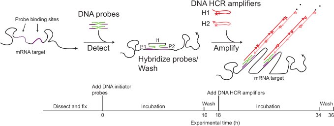
Figure 1: Graphical representation of the HCR-FISH workflow. DNA probes containing initiator sequences hybridize to a target RNA transcript. Fluorescently labeled DNA amplifier hairpins bind to the initiator sequence and undergo sequential polymerization to form stable double-stranded amplification chains. Direct fluorophore conjugation improves signal amplification and signal-to-noise ratio. Please click here to view a larger version of this figure.
Recent improvements to HCR-FISH have further increased its utility. Split initiator probe design improves amplifier specificity, and secondary antibodies conjugated to HCR initiators enable simultaneous immunohistochemistry and HCR-FISH18. In addition, expanded fluorophore options now permit multiplexed imaging of up to 10 genes simultaneously19,20.
Although initial proof-of-concept experiments for HCR-FISH were performed in zebrafish (Danio rerio), the method has gained widespread use across many systems, particularly in non-model organisms where antibodies are expensive or unavailable15. Probe synthesis also offers a faster alternative to antibody generation, which can require months of development. Consequently, HCR-FISH provides a rapid approach for validating candidate genes identified through single-cell RNA sequencing datasets21. Even in established genetic model organisms, HCR-FISH provides a valuable method for spatially mapping newly born neurons when GAL4 reporters or immunohistochemistry fail to adequately label specific cell populations.
This article describes an HCR-FISH protocol optimized for the third instar larval Drosophila optic lobe. The protocol adapts methods previously published in other organisms for use in the Drosophila visual system22. Fixation conditions commonly used for Drosophila immunohistochemistry were optimized for FISH applications. Probe and hairpin concentrations were increased to improve signal intensity and tissue penetration. PCR tubes were used instead of spot plates to reduce reagent consumption and simplify handling of small tissue samples. In addition, wash and hybridization times were shortened to accommodate the smaller size of the Drosophila brain relative to other insect systems commonly used for HCR-FISH.
Representative applications demonstrate how this approach can provide positional information for specific neuronal populations during development. These experiments indicate that certain transcripts, such as abrupt, can be detected earlier with HCR-FISH than with antibody staining or reporter lines. Potential challenges, troubleshooting strategies, and applications of HCR-FISH for studying visual system development in Drosophila are also discussed.
Access restricted. Please log in or start a trial to view this content.
Protocol
All Drosophila melanogaster experiments were performed in accordance with the City College of New York, Columbia University, and New York University guidelines for the care and use of research organisms. The research tools used in this protocol are listed in the Table of Materials.
1. Prepare buffers
NOTE: Prepare all reagents in nuclease-free tubes using filter tips.
NOTE: Wear gloves throughout the procedure. Clean work surfaces with RNase decontamination solution before beginning.
- Prepare nuclease-free solutions
- Prepare 1x PBS-T by diluting 20x PBS, Triton X-100, and glycine in nuclease-free water to final concentrations of 1x PBS, 1% (v/v) Triton X-100, and 1 mM glycine.
- Prepare 5x SSCT by diluting 20x SSC and Tween-20 in nuclease-free water to prepare 5x SSC containing 0.1% (v/v) Tween-20.
- Dilute 20x PBS in nuclease-free water to prepare 1x PBS.
2. Design probes
- To design probes commercially, select the isoform of interest for the species of interest and order probes matched to the desired amplifier using the vendor's web interface.
NOTE: Commercial HCR probe chemistries and initiator sequences are proprietary and under patent by Molecular Instruments.- Modify the target region in the web interface if transcript-specific or pre-mRNA detection is desired.
- Order approximately 20 probes for highly expressed transcripts. Use up to 40 probes for low-abundance transcripts in whole-mount Drosophila preparations.
NOTE: Order specialized oligos for shorter transcripts that accommodate fewer probe-binding sites.
- To design probes manually, download the FASTA sequence for the target transcript. Determine whether to target a single isoform or all isoforms of the gene.
NOTE: Prioritize constitutive exons or highly expressed isoforms identified from RNA sequencing datasets to improve signal.- If designing probes for pre-mRNA detection, design probes spanning intron-exon junctions. Design probes spanning splice junctions to detect mature transcripts. Use intronic probes to detect nascent transcripts within nuclei.
NOTE: Intronic probes may not colocalize with mature cytoplasmic transcripts and may require additional markers for cell identification. - Determine probe set size. Use a minimum of approximately 5 probes for short transcripts when larger probe sets cannot be designed. Use 20 probes for standard applications and approximately 40 probes for optimal sensitivity in whole-mount samples18,23.
- If designing your own probes, use 25nt sequences separated by a 2bp spacer and add split initiator sequences to the 5’ end of each oligo.
NOTE: Although first-generation split initiator sequences are publicly available, next-generation commercial initiator sequences are proprietary18. - If designing your own probes, validate the probe sequences. Ensure that probes within each set have similar melting temperatures. Confirm 100% identity between probes.
NOTE: Avoid strong G-rich regions and maintain similar GC content among probes24. Minimize repetitive sequences and evaluate off-target binding using BLAST25. - Use publicly available probe-design software when appropriate26,27.
NOTE: Open-source design tools may not reproduce the optimized performance of commercial probe-design pipelines.
- If designing probes for pre-mRNA detection, design probes spanning intron-exon junctions. Design probes spanning splice junctions to detect mature transcripts. Use intronic probes to detect nascent transcripts within nuclei.
3. Dissect, fix, and prehybridize brains
- Prewarm Probe Hybridization Buffer to 37 °C.
CAUTION: Probe Hybridization Buffer contains formamide. Handle under a chemical fume hood while wearing appropriate personal protective equipment. - Dilute each probe set to a final concentration of 1 µM using nuclease-free distilled water.
- Aliquot approximately 250 µL of nuclease-free PBS onto a dissecting dish.
- Transfer a wandering third instar larva into the PBS using forceps. Hold the abdomen with one pair of forceps and grasp the mouth hooks with a second pair. Pull the mouth hooks away from the body to isolate the brain and ventral nerve cord (VNC).
- Remove fat bodies and salivary glands while retaining the VNC. Transfer dissected brains into a tube containing 600 µL of ice-cold nuclease-free PBS.
NOTE: Keep optic lobes submerged throughout the dissection to prevent tissue drying. - Repeat dissection until approximately six optic lobes have been collected. Complete dissections within 20 min to minimize tissue degradation.
- Add 200 µL of 16% paraformaldehyde (PFA) to achieve a final concentration of 4% PFA. Invert the tube gently to mix and incubate for 15 min at room temperature.
CAUTION: PFA is toxic and mutagenic. Handle under a chemical fume hood while wearing gloves. - Remove fixative from the samples and deposit as designated chemical waste. Wash samples three times with 600 µL of cold nuclease-free PBS, using a dissecting dish to catch the wash solutions.
NOTE: Inspect wash solutions for tissue loss and recover any displaced brains under a dissecting microscope. - Transfer brains into a PCR tube containing 100 µL of prewarmed Probe Hybridization Buffer.
Mix gently by flicking the tube and incubate for 10 min at room temperature.
NOTE: Ensure that brains remain submerged and do not adhere to tube walls.
4. Hybridize probes
- Prepare a 4 nM probe solution by adding 0.4 pmol of each probe set to Probe Hybridization Buffer for a final volume of 100 µL.
NOTE: Increase probe concentration up to 16 nM if signal intensity is low in initial stains. - Remove prehybridization buffer without disturbing the tissue. Add diluted probe solution to each sample.
- Incubate samples at 37 °C on a nutator for 16–24 h.
5. Amplify probe signal
- Bring Amplification Buffer to room temperature. Prepare approximately 200 µL of Amplification Buffer per sample.
- Equilibrate wash buffer. Warm Probe Wash Buffer to 37 °C using a water bath.
CAUTION: Probe Wash Buffer contains formamide. Handle under a chemical fume hood. - Remove probe solution and store at -20 °C for reuse.
NOTE: Reuse probe solutions until staining intensity decreases. - Wash samples four times for 10 min each in preheated Probe Wash Buffer at 37 °C. Wash samples twice for 5 min each in room-temperature 5x SSCT.
- Add 100 µL of Amplification Buffer without hairpins. Incubate for 30 min at room temperature.
- Heat a thermocycler to 95 °C. Add 2 µL of each 3 µM hairpin stock to separate PCR tubes. Prepare one tube for each hairpin species.
NOTE: Increase amplifier concentration if staining produces sparse puncta or a low signal-to-noise ratio. - Incubate hairpins at 95 °C for 90 s. Protect tubes from light and incubate at room temperature for at least 30 min.
- Prepare amplification solution. Combine hairpins in Amplification Buffer to a final volume of 100 µL per sample.
- Remove Amplification Buffer from samples. Add hairpin-containing Amplification Buffer.
Incubate overnight at room temperature on a nutator protected from light (wrapped in foil). - Remove amplification solution. Wash samples twice for 5 min in 150 µL of 5x SSCT. Wash samples three times for 10 min in 150 µL of 5x SSCT on a nutator. Perform two brief washes in nuclease-free PBS to remove detergent.
6. Mount brains for imaging
- Adhere an imaging spacer sticker to a glass slide. Press gently with the bottom end of a forceps to remove trapped air bubbles.
- Bisect the brain through the ventral nerve cord using forceps.
- Place the brain in the center of the spacer well. Orient the tissue laterally with the optic lobe facing upward.
- Add sufficient antifade mounting medium (about 25 µL) to fill the well without introducing air bubbles. Distribute mounting medium evenly using forceps.
- Place a coverslip over the sample and seal with nail polish.
Access restricted. Please log in or start a trial to view this content.
Results
For scientists studying developmental neurobiology, HCR-FISH provides a robust method for visualizing gene expression patterns and determining the spatial positions of developing neurons within the Drosophila optic lobe.
In the Drosophila optic lobe (Figure 2A), neurons within the medulla neuropil arise from neural stem cells (neuroblasts) derived from a spatially patterned neuroepithelium called the Outer Proliferation Center (OPC) (
Access restricted. Please log in or start a trial to view this content.
Discussion
This study describes an adaptation of HCR-FISH for use in the Drosophila melanogaster larval optic lobe. Conventional in situ hybridization approaches often perform poorly in whole-mount larval brain tissue due to limited probe penetration and low signal intensity. The protocol presented here optimizes several critical steps to improve staining quality in the developing optic lobe. Fixation conditions commonly used for Drosophila immunohistochemistry were adapted to preserve tissue morphology w...
Access restricted. Please log in or start a trial to view this content.
Disclosures
The authors declare that they have no competing financial interests.
Acknowledgements
The authors thank Claude Desplan for providing access to Molecular Instruments HCR-FISH probes and the dpp-GAL4; UAS-Stinger fly line. The authors also thank Aneesh Acharya and Harry Choi from Molecular Instruments for technical feedback and discussions regarding HCR-FISH implementation. This protocol was adapted from previously published and unpublished HCR-FISH methodologies developed by Meg Younger22, Hunter Whitbeck, Bogdan Sieriebriennikov, Amanda Araujo Gomes Ferreira, Yen-Chung Chen, and Yanqiqi Zeng. Bogdan Sieriebriennikov was supported by the Human Frontier Science Program (LT000010/2020-L). Jennifer Malin was supported by the National Institutes of Health R00 grant EY032269.
Access restricted. Please log in or start a trial to view this content.
Materials
| Name | Company | Catalog Number | Comments |
|---|---|---|---|
| 10% Tween-20 | Bio-Rad Laboratories | 1662404 | NA |
| 20x PBS, pH7.4 | Growcells.com | MRGF-6396 | 2.74M NaCl, 53.7 mM KCl, 20.3 mM Na2HPO4, 29.4 mM KH2PO4 |
| 20x SSC | Quality Biological | 351-003-131 | 3M NaCl, 0.3M Sodium Citrate |
| 37 °C incubator | Fisher Scientific (Isotemp) | 15-015-2633 | NA |
| 37 °C water bath | Fisher Scientific | FSGPD15D | NA |
| Aluminum foil | Reynolds Wrap/Fisher Brand | 15-078-291 | NA |
| Antibody: Mouse-anti-Abrupt | DSHB | Abrupt; RRID: AB_528344 | Dilution: 1:20 |
| Antibody: Chicken anti-GFP | Millipore Sigma | Catalog# 06-896; RRID: AB-619712 | Dilution: 1:500 |
| Antibody: Rabbit anti-GFP | Life Technologies | #11122; RRID: AB_221569 | Dilution: 1:500 |
| Antibody: Rabbit anti-Dll | Genscript/Desplan lab | - | Dilution: 1:200 |
| Antibody: Rat anti-NCadherin | DSHB | DN-Ex #8, RRID: AB_528121 | Dilution: 1:50 |
| Antibody: Rabbit anti-SoxN | Genscript/Desplan lab | - | Dilution: 1:200 |
| Antibody: Guinea Pig anti-Traffic jam | Gift from D. Godt | - | Dilution: 1:2,000 |
| Antibody: Donkey anti-Rat 405 | Jackson ImmunoResearch | Code # 712-475-150; RRID: AB_2340680 | Dilution: 1:100 |
| Antibody: Donkey anti-Chicken 488 | Jackson ImmunoResearch | Code #703-545-155; RRID: AB_2340375 | Dilution: 1:200 |
| Antibody: Donkey anti-Mouse 488 | Jackson ImmunoResearch | Code #715-545-151; RRID: AB_2341099 | Dilution: 1:200 |
| Antibody: Donkey anti-Rabbit 488 | Jackson ImmunoResearch | Code #711-545-152; RRID: AB_2313584 | Dilution: 1:200 |
| Antibody: Donkey anti-Rabbit A555 | Thermo Scientific | Catalog #A-31572; RRID: AB_162543 | Dilution: 1:200 |
| Antibody: Donkey anti-Rat Cy3 | Jackson ImmunoResearch | Code #712-165-153; RRID: AB_2340667 | Dilution: 1:200 |
| Antibody: Donkey anti-Guinea Pig 647 | Jackson ImmunoResearch | Code #706-605-148; RRID: AB_2340476 | Dilution: 1:200 |
| Antibody: Donkey anti-Rat 647 | Jackson ImmunoResearch | Code #712-605-153; RRID: AB_2340694 | Dilution: 1:200 |
| Antifade mountant | Thermo Fisher Scientific | S36940 (SlowFade Gold Antifade Mountant) | Other slowfades will work as well, including ones with DAPI (S36936) |
| Confocal Microscope | Zeiss | LSM-880 | NA |
| Cover slips (22 x 30) | VWR | 16004-332 | NA |
| Fly: D. melanogaster: ab-GFP-FLAG | BDSC | 38626; RRID: BDSC_38626 | NA |
| Fly: D. melanogaster: Canton S | BDSC | 64349; RRID: BDSC_64349 | NA |
| Fly: D. melanogaster: dpp-Gal4 | BDSC | 1553; RRID: BDSC_1553 | NA |
| Fly: D. melanogaster: ham-GFP.FPTB | BDSC | 83660; RRID: BDSC_83660 | NA |
| Fly: D. melanogaster: yw; 10xUAS-myr-GFP; | BDSC | 32198; RRID: BDSC_32198 | NA |
| Fly: D. melanogaster: UAS-Stinger | BDSC | 84277; RRID: BDSC_84277 | NA |
| Fly: D. melanogaster: wit-KO-Gal4 | BDSC | 600136; RRID: BDSC_600136 | NA |
| Forceps | Dumont | #5 or #55s | NA |
| Glass slides | VWR | 16005-106 | NA |
| Glycine | Millipore Sigma | G7126-10MG | NA |
| HCR Amplification buffer | Molecular Instruments | HCR Gold RNA-FISH Kit | NA |
| HCR-Gold Amplifiers | Molecular Instruments | HCR Gold RNA-FISH Kit | NA |
| HCR HiFi Probes | Molecular Instruments | HCR Gold RNA-FISH Kit | NA |
| HCR Probe Hybridization Buffer | Molecular Instruments | HCR Gold RNA-FISH Kit | Contains formamide |
| HCR Probe Wash Buffer | Molecular Instruments | HCR Gold RNA-FISH Kit | Contains formamide |
| Nail polish (clear) | Electron Microscopy Sciences | 72180 | NA |
| Nuclease-free water (not DEPC-treated) | Thermo Fisher Scientific | AM9937 | NA |
| Nutating mixer | Corning/Fisher Scientific | 10-320-100 | NA |
| Paraformaldehyde, 16% (w/v) | Electron Microscopy Sciences | 15710 | Toxic |
| PCR Machine/Thermal Cycler | Bio-Rad | 1851148 (C1000 Touch Thermal Cycler) | NA |
| PCR pull-apart tube strips with dome cap strips (0.2 mL) | USA Scientific | #1402-2400 | NA |
| Petri dishes (150 x 15 mm; for making dissecting dishes) | Fisher Scientific | FB0875714 | NA |
| RNase ZAP | ThermoFisher Scientific | AM9780 | NA |
| Slide spacer stickers | SUNJin | IS016 (iSpacer stickers (0.2 mm deep)) | NA |
| Sylgard elastomer kit (for making dissecting dishes) | Fisher Scientific | NC9285739 | NA |
| Triton X-100 | Merck Millipore Sigma | X100 | NA |
References
- Zeng H, Sanes JR. Neuronal cell-type classification: challenges, opportunities and the path forward. Nat Rev Neurosci. 2017;18(9):530-46.
- Macosko EZ, Basu A, Satija R, Nemesh J, Shekhar K, Goldman M, et al. Highly parallel genome-wide expression profiling of individual cells using nanoliter droplets. Cell. 2015;161(5):1202-14.
- Brand AH, Perrimon N. Targeted gene expression as a means of altering cell fates and generating dominant phenotypes. Development. 1993;118(2):401-15.
- Coons AH, Kaplan MH. Localization of antigen in tissue cells: improvements in a method for the detection of antigen by means of fluorescent antibody. J Exp Med. 1950;91(1):1-13.
- Pardue ML, Gall JG. Chromosomal localization of mouse satellite DNA. Science. 1970;168(3937):1356-8.
- Gall JG, Pardue ML. Formation and detection of RNA-DNA hybrid molecules in cytological preparations. Proc Natl Acad Sci U S A. 1969;63(2):378-85.
- Singer RH, Ward DC. Actin gene expression visualized in chicken muscle tissue culture by using in situ. hybridization with a biotinylated nucleotide analog. Proc Natl Acad Sci U S A. 1982;79(23):7331-5.
- Bauman JG, Wiegant J, Borst P, van Duijn P. A new method for fluorescence microscopical localization of specific DNA sequences by in situ. hybridization of fluorochrome-labelled RNA. Exp Cell Res. 1980;128(2):485-90.
- Herrington CS, Burns J, Graham AK, Evans M, McGee JO. Interphase cytogenetics using biotin- and digoxigenin-labelled probes. I. Relative sensitivity of both reporter molecules for detection of HPV16 in CaSki cells. J Clin Pathol. 1989;42(6):592-600.
- Tautz D, Pfeifle C. A non-radioactive in situ hybridization method for the localization of specific RNAs in Drosophila. embryos reveals translational control of the segmentation gene hunchback. Chromosoma. 1989;98(2):81-5.
- Rudkin GT, Stollar BD. High-resolution detection of DNA-RNA hybrids in situ. by indirect immunofluorescence. Nature. 1977;265(5593):472-3.
- van de Corput MP, Dirks RW, van Gijlswijk RP, van de Rijke FM, Raap AK. Fluorescence in situ. hybridization using horseradish peroxidase-labeled oligodeoxynucleotides and tyramide signal amplification for sensitive DNA and mRNA detection. Histochem Cell Biol. 1998;110(4):431-7.
- Schröder C, Tautz D, Seifert E, Jäckle H. Differential regulation of the two transcripts from the Drosophila. gap segmentation gene hunchback. EMBO J. 1988;7(9):2881-7.
- Dirks RM, Pierce NA. Triggered amplification by hybridization chain reaction. Proc Natl Acad Sci U S A. 2004;101(43):15275-8.
- Choi HMT, Chang JY, Trinh LA, Padilla JE, Fraser SE, Pierce NA. Programmable in situ. amplification for multiplexed imaging of mRNA expression. Nat Biotechnol. 2010;28(11):1208-12.
- Femino AM, Fay FS, Fogarty K, Singer RH. Visualization of single RNA transcripts in situ. Science. 1998;280(5363):585-90.
- Trivedi V, Choi HMT, Fraser SE, Pierce NA. Multidimensional quantitative analysis of mRNA expression within intact vertebrate embryos. Development. 2018;145(1):dev156869.
- Choi HMT, Schwarzkopf M, Fornace ME, Acharya A, Artavanis G, Stegmaier J, et al. Third-generation in situ. hybridization chain reaction: multiplexed, quantitative, sensitive, versatile, robust. Development. 2018;145(12):dev165753.
- Schulte SJ, Fornace ME, Hall JK, Shin GJ, Pierce NA. HCR spectral imaging: 10-plex, quantitative, high-resolution RNA and protein imaging in highly autofluorescent samples. Development. 2024;151(4):dev202159.
- Schwarzkopf M, Liu MC, Schulte SJ, Ives R, Husain N, Choi HMT, et al. Hybridization chain reaction enables a unified approach to multiplexed, quantitative, high-resolution immunohistochemistry and in situ. hybridization. Development. 2021;148(22):dev199847.
- Ferreira AAG, Cordoba S, Rajesh R, Choi BJ, Desplan C. A developmental single-cell atlas of the Drosophila. visual system glia reveals cell type diversification and subcellular mRNA compartmentalization. bioRxiv. 2023. Available from: https://www.biorxiv.org/content/10.1101/2023.08.06.552169v2
- Herre M, Goldman OV, Lu TC, Caballero-Vidal G, Qi Y, Gilbert ZN, et al. Non-canonical odor coding in the mosquito. Cell. 2022;185(17):3104-23.e28.
- Olivares-Abril J, Joha J, Lee JY, Davis I. Optimization of hybridization chain reaction for imaging single RNA molecules in Drosophila. larvae. Fly (Austin). 2024;18(1):2409968.
- Burge S, Parkinson GN, Hazel P, Todd AK, Neidle S. Quadruplex DNA: sequence, topology and structure. Nucleic Acids Res. 2006;34(19):5402-15.
- Altschul SF, Gish W, Miller W, Myers EW, Lipman DJ. Basic local alignment search tool. J Mol Biol. 1990;215(3):403-10.
- Lovely AM, Duerr TJ, Stein DF, Mun ET, Monaghan JR. Hybridization chain reaction fluorescence in situ. hybridization (HCR-FISH) in Ambystoma mexicanum tissue. Methods Mol Biol. 2023;2562:109-22.
- Kuehn E, Clausen DS, Null RW, Metzger BM, Willis AD, Ozpolat BD. Segment number threshold determines juvenile onset of germline cluster expansion in Platynereis dumerilii. J Exp Zool B Mol Dev Evol. 2022;338(4):225-40.
- Hofbauer A, Campos-Ortega JA. Proliferation pattern and early differentiation of the optic lobes in Drosophila. melanogaster. Roux Arch Dev Biol. 1990;198(5):264-74.
- Li X, Erclik T, Bertet C, Chen Z, Voutev R, Venkatesh S, et al. Temporal patterning of Drosophila. medulla neuroblasts controls neural fates. Nature. 2013;498(7455):456-62.
- Hasegawa E, Kitada Y, Kaido M, Takayama R, Awasaki T, Tabata T, et al. Concentric zones, cell migration and neuronal circuits in the Drosophila. visual center. Development. 2011;138(5):983-93.
- Suzuki T, Kaido M, Takayama R, Sato M. A temporal mechanism that produces neuronal diversity in the Drosophila. visual center. Dev Biol. 2013;380(1):12-24.
- Zhu H, Zhao SD, Ray A, Zhang Y, Li X. A comprehensive temporal patterning gene network in Drosophila. medulla neuroblasts revealed by single-cell RNA sequencing. Nat Commun. 2022;13(1):1247.
- Konstantinides N, Holguera I, Rossi AM, Escobar A, Dudragne L, Chen YC, et al. A complete temporal transcription factor series in the fly visual system. Nature. 2022;604(7905):316-22.
- Konstantinides N, Kapuralin K, Fadil C, Barboza L, Satija R, Desplan C. Phenotypic convergence: distinct transcription factors regulate common terminal features. Cell. 2018;174(3):622-35.e13.
- Garcia-Alonso L, VanBerkum MF, Grenningloh G, Schuster C, Goodman CS. Fasciclin II controls proneural gene expression in Drosophila. Proc Natl Acad Sci U S A. 1995;92(23):10501-5.
- Simon F, Holguera I, Chen YC, Malin J, Valentino P, Njoo-Deplante C, et al. Spatial, temporal and Notch determination of terminal selector expression controls neuronal cell fate in the Drosophila. optic lobe. Nat Neurosci. 2026;29(5):1068-78.
- Malin JA, Chen YC, Simon F, Keefer E, Desplan C. Spatial patterning controls neuron numbers in the Drosophila. visual system. Dev Cell. 2024;59(9):1132-45.e6.
- Pinto-Teixeira F, Koo C, Rossi AM, Neriec N, Bertet C, Li X, et al. Development of concurrent retinotopic maps in the fly motion detection circuit. Cell. 2018;173(2):485-98.
- Erclik T, Li X, Courgeon M, Bertet C, Chen Z, Baumert R, et al. Integration of temporal and spatial patterning generates neural diversity. Nature. 2017;541(7637):365-70.
- Özel MN, Simon F, Jafari S, Holguera I, Chen YC, Benhra N, et al. Neuronal diversity and convergence in a visual system developmental atlas. Nature. 2021;589(7840):88-95.
- Marques GS, Teles-Reis J, Konstantinides N, Brito PH, Homem CCF. Asynchronous transcription and translation of neurotransmitter-related genes characterize the initial stages of neuronal maturation in Drosophila. PLoS Biol. 2023;21(5):e3002115.
- Pan Y, Rauskolb C, Irvine KD. Near-infrared nuclear markers for Drosophila. imaging. microPublication Biol. 2022;2022. Available from: https://www.micropublication.org/journals/biology/micropub-biology-000639
Access restricted. Please log in or start a trial to view this content.
Reprints and Permissions
