This protocol describes a standardized L5 lumbar laminectomy in aged C57BL/6J mice to establish a model of postoperative cognitive dysfunction while minimizing locomotor impairment, thereby supporting behavioral, histological, and mechanistic studies of perioperative neurocognitive disorders.
Method Article
L5 Lumbar Laminectomy in Aged C57BL/6J Mice to Model Postoperative Cognitive Dysfunction
* These authors contributed equally
In This Article
Summary
Abstract
Postoperative cognitive dysfunction (POCD) is a common neurological complication following surgery and anesthesia, particularly in older adults, and is associated with delayed recovery, prolonged hospitalization, and reduced quality of life. Orthopedic and spinal procedures are linked to a relatively high risk of POCD; however, preclinical models that closely reflect the perioperative features of spinal surgery remain limited. Commonly used models, including laparotomy and tibial fracture, may produce persistent locomotor impairment and prolonged peripheral inflammation that can confound the interpretation of cognitive behavioral outcomes. Here, this protocol describes a standardized, low-cost protocol for modeling POCD using L5 lumbar laminectomy under sevoflurane anesthesia in 18-month-old male C57BL/6J mice. The procedure is performed using routine surgical instruments and produces a controlled surgical injury while minimizing persistent motor deficits. Cognitive function is evaluated using the novel object recognition test, novel arm Y-maze, and Barnes maze, whereas hippocampal neuronal injury and neuroinflammatory changes are assessed by Nissl staining and Iba1 immunofluorescence, respectively. Representative results demonstrate preserved locomotor activity, impaired cognitive performance, reduced hippocampal neuronal integrity, and enhanced hippocampal neuroinflammation following surgery. This standardized and accessible model provides a clinically relevant platform for investigating the mechanisms underlying spine surgery-associated cognitive dysfunction and for evaluating candidate perioperative neuroprotective interventions.
Introduction
Postoperative cognitive dysfunction (POCD) is defined as a decline in learning and memory, attention, processing speed, and executive function after anesthesia and surgery1, predominantly affecting elderly patients, and its reported incidence appears to vary by surgical context and patient population2,3. POCD has been reported to occur frequently among older adults undergoing elective orthopedic surgery4. Clinically, POCD may persist for weeks to months after surgery and has been associated with prolonged hospitalization and increased mortality5,6,7. With ongoing population aging and rising surgical volume worldwide, POCD has become an important public health and perioperative care issue.
Pathophysiologically, POCD is considered a multifactorial syndrome triggered by surgical trauma and anesthetic exposure, with neuroinflammation, oxidative stress, autophagy dysregulation, and synaptic dysfunction being among the proposed mechanisms underlying neuronal dysfunction in cognition-relevant regions such as the hippocampus8. To dissect these mechanisms and evaluate candidate interventions, a range of preclinical POCD paradigms has been developed. Preclinical paradigms include anesthesia-only models, abdominal surgery–based models, and orthopedic injury models such as tibial fracture9. While these models have advanced the field, they may not fully recapitulate perioperative features relevant to spine surgery, and some may introduce confounds such as sustained locomotor deficits and prolonged tissue inflammation that complicate cognitive readouts and mechanistic interpretation.
Among available surgical strategies, we selected an L5 lumbar laminectomy model to investigate early postoperative cognitive impairment in a standardized and spine-relevant surgical context. This procedure provides a controlled, localized surgical insult under inhalational anesthesia, enabling investigation of postoperative neuroinflammatory responses and cognitive dysfunction without causing overt locomotor impairment. With refinement of key operative steps, this protocol offers a standardized, practical, and cost-effective method for investigating spine surgery–associated cognitive impairment and evaluating perioperative neuroprotective strategies.
Protocol
All animal experiments were performed in accordance with the Guide for the Care and Use of Laboratory Animals and approved by the Animal Care and Use Committee of Zhejiang University (Ethics approval number: ZJU20260153).
1. Preoperative preparation
- Experimental animals, group allocation, analgesia, and blinding
- Use 18-month-old male C57BL/6J mice weighing 30–34 g. House the mice under a 12 h light/dark cycle with free access to food and water.
- Allow the mice to acclimate to the housing conditions for at least 7 days before surgery. Randomly allocate the mice to the L5 laminectomy group or the control group after acclimation.
- Assign each mouse a coded identification number before experimental procedures. Maintain the group allocation code so that behavioral assessments can be performed blinded to treatment.
- Subject mice in the L5 laminectomy group to sevoflurane anesthesia followed by L5 lumbar laminectomy. Do not expose mice in the control group to sevoflurane anesthesia, surgical-site preparation, or surgical intervention.
- Administer meloxicam subcutaneously at 2 mg/kg to mice in both groups. Administer the first dose after anesthesia induction in the L5 laminectomy group and at the time point corresponding to the end of surgery in the control group.
- Administer subsequent doses of meloxicam subcutaneously at 2 mg/kg every 24 h for 48 h to mice in both groups. House, handle, and monitor both groups under the same conditions throughout the experimental period.
- Perform all behavioral assessments according to the same experimental schedule for both groups. Ensure that the investigator conducting the behavioral assessments remains blinded to group allocation.
- Sterilize all surgical instruments and disinfect the surgical workspace. Prewarm a heating pad to maintain body temperature during anesthesia. Prepare meloxicam for perioperative administration.
- Turn on the gas anesthesia machine and place the mouse in an induction chamber. Induce general anesthesia with 3–5% sevoflurane for 3–5 min while maintaining an oxygen flow rate of 1 L/min.
- Transfer the anesthetized mouse to the surgical platform. Maintain anesthesia with 1.5–3% sevoflurane delivered through an anesthesia mask at an oxygen flow rate of 0.5–0.8 L/min. Administer meloxicam subcutaneously at 2 mg/kg. The total anesthesia duration, measured from the initiation of sevoflurane induction, is approximately 4 h. Adjust the sevoflurane concentration as needed to maintain a stable surgical plane of anesthesia.
- Position the mouse prone with the lumbar spine facing upward. Secure the forelimbs gently with tape to stabilize the animal and provide unobstructed access to the L5 surgical field.
- Confirm an adequate depth of anesthesia by verifying the absence of the pedal withdrawal reflex. Apply ophthalmic ointment to prevent corneal drying.
- Remove hair from the midline lumbar region (approximately L3–S1) using a small-animal trimmer. Disinfect the exposed skin with 75% ethanol, then povidone-iodine, repeating the procedure 2–3 times to ensure aseptic conditions.
2. L5 lumbar laminectomy surgery
NOTE: In the present protocol, all surgical steps were performed under direct visualization without a stereomicroscope or magnifying loupes.
- Place the anesthetized mouse in the prone position on a prewarmed surgical platform. Secure the forelimbs with tape to stabilize the body and fully expose the lumbar region (Figure 1).
- Identify the L5 vertebral level using surface landmarks. Palpate the bilateral iliac crests and draw an imaginary transverse line connecting their highest points.
- Use the transverse line as an approximate landmark for the L5–L6 intervertebral space. Center the planned skin incision slightly cranial to this line to target the L5 lamina.
- Disinfect the surgical site. Make a midline longitudinal skin incision approximately 1.5–2.0 cm long over the predetermined L5 level to expose the L4–L6 region.
- Retract the skin edges with sterile forceps. Expose the dorsal fascia and identify the midline fascial raphe.
- Confirm the vertebral level after exposing the spine. Identify the lumbosacral junction and the most caudal lumbar vertebra.
- Identify L5 as the vertebra immediately cranial to L6 because C57BL/6 mice typically possess six lumbar vertebrae. Confirm the level by counting two vertebrae cranially from the lumbosacral junction.
- Create a small opening along the midline fascial raphe. Retract the paraspinal muscles bilaterally under direct visualization.
- Separate the muscles adjacent to the L5 spinous process to expose the underlying lamina. Extend the exposure cranially to the L4 spinous process and caudally to the L6 spinous process until all three spinous processes are clearly visible.
- Perform primarily blunt, bone-adjacent dissection using cotton swabs or fine forceps. Use sharp dissection only when necessary to release fascial adhesions and minimize bleeding.
- Maintain lateral retraction with cotton swabs to expose the L5 lamina and the L5–L6 interlaminar space completely. Reconfirm the vertebral level before proceeding.
- Gently grasp the L5 spinous process with forceps and elevate the spinous process and adjacent lumbar spine to an angle of approximately 30° relative to the surgical platform to widen the L5–L6 interlaminar space.
- Insert the closed tips of the ophthalmic scissors into the L5–L6 interlaminar space under direct visualization. Advance the scissors along a shallow plane immediately beneath the caudal edge of the L5 lamina.
- Open the scissors slightly to create the initial bony opening. Enlarge the laminectomy window by removing small bone fragments sequentially along the laminar margins. Restrict bone removal to the dorsal L5 lamina and avoid extending the bony window laterally into the bilateral facet joints.
- Avoid excessive insertion depth and excessive cutting force to prevent dural injury.
- Continue removing the lamina in a stepwise manner until the dura mater is clearly visible through the bony window.
- Inspect the surgical field carefully. Confirm the absence of active bleeding and verify that the dura mater remains intact.
- Irrigate the surgical site with warm sterile saline. Achieve hemostasis using gentle compression.
- Close the wound in layers. Close the skin with 6-0 nylon sutures and disinfect the incision site.
- After wound closure, continue sevoflurane anesthesia until the total anesthesia duration reaches approximately 4 h. Then discontinue sevoflurane administration and proceed to postoperative care.
3. Postoperative care
- At the time point corresponding to the end of surgery in the laminectomy group, administer meloxicam subcutaneously at 2 mg/kg to mice in the control group. Place each mouse in the laminectomy group on a temperature-controlled heating pad and maintain body temperature until the mouse regains consciousness and exhibits stable spontaneous breathing and motor activity.
- Return each mouse in the laminectomy group to its home cage after full recovery from anesthesia. Thereafter, monitor the animal until normal ambulation is observed. Administer subsequent doses of meloxicam subcutaneously at 2 mg/kg every 24 h for 48 h to mice in both groups.
- Inspect the surgical incision and suture integrity daily through postoperative day 3. Re-anesthetize the mouse and replace any loosened or broken sutures if necessary.
- Examine the incision daily for swelling, bleeding, discharge, or signs of local inflammation.
- Monitor body weight, grooming behavior, spontaneous activity, and food and water intake throughout the postoperative period.
- Humanely euthanize any mouse before the scheduled experimental endpoint if any of the following predefined humane endpoint criteria are met: a loss of ≥20% of preoperative body weight; complete anorexia for 24 h or food intake below 50% of the normal amount for 3 consecutive days; inability or marked reluctance to stand or access food or water persisting for 24 h after recovery from anesthesia; or a moribund condition characterized by marked depression accompanied by a body temperature below 37 °C.
4. Behavioral assessment of POCD
- Open Field test (Locomotor control; postoperative day 3)
- Conduct the open-field test in a 45 cm × 45 cm arena with walls 40 cm high under an illumination level of 20 lux. Position the video camera directly above the center of the arena so that the entire testing area is within the field of view.
- Place the mouse in the center of the arena and initiate video tracking immediately after placement. Allow the mouse to explore freely for 5 min. Analyze the entire 5-min session and calculate the total distance traveled and mean movement speed using the video-tracking system.
- Gait analysis
- Perform gait analysis on postoperative day 3 using an automated gait analysis system.
- Place each mouse at one end of the walkway and allow it to traverse the walkway freely. Record the paw prints during locomotion using the high-speed camera integrated into the gait analysis system.
- Record gait trials for each mouse and collect three valid runs with continuous forward movement across the walkway for subsequent analysis.
- Analyze the recorded paw-print data using the gait analysis software. Review the automatically identified paw prints and manually correct the classification of the right forepaw, left forepaw, right hindpaw, and left hindpaw when necessary before extracting gait parameters.
- Quantify stride length, stand time, swing speed, and print area, and calculate the mean value from the three valid runs for each mouse for statistical analysis.
- Barnes maze (Spatial learning and memory; postoperative days 4–6)
NOTE: Perform Barnes maze testing once daily for three consecutive days beginning on postoperative day 4. Conduct testing within the same daily time window to minimize circadian variability.- Position the escape box beneath the designated target hole. Maintain the same target location throughout the experiment.
- Place the mouse in the center of the maze to begin each trial. Allow the mouse to search for the escape box for up to 3 min. End the trial when the mouse enters the escape box.
- Guide the mouse gently to the target hole if it fails to locate the escape box within 3 min. Allow the mouse to remain briefly in the escape box to reinforce task learning.
- Record the escape latency and travel distance using the video-tracking system. For trials in which the mouse fails to enter the escape box within 3 min, assign an escape latency of 180 s. Include only the escape latency and travel distance recorded during the final session on day 3 in the statistical analysis.
- Novel object recognition (Recognition memory; postoperative days 3–4)
NOTE: Use the same arena and video-tracking system as described for the open-field test. Use the open-field session on postoperative day 3 to habituate the mice to the testing arena before novel object recognition training.- Place two identical cylindrical objects at symmetrical positions in the arena during the training session. Allow the mouse to explore freely for 5 min.
- After a 1 h retention interval, replace one familiar cylindrical object with a novel cube-shaped object. Counterbalance the position of the novel object across mice to minimize potential side preference. Allow the mouse to explore for 5 min.
- Record the exploration time for the familiar and novel objects using the video-tracking system. Include only mice with a total object exploration time of at least 3 s in the analysis.
- Calculate the preference index (PI) as:

Define Tnovel and Tfamiliar as the times spent exploring the novel and familiar objects, respectively, during the test session.
- Novel arm Y-maze (Spatial recognition memory; postoperative day 5)
- Conduct the test in a Y-shaped maze comprising three identical arms separated by 120°. Randomly block one arm with an opaque door and designate this arm as the novel arm.
- Randomly select one of the two open arms as the start arm. Designate the remaining open arm as the familiar arm. Place the mouse at the end of the start arm and allow it to explore the two accessible arms freely for 5 min.
- After a 1 h intertrial interval, remove the opaque door and return the mouse to the end of the same start arm.
- Allow the mouse to explore all three arms freely for 5 min.
- Define an arm entry as the tracked body center entering a predefined arm zone from the central zone. Record the number of entries and the time spent in each arm using the video-tracking system.
- Calculate the time-based novel arm preference index as:

Define Tnovel, Tstart, and Tfamiliar as the time spent in the novel, start, and familiar arms, respectively. - Calculate the entries-based preference index as:

Define Enovel as the number of entries into the novel arm, and Etotal as the total number of entries into all three arms during the test trial.
5. Tissue processing, Nissl staining, and immunofluorescence staining
- Tissue processing
- At the designated experimental endpoint, deeply anesthetize the mouse with 3% sevoflurane delivered at an oxygen flow rate of 2 L/min.
- Confirm an adequate depth of anesthesia by the absence of the pedal withdrawal reflex. While maintaining deep anesthesia and before cardiac arrest, perform transcardial perfusion with 20 mL of phosphate-buffered saline (PBS) to remove intravascular blood.
- Immediately perfuse the mouse with 20 mL of 4% paraformaldehyde (PFA).
- Dissect the whole brain carefully. Immerse the brain in 4% PFA for 24 h at 4 °C for postfixation.
- Transfer the brain to 30% sucrose until the tissue sinks completely.
- Embed the brain in optimal cutting temperature (OCT) compound.
- Freeze the embedded tissue rapidly and store it at −80 °C until cryosectioning.
- Nissl staining
- Bring 16 µm cryosections to room temperature. Air-dry the slides for 20 min.
- Rinse the slides in PBS for 2 min.
NOTE: Ensure that tissue sections remain firmly attached to the slides before staining. - Cover the sections completely with toluidine blue staining solution. Incubate for 30 min at 55 °C.
NOTE: Optimize the staining time empirically for 16 µm sections to prevent overstaining. - Rinse the slides briefly in distilled water for 10–30 s.
- Differentiate the sections sequentially in 95%, 85%, and 75% ethanol for 2 min each.
NOTE: Adjust the differentiation time if necessary to optimize staining intensity. - Clear the slides twice in fresh xylene for 5 min each.
NOTE: Perform all xylene steps in a chemical fume hood. - Mount the sections with neutral balsam. Place a coverslip gently over each section.
- Image the stained sections using a light microscope with a 20× objective. Identify the CA1 region based on its characteristic compact pyramidal cell layer in the coronal hippocampal section. For each mouse, analyze one coronal section and acquire two non-overlapping fields from the CA1 region of each hippocampal hemisphere.
- Manually count morphologically intact Nissl-positive neurons and Nissl bodies using the cell-counting plugin in image-analysis software. Identify intact neurons by an intact soma, a round nucleus with a visible nucleolus, and abundant cytoplasmic Nissl substance; identify Nissl bodies as intensely basophilic granular or clumped structures within the neuronal cytoplasm.
- Average the values obtained from all analyzed fields to generate a single value per animal. Ensure that the observer performing the counts remains blinded to group allocation.
- Iba1 immunofluorescence staining
- Allow the brain sections to equilibrate to room temperature before staining.
- Immerse the slides in sodium citrate antigen retrieval solution and perform antigen retrieval in an 80 °C water bath for 25 min. After antigen retrieval, allow the slides and antigen retrieval solution to cool naturally to room temperature.
- Wash the sections three times with phosphate-buffered saline (PBS) for 3 min each.
- Permeabilize the sections with 0.2% Triton X-100 in PBS for 30 min at room temperature, followed by three washes with PBS for 3 min each.
- Block nonspecific binding by incubating the sections with 3% bovine serum albumin (BSA) in PBS for 1 h at room temperature in a humidified chamber.
- Wash the sections three times with PBS for 3 min each.
- Dilute the primary antibody against ionized calcium-binding adaptor molecule 1 (Iba1; 1:100) in blocking solution (3% BSA in PBS) and incubate the sections with the diluted primary antibody overnight at 4 °C in a humidified chamber.
- Wash the sections three times with PBS for 3 min each.
- Incubate the sections with 488-conjugated goat anti-rabbit IgG (H+L) (1:1000) for 90 min at room temperature in the dark.
- Wash the sections three times with PBS for 3 min each in the dark.
- Counterstain the nuclei with DAPI (1:8000) for 5 min in the dark.
- Wash the sections three times with PBS for 3 min each in the dark.
- Mount the sections with antifade mounting medium, apply coverslips, and protect the sections from light until imaging.
- Acquire fluorescence images of the hippocampal CA1 region using a confocal fluorescence microscope with a 20× objective. Use identical image-acquisition settings for all sections included in quantitative comparisons. For each mouse, analyze one coronal section and acquire two non-overlapping fields from the CA1 region of each hippocampal hemisphere.
- Quantify Iba1 mean fluorescence intensity in the hippocampal CA1 region with identical threshold settings across all images. Average the values from all fields to obtain a single value per animal.
6. Statistical analysis
- Perform all statistical analyses using statistical analysis software. Present the data as the mean ± standard deviation (SD).
- Assess data normality using the Shapiro–Wilk test. Assess the equality of variances using the F test.
- Compare the control and L5 laminectomy groups using an unpaired two-tailed Student’s t-test when the variance assumption is met. Use Welch’s t-test when the variance assumption is not met.
- Define n as the number of animals included in each analysis. Consider a P value < 0.05 statistically significant.
Results
All mice survived to the scheduled experimental endpoint, none met the predefined humane endpoint criteria, and all were included in the final analyses. Motor function was evaluated on postoperative day 3 using open-field and gait analyses. Neither total distance traveled nor average speed in the open-field test differed between groups, indicating comparable spontaneous locomotor activity (Figure 2A). Gait analysis likewise showed no significant between-group differences in stride length, stand time, swing speed, or print area (Figure 2B). Together, these findings indicate that the L5 laminectomy procedure did not produce overt motor impairment sufficient to account for the subsequent differences in cognitive-behavioral performance.
Cognitive performance was evaluated using the novel object recognition (NOR) test, novel-arm Y-maze, and Barnes maze. In the NOR test, the L5 laminectomy group showed a significantly lower novel-object preference index (PI) than the control group (Figure 3A). In the novel-arm Y-maze, the L5 laminectomy group showed significantly lower PIs for both time spent in and entries into the novel arm (Figure 3B). In the Barnes maze, the L5 laminectomy group showed longer escape latency and greater distance traveled on day 3 of Barnes maze testing (Figure 3C). Together, these behavioral findings indicate impaired postoperative cognitive performance following L5 laminectomy.
In the hippocampal CA1 region, the L5 laminectomy group showed significantly fewer Nissl-positive neurons and significantly fewer Nissl bodies than the control group, indicating neuronal structural damage (Figure 4A, B). In addition, Iba1 mean fluorescence intensity was significantly increased in the L5 laminectomy group, consistent with increased microglial activation (Figure 4C, D).
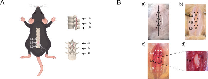
Figure 1: The L5 lumbar laminectomy procedure for postoperative cognitive dysfunction (POCD) modeling in mice. (A) Illustration of the surgical target at the L5 lamina and the exposure range spanning the adjacent vertebral levels (L4–L6). (B) Intraoperative images illustrating key steps of the L5 lumbar laminectomy procedure. (a) The lumbar region after shaving, with the spinal midline identified; arrows indicate the direction of the planned skin incision. (b) Midline skin incision over the lumbar spine. (c) Incision of the fascia with stepwise separation of the paraspinal muscles to expose the spinous processes and further clear the tissue around the L5 spinous process and lamina to fully expose the L5 lamina. (d) L5 laminectomy after removal of the L5 lamina. Please click here to view a larger version of this figure.
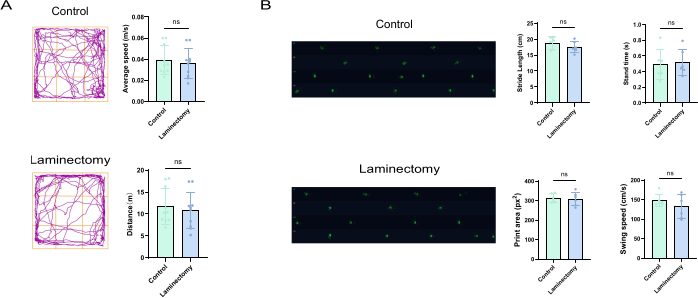
Figure 2: Assessment of locomotor activity and gait after L5 lumbar laminectomy. (A) Open-field test showing total distance traveled and average speed (n = 10). (B) Gait analysis showing stride length, stand time, swing speed, and print area (n = 6). Data represent the mean ± SD. Statistical significance is indicated as ns, not significant. Abbreviations: Control = control group; Laminectomy = L5 laminectomy group. Please click here to view a larger version of this figure.
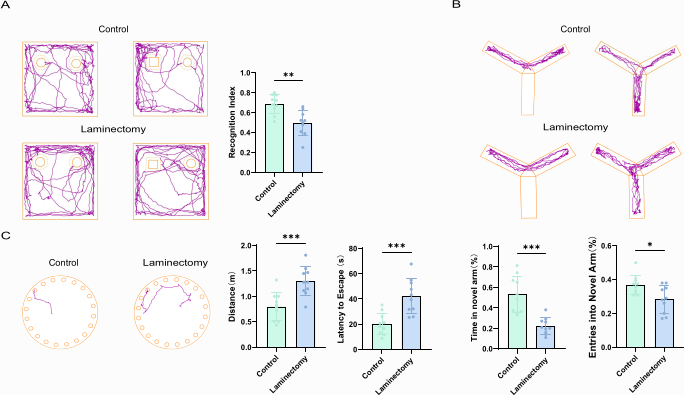
Figure 3: Cognitive behavioral assessments after L5 lumbar laminectomy. (A) Novel object recognition test (n = 10). (B) Novel arm Y-maze test (n = 10). (C) Barnes maze test (n = 10). Data represent the mean ± SD. Statistical significance is indicated as *P < 0.05, **P < 0.01, and ***P < 0.001. Abbreviations: Control = control group; Laminectomy = L5 laminectomy group. Please click here to view a larger version of this figure.
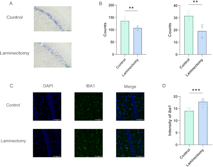
Figure 4: Histological assessment of the hippocampal CA1 region after L5 lumbar laminectomy. (A) Representative images of Nissl staining in the hippocampal CA1 region. (B) Quantification of Nissl-positive neurons (left) and Nissl bodies (right) (n = 5). (C) Representative images of Iba1 immunofluorescence in the hippocampal CA1 region. Scale bar = 100 µm. (D) Quantification of Iba1 mean fluorescence intensity (n = 6). Data represent the mean ± SD. Statistical significance is indicated as **P < 0.01, and ***P < 0.001. Abbreviations: Control = control group; Laminectomy = L5 laminectomy group. Please click here to view a larger version of this figure.
Discussion
Postoperative cognitive dysfunction has been reported in older adults undergoing spinal surgery10. Experimental evidence further indicates that aging increases susceptibility to surgery-associated cognitive impairment and neuroinflammatory responses11,12. The present protocol combines approximately 4 h of sevoflurane anesthesia with L5 laminectomy in aged mice to model early postoperative cognitive impairment. Its principal methodological value lies in providing a practical, accessible procedure for investigating behavioral and hippocampal histological changes following spinal surgery.
Preclinical POCD models include anesthesia-only exposure, abdominal surgery, and orthopedic injury paradigms13. Anesthesia-only paradigms permit evaluation of anesthetic effects in the absence of surgical trauma14, but do not reproduce the combined exposure to anesthesia and tissue injury. Abdominal surgery and tibial fracture models incorporate surgical injury and have been used to investigate surgery-associated cognitive impairment15,16. However, their operative sites and tissue-injury patterns differ from those of spinal procedures. Because the type and intensity of surgical trauma can affect postoperative cognitive outcomes and central inflammatory responses9, the surgical context should be considered when selecting a preclinical model. The present L5 laminectomy protocol, therefore, provides a spine surgery–related experimental paradigm for examining early postoperative cognitive impairment in aged mice.
The key technical challenge in this protocol is removing sufficient bone from the L5 lamina to expose the dura clearly while avoiding dural injury. During a laminectomy under direct visualization, careful control of instrument depth, cutting force, and the insertion angle of the ophthalmic scissors is required. The bone should be removed stepwise using small, controlled cuts along the margins of the lamina. Grasping the L5 spinous process with forceps and elevating the operative lumbar segment to an angle of approximately 30° relative to the surgical platform widens the L5–L6 interlaminar space. This maneuver permits shallow insertion of the scissor tips beneath the bony edge and reduces the risk of dural contact. Incomplete bone removal may limit surgical exposure, whereas overly aggressive cutting or excessive insertion depth may cause dural injury. Uncontrolled bleeding may obscure the operative field, impair postoperative recovery, and increase variability in behavioral outcomes. When the interlaminar space is insufficient for safe insertion of the scissor tips, the position and elevation angle of the operative lumbar segment should be readjusted rather than increasing the insertion depth. If bleeding obscures the operative field, bone removal should be paused until adequate visualization has been restored, after which any remaining bone should be removed incrementally.
Paraspinal muscle separation and L5 laminectomy may contribute to transient postoperative pain, and mechanical hypersensitivity has been documented after experimental laminectomy in mice17. Meloxicam was administered according to a predefined analgesic regimen. Postoperative pain-related facial expressions in mice have been reported to persist for approximately 36–48 h18. The 48 h analgesic regimen was intended to cover this early postoperative period, and cognitive behavioral testing was initiated on postoperative day 3, after completion of the analgesic regimen, to reduce the potential influence of immediate postoperative pain. The open-field test has been used in experimental POCD studies to assess spontaneous locomotor activity and to determine whether reduced locomotion may confound cognitive-behavioral performance19,20. Accordingly, open-field testing and gait analysis were performed on postoperative day 3, prior to cognitive assessment. No significant between-group differences were observed in open-field locomotor activity or gait parameters, suggesting that the subsequent cognitive-behavioral differences were unlikely to be explained primarily by overt motor impairment. However, because spontaneous postoperative pain and wound discomfort were not directly assessed, their residual effects on behavioral performance cannot be entirely ruled out. Future studies could incorporate direct assessments of spontaneous postoperative pain.
Overall, this protocol provides a practical experimental approach for investigating early postoperative cognitive impairment in aged mice following L5 laminectomy. It can be used to investigate the behavioral outcomes and underlying mechanisms of spine surgery–associated cognitive impairment, as well as to evaluate interventions aimed at preventing or reducing postoperative cognitive impairment. Validation in larger cohorts and at independent laboratories would further establish the model's stability and reproducibility.
Disclosures
The authors declare no competing interests.
Acknowledgements
We thank Sanhua Fang from the core facility platform of Zhejiang University School of Medicine, Yanhong Chen, and Xuliang Zhang from the animal center of Zhejiang University for their technical support. This study was funded by the following funds: National Natural Science Foundation of China (Nos. 82575196, 82074535, and U23A20508).
Materials
| Name | Company | Catalog Number | Comments |
|---|---|---|---|
| C57BL/6J male mice | Suzhou Industrial Park Ai'er Maite Technology Co., Ltd. | SYXK2023-0005 | Animal model |
| ANY-maze Video Tracking Software | Stoelting Co., USA | 6000 | Behavioral video tracking and analysis |
| Erythromycin ophthalmic ointment | Beijing Twinluck Pharmaceutical Co,. Ltd. | H11021270 | Prevent eye dry |
| Fine forceps | NA | NA | Clip the skin |
| Heating pad | NA | NA | Temperature-controlled |
| Iba1/AIF-1 (E4O4W) Rabbit Monoclonal Antibody | Cell Signaling Technology | 17198 | Iba1 immunofluorescence staining |
| Inhalant anesthesia equipment | RWD Life Science Co., Ltd. | R581S | Anesthesia |
| MGT-PR | Anhui Zhenghua Biological Instrument Equipment Co., Ltd., China | MJY2025112704 | Gait analysis |
| Micro Ophthalmic scissors | NA | NA | Bone removal |
| Nissl staining solution | Haoke Biotechnology Co., Ltd. | HK2033 | Nissl staining |
| Scissors | Shang Shou Jin Zhong Surgical Instruments Jiangsu ITD.,Corp | Y00030 | Skin incision |
| Sevoflurane | Shanghai Hengrui Pharmaceutical Co,. Ltd. | 25062331 | Anesthesia |
| Single Use Surgical Drape | Jiangxi Zhizheng Medical Equipment Co., Ltd. | 20212140221 | Platform for surgical operation |
| Small animal trimmer | Yiwu Leiben Trading Co., Ltd. | 2504KFZB3592-03310 | Hair removal |
| Surgical Suture Needles with Thread | Shanghai Pudong Jinhuan Medical Products Co., LTD. | F603 | Suture skin |
| Veet hair removal cream | RECKITT BENCKISER (India) Ltd | NA | Hair removal |
References
- Zhao Q, Wan H, Pan H, Xu Y. Postoperative cognitive dysfunction-current research progress. Front Behav Neurosci. 2024;18:1328790.
- Kong H, Xu LM, Wang DX. Perioperative neurocognitive disorders: A narrative review focusing on diagnosis, prevention, and treatment. CNS Neurosci Ther. 2022;28(8):1147-1167.
- Monk TG, et al. Predictors of cognitive dysfunction after major noncardiac surgery. Anesthesiology. 2008;108(1):18-30.
- Li X, et al. Risk factors for postoperative cognitive decline after orthopedic surgery in elderly Chinese patients: A retrospective cohort study. Clin Interv Aging. 2024;19:491-502.
- Krenk L, et al. Cognitive dysfunction after fast-track hip and knee replacement. Anesth Analg. 2014;118(5):1034-1040.
- Yang X, et al. Identification of individuals at risk for postoperative cognitive dysfunction (POCD). Ther Adv Neurol Disord. 2022;15:17562864221114356.
- Suraarunsumrit P, et al. Outcomes associated with postoperative cognitive dysfunction: A systematic review and meta-analysis. Age Ageing. 2024;53(7):afae160.
- Lin X, et al. The potential mechanism of postoperative cognitive dysfunction in older people. Exp Gerontol. 2020;130:110791.
- Lu B, et al. Effects of different types of non-cardiac surgical trauma on hippocampus-dependent memory and neuroinflammation. Front Behav Neurosci. 2022;16:950093.
- Park S, et al. Preoperative mild cognitive impairment as a risk factor of postoperative cognitive dysfunction in elderly patients undergoing spine surgery. Front Aging Neurosci. 2024;16:1292942.
- Xu Z, et al. Age-dependent postoperative cognitive impairment and Alzheimer-related neuropathology in mice. Sci Rep. 2014;4:3766.
- Zhang ZJ, et al. Aging alters Hv1-mediated microglial polarization and enhances neuroinflammation after peripheral surgery. CNS Neurosci Ther. 2020;26(3):374-384.
- Eckenhoff RG, et al. Perioperative neurocognitive disorder: State of the preclinical science. Anesthesiology. 2020;132(1):55-68.
- Guo LY, et al. Cognitive deficits after general anaesthesia in animal models: A scoping review. Br J Anaesth. 2023;130(2):e351-e360.
- Qiu LL, et al. NADPH oxidase 2-derived reactive oxygen species in the hippocampus might contribute to microglial activation in postoperative cognitive dysfunction in aged mice. Brain Behav Immun. 2016;51:109-118.
- Cibelli M, et al. Role of interleukin-1beta in postoperative cognitive dysfunction. Ann Neurol. 2010;68(3):360-368.
- Yamamoto S, et al. Intraoperative spinal cord stimulation mitigates central sensitization after spine surgery in mice. Spine (Phila Pa 1976). 2023;48(11):E169-E176.
- Matsumiya LC, et al. Using the mouse grimace scale to reevaluate the efficacy of postoperative analgesics in laboratory mice. J Am Assoc Lab Anim Sci. 2012;51(1):42-49.
- Sun L, et al. Activation of cannabinoid receptor type 2 attenuates surgery-induced cognitive impairment in mice through anti-inflammatory activity. J Neuroinflammation. 2017;14(1):138.
- Zhan G, et al. Anesthesia and surgery induce cognitive dysfunction in elderly male mice: The role of gut microbiota. Aging (Albany NY). 2019;11(6):1778-1790.
Reprints and Permissions
This article has been published
Video Coming Soon