To study Mycobacterium tuberculosis mutations for gene function and drug resistance, two targeted site-directed mutagenesis techniques for introducing changes into an isogenic background, two-step allelic exchange (AE) with a suicide vector, or recombineering with ssDNA oligonucleotides, are described and compared. Mutagenesis is confirmed via PCR and sequencing of the amplicon.
Method Article
Site-Directed Mutagenesis in Mycobacterium tuberculosis
* These authors contributed equally
In This Article
Summary
Abstract
Precise manipulation of the mycobacterial genome is required to study the effect of specific mutations without confounding influences from the diverse genetic backgrounds in clinical isolates. This is particularly important for interrogating gene function and drug resistance mechanisms, where background mutations may enhance or suppress the effects of a mutation of interest. Here, two targeted site-directed mutagenesis approaches, two-step allelic exchange using a suicide vector and oligonucleotide-mediated recombineering, are described and compared for introducing defined mutations into an isogenic Mycobacterium tuberculosis background.
Two-step allelic exchange uses a suicide vector lacking a mycobacterial origin of replication and carrying a mutated DNA fragment, selectable markers such as sacB for sucrose sensitivity, lacZ for blue-white screening, and antibiotic resistance genes (kan or hyg). The process involves generating the mutated fragment via a multi-step polymerase chain reaction (PCR) or synthesis, cloning it into the vector, electroporating it into M. tuberculosis, and selecting single-crossover (SCO) integration events using antibiotic resistance and blue-white colony screening. This is followed by counter-selection with sucrose and X-gal to eliminate bacteria carrying the wild-type sequence. This method ensures stable, unmarked mutations but is time-consuming and labor-intensive.
Recombineering uses a recombinase-expressing plasmid, such as pJV75amber expressing Che9c gp61 recombinase under an inducible acetamidase promoter, together with a single-stranded DNA (ssDNA) oligonucleotide to introduce precise mutations. Transformants are selected on appropriate antibiotics and confirmed by PCR and Sanger sequencing. This approach is simpler, faster, and more cost-effective, but may be limited by low transformation efficiency, the need for fresh competent cells, and variable efficiency depending on the genomic region and oligonucleotide design.
Together, two-step allelic exchange and recombineering offer complementary strategies for introducing defined mutations into M. tuberculosis. Both methods enable experimental validation of mutations, such as variants associated with gene function and drug resistance.
Introduction
The study of genomic mutations enables interrogation of gene function and drug resistance using a variety of methods. However, the complex genetic background of clinical isolates harboring a mutation of interest often complicates the interpretation of results, as numerous other mutations may interact, leading to synergistic or antagonistic effects1. Although artificial intelligence (AI)-based approaches can predict resistance, they cannot conclusively prove it or determine downstream effects2,3.
Fluctuation assays, which rely on spontaneous mutations, can effectively select for resistance under known stressors such as antimycobacterial drugs4,5. However, this untargeted strategy requires extensive screening and offers no guarantee of recovering a specific mutation, e.g., novel mutations in drug-resistance-associated genes observed in clinical isolates.
Several targeted approaches have been developed, among which site-directed mutagenesis encompasses a suite of molecular techniques used to introduce precise single-nucleotide substitutions (point mutations) into a gene of interest6. The general workflow involves: (1) generating a donor DNA fragment or plasmid containing the gene of interest with the desired point mutation(s), (2) introducing this DNA into the bacterium, (3) delivering the mutated sequence to the chromosomal target locus, (4) promoting incorporation of the mutation via recombination, and (5) screening for recombinant mutants.
In this publication, two distinct site-directed mutagenesis strategies for targeted manipulation of M. tuberculosis genes are described, their workflows compared, and the advantages and disadvantages of each discussed. The methods exploit either the mycobacterium's natural homologous recombination (HR) repair pathway, also referred to here as two-step Allelic Exchange (AE) with a suicide vector7, or a plasmid-encoded phage-derived recombineering system8.
The two-step AE method incorporates a suicide delivery vector into the chromosomal DNA of an organism by HR. This suicide delivery vector contains a flexible cassette which forms part of the pNIL/pGOAL vector series and has been well discussed7. It provides high precision and stable integration for complex edits, including gene knockouts and large insertions. The core principles of AE using suicide vectors and recombineering have been established in prior protocols for mycobacteria9,10, including HR-based gene knockouts and introducing point mutations7. Limitations of AE include potential in vitro antibiotic stress from multiple selection markers, risk of spontaneous mutations during serial passages, labor-intensive cloning steps, and a two-step selection process that can span 8–12 weeks.
In contrast, recombineering leverages the Che9c gp61 recombinase from pJV75amber and single-stranded DNA (ssDNA) oligonucleotides8,11, to facilitate site-directed mutagenesis and screening. The episomal plasmid pJV75amber expresses the Che9c gp61 recombinase (a single-stranded DNA annealing protein homologous to E. coli RecT) under the control of an inducible acetamidase promoter8. The recombinase protein mediates ssDNA recognition, annealing, and allelic exchange between an exogenously synthesized ssDNA oligonucleotide template containing a point mutation and the endogenous homologous DNA strands8.
Recombineering eliminates cloning steps, reducing the timeline to 4–6 weeks, making it ideal for rapid validation of drug resistance variants in clinical isolates. However, recombineering may exhibit variable efficiency in certain genomic regions or result in off-target (illegitimate) recombination due to the use of short homology arms, requiring repeated rounds of experimentation. It may further require extensive screening of colonies in the absence of built-in counter-selection.
Presenting both methods side-by-side enables researchers to select one or the other based on project needs: AE for robust, unmarked edits in essential genes versus recombineering for speed and simplicity in high-throughput mutagenesis. Ultimately, these approaches facilitate studies on gene function, epistasis, and resistance mechanisms without confounding genetic backgrounds. We highlight the suitability of AE for complex edits, the speed of recombineering, and the labor intensity of AE. Further, recombineering shows genomic region dependency, which is not emphasized in prior single-method protocols8. Here, targeted adaptations and improvements are provided for site-directed mutagenesis in M. tuberculosis.
All experiments involving the manipulation of live M. tuberculosis must be conducted by trained personnel in a BSL3 facility, following institutional biosafety protocols and risk assessments. The facility should be equipped with controlled access, negative-pressure airflow, and a HEPA-filtered exhaust system, and work should be conducted in certified biological safety cabinets (Class II). Additionally, personal protective equipment (PPE), such as a powered air-purifying respirator (PAPR) or N95 respirators, gloves, coveralls or gowns, and eye protection, should always be worn while inside a BSL-3 facility. All waste, including culture media and disposables, must be autoclaved or chemically decontaminated before removal from the BSL3.
Prior to removal from the BSL3 for downstream analyses such as PCR, cultured materials including cell pellets should be heat-inactivated at 80 °C for 60 min. Inactivated samples are then double-bagged, surface-decontaminated with a suitable detergent and 70% ethanol, and transported to a BSL2 for molecular work. These safety measures are important to minimize the risk of aerosol formation during procedures such as centrifugation or pipetting, which may result in accidental exposure.
UV-protective goggles or a face shield, and gloves should be worn during UV exposure; exposures should only be conducted in a closed UV irradiator system to prevent skin and eye damage.
Protocol
Method A: Two-step allelic exchange (AE)
NOTE: Homologous recombination provides an effective method for introducing point mutations into the M. tuberculosis gene of interest. The technique consists of five phases: 1) generating and amplifying an insert sequence; 2) constructing a suicide delivery vector; 3) delivering the mutant DNA to the mycobacterial genome; 4) transformation of the plasmid carrying the mutant allele to generate a co-integrant; and 5) counter-selecting for the resolution of the co-integrant. While this method can be applied to construct deletion mutants, the focus here is on generating single-nucleotide variant strains.
- Generation of a dsDNA fragment for homologous recombination
NOTE: When investigating the effect of a single-nucleotide change using AE, three options exist to generate the fragment or gene of interest harboring the single-nucleotide variant (SNV). (1) Amplify a genomic region containing an SNV of interest from a clinical strain by polymerase chain reaction (PCR) and clone it into a suitable vector. (2) Purchase a custom-designed oligonucleotide synthesized to carry the point mutation of interest. (3) Use a three-step PCR method (described below) to introduce the point mutation into the fragment or gene of interest. This overlap PCR introduces a single nucleotide change precisely at the targeted position in a genomic sequence. It requires two primer sets and involves three steps: 1) PCR amplification of the upstream and downstream regions separately, 2) annealing the two overlapping fragments, and 3) amplifying the full-length DNA fragment using the outer primers.- Design the outer primers to bind at the 5' and 3' ends of the upstream and downstream regions of the gene, respectively. Include in these outer primers a 20-base-pair (bp) homology region to the gene, restriction enzyme sites, and an additional 6-bp non-coding sequence overhang at each primer end to allow direct restriction enzyme digestion during cloning experiments. Ensure the restriction sites are present in the vector but not in the gene of interest (Table of Materials).
- Design the central primers to be complementary and carry the SNV of interest. Allow the complementary forward and reverse primers to overlap in the middle of the upstream and downstream regions, incorporating the nucleotide base change into the center of the dsDNA fragment.
- Use genomic dsDNA from M. tuberculosis H37Rv as template DNA in a 25 µL PCR reaction mix (Figure 1) using a high-mismatch proofreading Taq polymerase. Set up the separate reactions as follows: Add 5 µL of GC Buffer (final 1×, recommended for GC-rich M. tuberculosis DNA), 0.5 µL of 10 mM dNTPs (final 200 µM each), 1.25 µL of 10 µM forward primer (final 0.5 µM), 1.25 µL of 10 µM reverse primer (final 0.5 µM), 0.25 µL of Polymerase (final 1 U), 1 µL of template DNA (50–100 ng), 0.75 µL of 100% DMSO (final 3% for GC-rich templates), and nuclease-free water to 25 µL final volume. Use primers A and B in one reaction, and C and D in the other (Figure 1A).
- Amplify two DNA fragments separately: the upstream region with primers A and B, and the downstream region with primers C and D (Figure 1A). Optimize the cycling conditions according to the primers used and length of the amplicon, starting with initial denaturation at 98 °C for 30 s; 30 cycles of denaturation at 98 °C for 10 s, annealing at Tm (typically 55–65 °C for these primers) for 30 s, extension at 72 °C for 30 s per kilobase (kb); final extension at 72 °C for 5 min. Hold at 4 °C.
- Anneal and extend the upstream and downstream regions by combining 5 µL of each generated PCR product (from step 1.4) into a new 25 µL reaction (adjust component proportions proportionally, omitting addition of primers) (Figure 1B). Use the same cycling conditions but reduce the number of cycles to 10–15.
- To remove excess primers from the first-step PCR products before the third step, dilution (1:10 to 1:50) is often sufficient, but for higher purity, use commercial column-based cleanup kits or gel extraction if non-specific bands are present.
- In the last amplification step, add 2 µL from the overlapping PCR product (from step 1.5) to a new 25 µL reaction with primers A and D (final 0.5 µM each) (Figure 1C). Use the same cycling conditions as in step 1.4.
NOTE: Remember to adjust the annealing Tm and extension time for the new primer combination (primer A and D) and extended fragment length. - Verify amplification by agarose gel electrophoresis; expect a single band of the predicted size, with no smears.
NOTE: Do not discard gel. Used in step 2.1.
- Vector construction for sequencing SNV-containing dsDNA fragment
NOTE: This section describes the second step of AE. The generated fragment of interest is cloned into a sequencing vector to enable plasmid propagation and obtain multiple copies for more efficient cloning in subsequent steps.- Prepare chemically competent E. coli cells according to the method of Green and Rogers12.
- Excise the PCR-amplified dsDNA fragment containing the SNV of interest from step 1.8 from the gel using a clean scalpel blade and purify using a commercial gel and PCR cleanup kit according to the manufacturer's protocol. Measure the DNA concentration using spectrophotometry; aim for 50–100 ng/µL. Observe a clear solution post-purification.
- Ligate the linear DNA fragment into a sequencing vector (e.g., pJET1.2) using a suitable ligase. Perform ligations in a 10 µL reaction: add 1 µL of 10× ligase buffer, 50 ng of vector, 150 ng of insert (3:1 molar ratio), 1 µL of DNA ligase (400 U/µL), and water to 10 µL; incubate at 4 °C overnight and inactivate by incubating for 10 min at 65 °C.
- Transform the plasmid construction into chemically competent E. coli cells by adding 5 µL of ligated plasmid DNA (see step 2.3) to 100 µL of competent cells and gently mix by tapping. Incubate the mixture on ice for 30 min. Heat-shock the cells at 42 °C for 90 s, then immediately return the tube to ice for 2 min. Add 900 µL of Luria-Bertani (LB) medium and incubate the cells at 37 °C with shaking (200 rpm) for 1 h to allow recovery.
- Harvest cells by centrifugation (3,000 × g for 10 min) and discard the LB media. Resuspend recombinant cells in 100 µL fresh LB media, plate 50 µL on LB agar containing the appropriate antibiotics, and incubate statically at 37 °C overnight. Expect 50–500 colonies per plate.
- Screen colonies for the presence of the DNA fragment using colony PCR by picking 1–10 single colonies and adding each colony's cells to a PCR reaction, as described in step 1.3 with primers A and D. The PCR is run as described in step 1.4.
- Select PCR-positive colonies (usually 5–10) and culture overnight in 5 mL of LB media with antibiotics at 37 °C with shaking at 200 rpm.
- Prepare a glycerol -80 °C freezer stock of each overnight culture. Extract plasmid DNA using a commercial miniprep DNA purification kit and perform Sanger sequencing to confirm the dsDNA fragment sequence and the presence of the SNV. Additionally, use restriction enzyme (RE) mapping to ensure that the correct-sized fragment is incorporated into the sequencing plasmid. Verify by gel electrophoresis; expect fragments matching the predicted sizes.
- Constructing the suicide delivery vector
NOTE: This section describes the final preparation of the suicide delivery vector. The two widely used suicide delivery vectors are p1NIL and p2NIL, differing only in the restriction enzyme cut sites in their multiple cloning sites. This protocol refers to p2NIL (Table of Materials). To aid in vector construction, consult plasmid maps of p2NIL and pGOAL19 for unique restriction sites.- Perform RE digestion to remove the verified SNV-containing DNA fragment from step 2.8, from the sequencing vector, using the appropriate enzymes (e.g., HindIII and KpnI). In a separate reaction, digest the suicide delivery vector (e.g., p2NIL) with the same enzymes. Perform both digests at a large scale (1 µg DNA) in a 50 µL reaction: add 10 µL of 10× buffer, 1 µL of each enzyme (10 U/µL), 1 µg of DNA, and nuclease-free water to a final volume of 50 µL; incubate at 37 °C for 1 h. Heat-inactivate if required as per the manufacturer's recommendation.
- Separate the DNA fragments by agarose gel electrophoresis. Observe clear bands at expected sizes. Excise the linearized p2NIL vector and the SNV-containing DNA fragment from the agarose gel and purify using a commercial gel and PCR cleanup kit.
- Measure the DNA concentrations using spectrophotometry and calculate the volume of DNA needed to achieve a 3:1 insert-to-vector molar ratio using 50 ng of p2NIL vector DNA. Perform ligations in a 10 µL reaction: add 1 µL of 10× ligase buffer, 50 ng of vector, 150 ng of insert (3:1 molar), 1 µL of T4 DNA ligase (400 U/µL), and water to 10 µL; incubate at 4 °C overnight.
- Transform the ligated vector with the insert into competent E. coli cells (see step 2.4). Plate the transformation on LB agar with 50 μg/mL Kanamycin (Kan). Incubate at 37 °C overnight.
- Subject the resulting colonies (p2NIL with the SNV-containing DNA fragment, hereafter p2NIL_insert) to colony PCR (see step 2.6) and agarose gel electrophoresis to confirm the presence of the SNV-containing DNA fragment. Run the PCR as described in step 1.3, using primers A and D as optimized in step 1.7.
- Inoculate a colony containing the correctly constructed p2NIL_insert vector into 10 mL of LB media with Kan at 37 °C, shaking at 200 rpm overnight. Prepare freezer stocks (use 1–2 mL of culture) and perform miniprep to isolate p2NIL_insert.
- Introduce selectable markers from the pGOAL vector series (pGOAL17 or pGOAL19) into p2NIL_insert by digesting both pGOAL and p2NIL_insert with PacI restriction enzyme in separate 50 µL reactions (see step 3.1).
- Separate the resulting fragments by agarose gel electrophoresis, excise fragments of interest, and purify using a commercial gel and PCR cleanup kit.
- Dephosphorylate the linearized p2NIL_insert to prevent self-ligation of the 5' and 3' ends by alkaline phosphatase treatment: add 1 unit/µg of fragment DNA in a 20 µL reaction with 2 µL of 10× buffer and water; incubate for 10 min at 37 °C, followed by heat-inactivation for 2 min at 75 °C.
NOTE: When required to prevent self-ligation, vector DNA is de-phosphorylated using 1 unit/µg of rAPid alkaline phosphatase and incubated to heat-inactivate. - Ligate the pGOAL selectable marker cassette into the dephosphorylated p2NIL_insert to generate p2NIL_insert_SM in a 10 µL reaction (see step 2.3) and transform into chemically competent E. coli cells (see step 2.4).
- Plate cells on LB agar with the appropriate antibiotic (50 µg/mL Kan and/or 200 µg/mL Hyg) and 50 µg/mL X-gal to confirm the functionality of the selection markers. Incubate at 37 °C overnight; expect blue colonies indicating LacZ activity.
- Confirm sucrose sensitivity by streaking individual blue colonies on LB agar containing antibiotics, with and without 2% sucrose, to verify sucrose sensitivity.
- Confirm the final construct of the suicide delivery vector by RE mapping. Verify expected fragment patterns by purifying the plasmid from E. coli. Perform RE plasmid mapping to compare the pattern of restriction to that predicted by the plasmid's sequence file.
- Generation of M. tuberculosis harboring SNV through homologous recombination
NOTE: A suicide delivery vector is introduced into genomic DNA through homologous recombination, followed by a two-step selection method to select for, and then against, the selectable markers. The following steps are performed under Biosafety Level 3 conditions.
NOTE: Step 1 - 11 describes selection of a co-integrant M. tuberculosis strain.- Prepare electrocompetent M. tuberculosis cells by harvesting cells at an OD600 of 0.6–1.0 and washing three times in 1/50 volume of cold 10% glycerol, followed by resuspension in 10% glycerol. Cell concentration should be around 1011 cells per mL, and may be stored at -80 °C10.
- Place 10 µg of the plasmid DNA in a Petri dish in a UV irradiator system and expose to UV light until the energy reaches 100 mJ/cm2.
- Add 10 µg UV-pretreated plasmid DNA or 6 µL of Tris-EDTA (TE) buffer (negative control) to 400 µL of electrocompetent M. tuberculosis cells in separate 1.5 mL microcentrifuge tubes. Mix gently by pipetting.
- Transfer the mixture into a 0.2 cm electroporation cuvette and electroporate with a single pulse of 2.5 kV, 25 µF, and 1,000 Ω resistance. Monitor the time constant (optimum 15–25 ms). Discard if arcing occurs.
- Resuspend the electroporated cells in 600 µL of 7H9-OADC medium and transfer cells to sterile 2 mL screw cap tubes (with o-ring).
- Incubate transformed cells for 24–48 h at 37 °C.
- Harvest cells by centrifugation at 3,000 × g for 10 min at room temperature. Expect a visible pellet. Remove media from pelleted cells.
- Streak cells on 7H11-OADC agar plates containing the appropriate antibiotics (20 µg/mL Kan and/or 50 µg/mL Hyg) and 50 µg/mL X-gal.
NOTE: Add X-gal to cooled 7H11-OADC agar (50 °C) before pouring plates. X-gal may degrade over extended 37 °C incubations (3–4 weeks) required for M. tuberculosis, potentially reducing blue-white screening sensitivity due to thermal instability. Store pre-poured plates at 4 °C in the dark for up to 2 weeks to minimize degradation. If faint coloration is observed, consider the X-gal underlay method: Pour plain 7H11-OADC agar plates, solidify, then lift the agar slightly and add 200–500 µL of X-gal solution (20 mg/mL in DMF) under the agar before resealing and incubating. This reduces direct heat exposure during storage and incubation. - Incubate at 37 °C for 21 days. Colonies that have successfully undergone the first AE appear blue, are sucrose-sensitive, and antibiotic-resistant (Figure 2). The single crossover (SCO) M. tuberculosis strains contain both the wild-type and SNV-containing regions.
- Pick SCO colonies and inoculate 5 mL of 7H9-OADC supplemented with 0.05% Tween 80 (from here, 7H9-OADC-Tween) and the appropriate antibiotic. Incubate at 37 °C for 7 days.
- To confirm sucrose-sensitivity, streak each culture on a 7H11-OADC agar plate containing 2% sucrose. Incubate at 37 °C for 21 days. Expect SCO cultures not to grow on sucrose. Continue to use the SCO cultures from step 4.10 that displayed sucrose sensitivity.
NOTE: Step 12-18 describes selection of the resolution of the co-integrant M. tuberculosis strain. - Sub-culture the SCO colonies into 5 mL of 7H9-OADC-Tween (without antibiotics) and incubate for 7 days at 37 °C to select for the resolution of the co-integrant (DCO).
- Streak 100 µL on 7H11-OADC agar plates with 2% sucrose and X-gal (50 µg/mL). Incubate at 37 °C for 21 days. Although it should still be confirmed, expect white colonies that are antibiotic-sensitive and sucrose-resistant if the second allelic exchange step was successful (Figure 2).
- Pick 10–30 DCO single colonies and inoculate them in 5 mL of 7H9-OADC-Tween (without antibiotic). Incubate for 7 days at 37 °C.
- To confirm resolution of the integrated vector backbone, perform patch tests on kanamycin and, where applicable, hygromycin (for pGOAL19 constructs). Additionally, conduct sucrose counter-selection patch tests depending on the vector used (pGOAL17 or pGOAL19): streak colonies onto 7H11-OADC agar plates containing either 50 µg/mL Hyg or 2% sucrose, incubate at 37 °C for up to 21 days. Colonies that have resolved the integrated vector backbone will grow on the plate containing 2% sucrose, but not on the plates containing hygromycin.
- Perform colony PCR by heating a 200 µL aliquot of the DCO M. tuberculosis cultures at 80 °C for 60 min. Centrifuge boiled cultures at 15,000 × g for 15 min at room temperature to form a pellet; use the supernatant (containing DNA) for PCR amplification. Perform PCR setup as described in step 1.3, with primers A and D. Run PCR as described in step 1.4.
- Separate the resulting fragments by agarose gel electrophoresis. Excise fragments of interest and purify using a commercial gel and PCR cleanup kit according to the manufacturer's protocol.
- Sanger sequence the PCR product to confirm the presence of the SNV. Analyze the output by mapping the sequenced DNA fragment to the M. tuberculosis H37Rv reference genome (GenBank NC_000962.2). Additionally, perform whole-genome sequencing of the progenitor and mutant strains to ensure no off-target mutations were acquired during culturing.
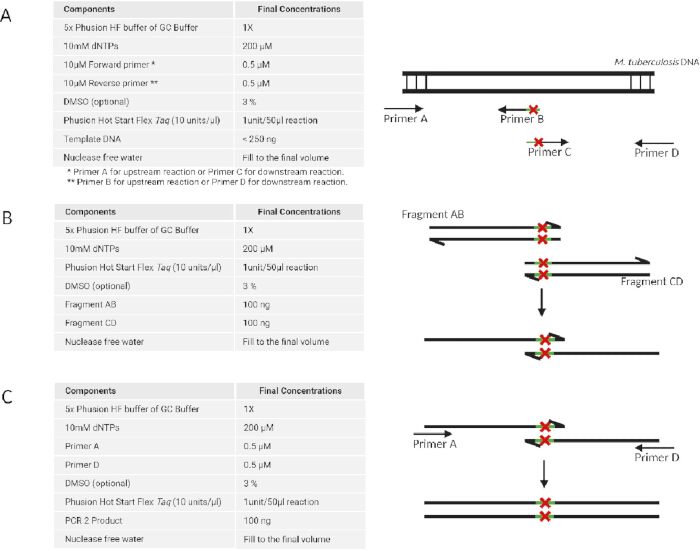
Figure 1: Generation of dsDNA fragments harboring a single nucleotide variant using a multi-step PCR method. Generating the DNA fragment involves three steps: (A) PCR amplification of the upstream and downstream regions separately using two primer sets. (B) The upstream and downstream regions are annealed to form a single overlapping fragment. (C) Further amplification of the full-length DNA fragment is done using the outer primers. Please click here to view a larger version of this figure.
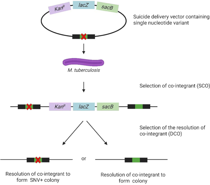
Figure 2: Construction of an M. tuberculosis strain harboring an SNV for homologous recombination. Created in BioRender (https://BioRender.com). Please click here to view a larger version of this figure.
Method B: Recombineering
NOTE: Recombineering begins with electroporation of M. tuberculosis with three components: the recombineering plasmid pJV75amber, the 63 bp ssDNA oligonucleotide JCV198 (to restore hygromycin resistance on the plasmid), and a 63 bp ssDNA oligonucleotide carrying the desired point mutation. Transformants are then screened by colony PCR and Sanger sequencing. The composition for restriction enzyme digestion of pJV75amber is outlined in Table 1. The following steps are performed under Biosafety Level 3 conditions:
| Reagents | Tube A (µL) | Tube B (µL) | Tube C (µL) | Tube D (µL) |
| 10x Buffer B | 2 | - | 2 | 2 |
| 10x Buffer M | - | 2 | - | - |
| Nuclease-free water | 16 | 15 | 15 | 13 |
| pJV75 amber DNA aliquots | 2 | 2 | 2 | 2 |
| Dra1 Enzyme | - | 1 | - | 2 |
| EcoRv Enzyme | - | - | 1 | 1 |
| Final Reaction volume | 20 | 20 | 20 | 20 |
Table 1: Restriction digestion: pJV75amber DNA.
- M. tuberculosis transformation with pJV75amber
- Prepare electrocompetent M. tuberculosis cells as described in Method A step 4.1.
- Add 100 ng of pJV75amber plasmid DNA or 5 µL of nuclease-free water (negative control) to 95 µL of electrocompetent M. tuberculosis cells in separate 1.5 mL microcentrifuge tubes. Mix gently by pipetting.
- Transfer into two separate electroporation cuvettes (0.2 cm gap) and incubate at room temperature for 30 min. Ensure the cell suspension settles to the bottom of the cuvette (tap gently to remove any bubbles, if needed).
- Place the cuvette into the sample chamber of the electroporator and apply the pulse at a resistance of 1000 Ω, a voltage of 2.5 kV, and a capacitance of 25 µF. Monitor the time constant (optimum 15–25 ms). Discard if arcing occurs.
- Immediately add 900 µL of 7H9-OADC-Tween and mix by pipetting up and down 10 times.
- Transfer the cells (1.0 mL total volume) into two 50 mL tubes, one each for the sample and the control, and incubate for 48 h at 37 °C.
- Harvest cells by centrifugation at 3,000 × g for 15 min at 37 °C. Expect a visible pellet. Remove media from pelleted cells. Resuspend in 200 µL of 7H9-OADC-Tween.
- Plate 100 µL of culture on 7H11-OADC agar plates containing 25 µg/mL Kan.
- Incubate the plates for 21 days at 37 °C.
- Colony PCR-test to confirm pJV75amber
NOTE: Refer to the general colony PCR protocol (see Method A, step 2.6) with the following M. tuberculosis-specific modifications:- Pick single colonies of the pJV75amber-transformed bacteria into 15 mL microcentrifuge tubes with 7H9-OADC-Tween and Kan (25 µg/mL).
- Incubate at 37 °C for 7 days.
- Transfer 200 µL of the culture into 1.5 mL centrifugation tubes, storing the remainder at 4 °C, or as 50% glycerol stocks at -80 °C for use in Method B, step 3.1.
- Release plasmid DNA from M. tuberculosis cells by heating the culture for 60 min at 80 °C.
- Pellet cells by centrifugation at 16,000 × g for 15 min at room temperature.
- Perform PCR using forward primer (KANR_F) and reverse primer (KANR_R): add 12.5 μL Taq master mix, 1.25 μL of each primer (final 0.5 μM), 1 μL of template DNA (supernatant from Method B, step 2.5), water to 25 μL. Use cycling conditions optimized for the kanamycin gene (aph) primer set: initial denaturation at 95 °C for 3 min; 35 cycles of 95 °C for 30 s, 55 °C for 30 s, and 72 °C for 45 s; final extension at 72 °C for 5 min. Hold at 4 °C.
- Analyze on a 1% agarose gel at 150 V for 45 min. Colonies that produce the expected 700 bp aph band are confirmed to contain pJV75amber; select these for downstream recombineering.
- Recombineering electrocompetent M. tuberculosis transformed with pJV75amber
- Using the single colony cultures, with confirmed pJV75amber, from step 2.2 (Method B), inoculate 20 mL 7H9-OADC-Tween containing Kan (25 μg/mL), and 0.2 % succinate.
- Incubate at 37 °C until OD600 reaches 0.6 (mid-log phase growth) to prepare competent M. tuberculosis cells (see Method A, step 4.1).
- Add acetamide to a final concentration of 0.2%.
- Incubate at 37 °C overnight.
- Set out labeled electroporation cuvettes (0.2 cm gap), each containing 100 µL induced, competent M. tuberculosis cells, and add ssDNA oligonucleotides for recombineering as outlined in Table 2:
- Sample: Add 1 µg of JCV198 ssDNA oligonucleotide and 1 µg of ssDNA recombineering oligonucleotide with the sequence of interest.
- Positive control: Add 1 µg of JCV198 ssDNA oligonucleotide.
- Negative control: Add 2 µL of nuclease-free water (no DNA).
- Mix by pipetting gently.
- Electroporate and recover cells as described in Method B, steps 1.3 - 1.7.
- Plate 100 µL of transformation cultures on 7H11-OADC agar plates with and without Hyg (50 µg/mL).
- Incubate the plates for 21 days at 37 °C.
- Screen for recombinant M. tuberculosis to confirm the inserted point mutation
- Pick single colonies of recombinant M. tuberculosis cells and inoculate them in 5 mL of 7H9-OADC-Tween with Hyg (50 µg/mL).
- Incubate at 37 °C for 7 days.
- Transfer 200 µL aliquots of each overnight culture into sterile 1.5 mL microcentrifuge tubes. Store the remaining cultures at 4 °C (or prepare glycerol stocks) until PCR and Sanger sequencing identify the colonies carrying the SNV of interest. Heat-kill by heating at 80 °C for 1 h centrifuge at 16,000 × g for 5 min at room temperature to pellet debris.
- Perform PCR and Sanger sequencing on the supernatant to confirm the presence of the SNV of interest. Verify by mapping to reference; expect the SNV without additional mutations.
- To expedite colony PCR confirmation of pJV75amber in M. tuberculosis (see Method B, step 2), consider a direct lysis method using Chelex-100:
- Pick a single colony (retaining some live material) directly into 100 µL of 5% Chelex-100 suspension in a 1.5 mL tube, vortex to resuspend, boil at 100 °C for 10 min, centrifuge at 16,000 × g for 5 min, and use 1–2 µL of the cleared supernatant as PCR template.
NOTE: This bypasses the 7-day liquid culture step, reducing the overall timeline from 1 week to a day while still yielding sufficient plasmid DNA for aph amplification. - Monitor for potential PCR inhibition or false negatives due to incomplete lysis in mycobacteria, due to the thick mycolic acid cell wall.
- If results are inconsistent, test both the Chelex direct-lysis method and the heat-lysis approach in parallel on the same set of colonies to compare sensitivity and reliability.
- Pick a single colony (retaining some live material) directly into 100 µL of 5% Chelex-100 suspension in a 1.5 mL tube, vortex to resuspend, boil at 100 °C for 10 min, centrifuge at 16,000 × g for 5 min, and use 1–2 µL of the cleared supernatant as PCR template.
| Samples | Competent M. tuberculosis|pJV75amber | JCV198 | ssDNA of interest | Nuclease free water |
| A: sample | 100 μL | 1 μL | 1 μL | n/a |
| B: positive control | 100 μL | 1 μL | n/a | 1 μL |
| C: negative control | 100 μL | n/a | n/a | 2 μL |
Table 2: Experimental setup for transformation with ssDNA oligonucleotides.
Results
Two-step allelic exchange
PCR amplification of the rv0678 A97G DNA fragment flanked by restriction enzyme sites
The upstream and downstream regions of M. tuberculosis H37Rv rv0678 (1413 bp each) were amplified by PCR. This step ensured incorporation of an c.97A>G single nucleotide change in rv0678 (Thr33Ala), as well as restriction sites to allow cloning into the p2NIL vector. Bands corresponding to the expected fragment size, approximately 1400 bp, were confirmed by agarose gel electrophoresis (Figure 3). DNA bands were purified from the gel, yielding 67.9 ng/µL and 40.50 ng/µL of the upstream and downstream DNA fragments, respectively.
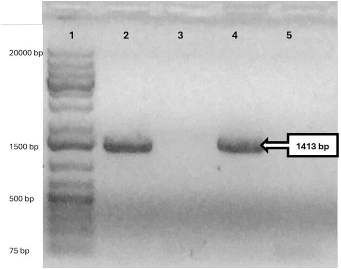
Figure 3: PCR amplified upstream (1413 bp) and downstream (1413 bp) regions of rv0678 for the construction of the suicide delivery vector. A 0.5% agarose gel was used to confirm successful amplification of the rv0678 upstream and downstream fragments. Lane 1: 1 kb DNA ladder; lane 2: PCR product of rv0678 upstream region; lane 3: negative control for the amplification of rv0678 upstream region; lane 4: PCR product of rv0678 downstream region; lane 5: negative control for the amplification of rv0678 downstream region. Please click here to view a larger version of this figure.
The full-length rv0678 fragment containing the central c.97A>G nucleotide change was generated by overlap PCR of the upstream and downstream fragments, yielding a single 2825 bp fragment, confirmed by gel electrophoresis (Figure 4).
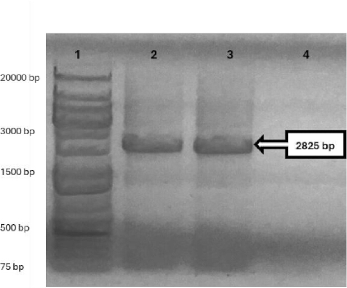
Figure 4: Overlap PCR of the full-length rv0678 DNA fragment (2825 bp). A 0.5% agarose gel was used to confirm the successful overlap of the upstream and downstream regions to generate the full-length rv0678 DNA fragment containing the A97G mutation. Lane 1: 1 kb DNA fragment; lane 2 and 3: full-length rv0678 fragment; lane 4: negative control. Please click here to view a larger version of this figure.
The PCR products were purified using a column to yield 126.6 ng/µL of purified rv0678 DNA, which was then ligated into the sequencing plasmid vector. Sanger sequencing of plasmid DNA (pJET1.2_rv0678) isolated from E. coli single colonies (ampR) recovered from transformation plates confirmed an E. coli colony of pJET1.2_rv0678 containing the desired mutation ACC > GCC at nucleotide position 97. No off-target mutations were present within the gene. Double restriction enzyme (HindIII and KpnI) digestion was used to excise the DNA insert from circular vector pJET1.2_rv0678. This was separated on by gel electrophoresis, confirming the correct sizes of the pJET1.2 vector (2974 bp) and the rv0678 insert (2825 bp). The insert was purified from excised gel slices and ligated into p2NIL (4418 bp) that had been digested with HindIII and KpnI (Figure 5) to create p2NIL_rv0678 (7243 bp); the construct was confirmed by restriction mapping.
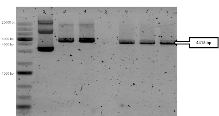
Figure 5: Restriction digestion analysis of p2NIL plasmid DNA (4753 bp). A 1% agarose gel confirmed successful double digestion of p2NIL with HindIII and KpnI, yielding a larger vector fragment (4418 bp), carrying the aph cassette and a smaller fragment (335 bp), not visible due to electrophoresis conditions, in lanes 6–8. Controls: lane 5, no DNA material; lane 2, undigested circular p2NIL; lane 3, p2NIL linearized with HindIII (4753 bp); lane 4, p2NIL linearized with KpnI (4753 bp). All fragments were resolved relative to lane 1, a 1 kb DNA ladder. Please click here to view a larger version of this figure.
To construct the final suicide vectors, p2NIL_rv0678, isolated from E. coli, was linearized with the PacI high-fidelity (HF) enzyme and dephosphorylated to prevent recircularization. The flexible marker cassette DNA fragment from pGOAL19 (7949 bp; lacZ, sacB, and hph) was also excised by PacI-HF restriction enzyme digestion. DNA fragments were purified from the excised gel slices. The purified pGOAL19 (7949 bp; lacZ, sacB, and hph) DNA fragment was ligated into linear p2NIL_rv0678 to construct p2NIL_rv0678_pGOAL19 (15182 bp).
The p2NIL_rv0678_pGOAL19 E. coli transformants were selected on plates containing X-gal, Kan, and Hyg. Blue colonies were patched onto plates containing sucrose to confirm functionality of the sacB gene. The plasmid DNA p2NIL_rv0678_pGOAL19 isolated from E. coli single colonies was mapped by restriction enzyme digestion (Figure 6). These suicide plasmid vectors were used for AE.
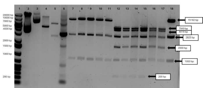
Figure 6: Restriction digestion analysis of the suicide plasmid vector p2NIL_rv0678_pGOAL19 (15,182 bp). Digestion with HindIII, KpnI, or PacI (alone or in combination) of plasmid DNA isolated from E. coli single colonies confirmed the expected fragmentation pattern into the p2NIL backbone (4,418 bp), pGOAL19 cassette (7,949 bp), and rv0678 insert (2,825 bp). Lane 1: 1 kb DNA ladder. Controls include: undigested circular p2NIL (lane 2) and pGOAL19 (lane 3); p2NIL linearized with KpnI (lane 4, 4,753 bp); pGOAL19 linearized with PacI (lane 5, 7,949 bp and 2,486 bp); and PCR amplification of the rv0678 insert from p2NIL_rv0678_pGOAL19 (lane 6, 2,825 bp). p2NIL_rv0678_pGOAL19 linearized with HindIII (15,182 bp) is shown in lanes 7–11 and 19. Triple restriction digestion with HindIII, KpnI, and PacI of plasmids from six independent E. coli colonies is shown in lanes 12–18. Please click here to view a larger version of this figure.
Following the transformation of M. tuberculosis with p2NIL_rv0678_pGOAL19, SCO transformants were selected on Middlebrook 7H10 agar plates containing X-gal, Kan and Hyg. Colony PCR and Sanger sequencing of M. tuberculosis single colonies confirmed p2NIL_rv0678_pGOAL19 (Figure 7).

Figure 7: Colony PCR and Sanger sequencing confirmation of the A97G mutation in rv0678. Colony PCR products from E. coli single colonies carrying p2NIL_rv0678_pGOAL19 were sequenced using rv0678-specific primers. Sanger sequencing chromatograms (alignments) show the introduced variant rv0678_97 (GAC > GGC), corresponding to the A97G mutation) in the cloned insert, 6 colonies aligned to the reference M. tuberculosis H37Rv rv0678 sequence visualized in BioEdit. Please click here to view a larger version of this figure.
To select for transformants that have lost the suicide plasmid vector in the second crossover event, the selected blue single colonies were cultured in 7H9-OADC media containing Kan and then sub-cultured in the absence of Kan. The inoculums were plated on 7H11-OADC agar containing 2% sucrose and X-gal (without Hyg) to select for the rv0678 mutant, representing the resolution of the co-integrant. White single colonies were recovered from plates after 16 days. Some blue colonies were present and indicate partial resolution of the co-integrant. Sucrose-resistant white single colonies of M. tuberculosis picked from 7H11-OADC agar plates and screened by Sanger sequencing were confirmed to harbour the rv0678 Thr33Ala point mutation c.97A>G (Figure 8).
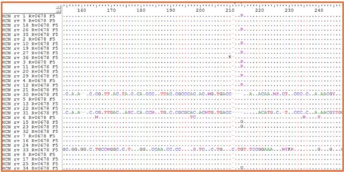
Figure 8: PCR amplification and Sanger sequencing confirmation of the rv0678 A97G mutation in M. tuberculosis. Colony PCR products from sucrose-sensitive white single colonies (selected on sucrose, X-GAL, and/or kanamycin) were sequenced using rv0678-specific primers. Sanger sequencing data aligned to the reference M. tuberculosis H37Rv rv0678 sequence confirmed the presence of the desired rv0678_97 variant (GAC > GGC, Thr33Ala; 4 colonies with pure mutant "G" background and 8 colonies with mixed wild-type and mutant "R" background) in the selected colonies visualized in BioEdit. Please click here to view a larger version of this figure.
RECOMBINEERING
Transformation of M. tuberculosis with pJV75amber
The plasmid pJV75amber used for recombineering was isolated from DH5α E. coli, and its concentration was quantified using a spectrophotometer to be 180.7 ng/µL. Transformation of electrocompetent M. tuberculosis H37Rv with 2 µg of plasmid DNA was performed to generate M. tuberculosis (pJV75amber) expressing gp61 recombinase protein required for recombineering of the ssDNA oligonucleotide carrying the desired nucleotide change. Transformant recombinant M. tuberculosis cells were plated on 7H11-OADC agar plates. The cells were used for PCR amplification of the aph gene (700 bp) in pJV75amber (Figure 9). Isolation of 20 white single colonies of transformed M. tuberculosis confirmed the presence of the pJV75amber vector. An M. tuberculosis (pJV75amber) single colony was chosen for subsequent recombineering experiments.
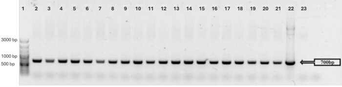
Figure 9: Colony PCR of M. tuberculosis (pJV75amber) single colonies with aph primers from 20 single colonies. Lane 1: 100 bp plus DNA ladder; lane 2–21: PCR amplification of aph from single colonies; lane 22: pJV75amber DNA; lane 23: negative control. Please click here to view a larger version of this figure.
Recombineering of atpE A196G (Ile66Val) in M. tuberculosis
Electrocompetent recombinant M. tuberculosis (pJV75amber) cells, co-transformed with a ssDNA oligonucleotide carrying a single nucleotide change atpE c.196A>G (resulting in Ile66Val mutation) and JVC198 ssDNA oligonucleotides, were selected on Hyg. Hyg resistant colonies were screened by PCR amplification of the atpE gene and Sanger sequencing to confirm that it has undergone recombineering. Only 2/33 single colonies screened were mutated, of which one (C11_atpE) had a mixed population of wild type (CAT) and mutant (CGT) alleles. However, the second colony (C14_atpE) had a pure atpE c.196A>G mutant population (Figure 10).
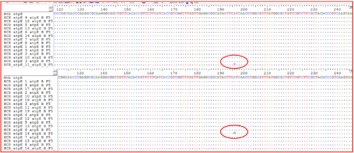
Figure 10: PCR amplification and Sanger sequencing of the atpE gene in M. tuberculosis single colonies. Colony PCR products were sequenced using atpE-specific primers and aligned to the reference M. tuberculosis H37Rv atpE sequence. The sequencing chromatograms (alignments) revealed: (top panel) one colony (out of 14 screened) with a mixed population showing both wild-type (A) and mutant (G) alleles at position 196 (C11_atpE); (bottom panel) one colony (out of 19 screened) with a pure mutant population carrying the atpE A196G substitution Ile66Val (C14_atpE) visualized in BioEdit. Please click here to view a larger version of this figure.
Discussion
Genetic manipulation in M. tuberculosis is technically challenging due to the organism's lipid-rich cell envelope, slow growth, and relatively inefficient uptake of foreign DNA. These features make transformation, colony recovery, and molecular confirmation slower and less predictable than in many other bacterial systems. In addition, the generation of large dsDNA fragments and the construction of plasmid-based delivery vectors are laborious and require multiple quality-control steps. The protocols described here address these challenges by presenting two complementary approaches for introducing defined point mutations into an isogenic M. tuberculosis background: two-step AE using a suicide vector and oligonucleotide-mediated recombineering.
The critical feature of the two-step AE protocol is the efficient recombination, but it is limited by the need for restriction-enzyme cloning into multiple cloning sites and the use of UV pre-treated plasmid DNA13 to stimulate HR and increase AE14. Although UV exposure may induce unwanted variation within the construct, the likelihood of generating SNV is reported to be minimal15,16. The two-step strategy of SCO and DCO events, which relies on multiple selection and counter-selection markers7,10, may impose in vitro antibiotic stress on M. tuberculosis17. While the use of additional markers can improve recovery of a pure mutant population, serial passages risk in vitro evolution, producing strain populations with distinct genetic backgrounds13,15, that may confound the interpretation of the introduced point mutant18. Transformation efficiency and recombination inefficiency limit SCO events, whereas DCO events are limited only by recombination efficiency and occur more readily19. Additionally, restriction-enzyme digestion and cloning into a multiple cloning site remain time-consuming and labor-intensive.
Recombineering using ssDNA oligonucleotides is efficient for introducing point mutations into M. tuberculosis. The short homology arms of the targeting ssDNA oligonucleotide enable direct chemical synthesis, thereby eliminating the multiple steps of restriction cloning and PCR amplification required for homologous recombination20. Recombineering relies exclusively on a curable plasmid that can self-replicate outside the chromosome in M. tuberculosis to induce mutagenesis21,22. Further, ssDNA oligonucleotides are easily synthesized. Recombineering is thus simpler, faster, and more cost-effective than homologous recombination because it requires fewer plasmids and culturing steps. However, depending on the targeted genomic region, recombineering may be less efficient at achieving site-directed mutagenesis and may require multiple attempts. Although the overall recombineering efficiency with ssDNA is reported to be relatively high compared with other techniques, the absolute number of cells carrying the intended change remains arguably low20,23. Challenges include unintended (illegitimate) recombination at loci sharing sequence similarity outside the region of homology, caused by the strand bias and relatively short homology of the exogenous donor ssDNA substrate. The HygR co-selection approach, used to screen for mutants without additional antibiotic selection24,25, may cause polar effects downstream of the operon, resulting in undesired mutations in the genetic background26. Recombineering efficiency can also vary by target, as illustrated in Figure 10 (2/33 colonies positive for atpE A196G), potentially due to suboptimal ssDNA oligo homology arms (<50 nt), secondary structures that impede annealing, insufficient recombinase induction, or competing wild-type repair pathways. Furthermore, recombineering requires screening many single colonies to confirm the desired variant, which can be time-consuming. In M. tuberculosis, uptake of plasmid DNA and ssDNA oligonucleotides remains inefficient, likely due in part to the lipid-rich mycobacterial cell envelope. Therefore, the advantage of the HygR selectable oligonucleotide is not that it increases transformation efficiency in absolute terms, but that it enables co-selection for the small proportion of cells that have taken up DNA during electroporation. This enriches the screened population for DNA-recipient cells, within which the desired chromosomal mutation can then be identified by PCR and sequencing. However, HygR selection does not itself confirm incorporation of the target-site mutation, and multiple HygR colonies must still be screened to identify confirmed mutants.
It should also be noted that the ssDNA oligonucleotide carrying the SNV creates a mismatch in the replication fork. The M. tuberculosis NucS mismatch repair system may recognize and repair this mismatch, thereby inhibiting incorporation of the intended mutation. To maximize efficiency, investigators should avoid mismatches that create G-G, G-T, or T-T base pairs, which are efficiently repaired by NucS, also known as EndoMS, a non-canonical mismatch repair (MMR) endonuclease involved in DNA mismatch repair27,28.
The protocols presented here advance tuberculosis research by enabling precise mutagenesis in controlled genetic backgrounds, thereby addressing limitations inherent in studies using clinical isolates. Relative to previous methods7,8,29,30, this publication provides M. tuberculosis-specific optimizations, including adjusted electroporation parameters (2.5 kV, 25 µF, 1000 Ω) that achieve >108 CFU/µg transformation efficiency and streamlined oligonucleotide design 63 bp for recombineering and 25 kb for recombination to minimize secondary structures and off-target effects. These adaptations shortened experimental timelines for recombineering, completed in 6 weeks compared to 12 weeks for AE, and improved reproducibility, as demonstrated by successful mutagenesis of bedaquiline-resistance loci in rv0678 and atpE. The novelty of this work lies in the direct side-by-side comparison of the two approaches, integration of modern tools such as whole-genome sequencing for variant confirmation and AI for mutation prediction, and visual documentation, all of which facilitate adoption in resource-limited settings.
To optimize the described methods and address common pitfalls, several modifications can be made that apply to both HR and recombineering. To improve electroporation efficiency, which is often a bottleneck in M. tuberculosis transformation due to its thick cell wall, prepare electrocompetent cells by glycine treatment (0.2–0.5 M for 24–48 h during growth) to weaken the cell wall and enhance DNA uptake. Additionally, use ultra-high-field electroporation (12.5–25 kV/cm) with optimized cuvettes (0.1–0.2 cm gap) and chilled buffers to achieve transformation efficiencies exceeding 105 CFU/µg DNA, minimizing arcing by desalting DNA via ethanol precipitation or column purification. Monitor cell density at OD600 0.6–0.8 for mid-log phase harvest and avoid excessive DNA (limit to 1–10 µg) to prevent saturation and reduced viability. If low colony yields persist, incorporate recovery incubation for 48 h post-electroporation to allow expression of resistance markers31,32.
Other common issues include low PCR yield in overlap extension. This can be optimized by using high-fidelity polymerases with GC enhancers (3–5% DMSO) and gradient annealing temperatures (55–65 °C). Antibiotic stress during selection can be mitigated by employing markerless systems, such as Cre-loxP, for excision after integration. Cre-loxP is a site-specific recombination system in which the Cre recombinase enzyme recognizes 34-bp loxP DNA sequences and catalyzes precise excision of the DNA segment flanked by two loxP sites, enabling conditional gene knockout in cells33.
For HR, extend homology arms to 1–2 kb in suicide vectors to favor legitimate over illegitimate events and thereby avoid off-target integrations. Use replicative plasmids with inducible recombinases (RecET systems) to enhance specific recombination rates while suppressing non-specific events. For UV pretreatment in HR, calibrate exposure to 100–150 mJ/cm2 to stimulate recombination without inducing excessive mutations, and verify constructs via whole-genome sequencing post-mutagenesis to detect any unintended changes9,34,35.
For recombineering, optimize oligo design with 63 - 90 nt homology arms, avoiding GC-rich mismatches, and use software such as OligoEvaluator for stability checks. Adjust acetamide induction to 0.2–0.5% for 4–6 h post-electroporation to balance expression without toxicity. To reduce strand bias and off-target annealing, design ssDNA oligonucleotides with 50–100 nt homology targeting the lagging strand and incorporate phosphorothioate modifications at the oligo ends to protect against exonuclease degradation. If illegitimate recombination occurs frequently, increase selection pressure via higher antibiotic concentrations or dual markers, or repeat transformations with fresh competent cells to boost yield. These steps can improve rates to >20% for challenging genomic loci. Extensive colony screening can be streamlined by pooling colonies for multiplex PCR or using fluorescence-based reporters if compatible with the target gene36.
The methods described here provide foundational tools for M. tuberculosis genetics, with potential extensions to emerging technologies. Site-directed mutagenesis can be used to validate identified drug resistance mutations35 via next-generation sequencing37, enabling rapid phenotyping of novel variants in clinical isolates. Future studies could integrate these with CRISPR-Cas systems for multiplex editing, such as CRISPR-guided mutagenic DNA polymerases, to diversify the repertoire of drug-target genes and accelerate the discovery of resistance mechanisms. Exploiting M. tuberculosis's endogenous Type III CRISPR for defense against phages or plasmids could lead to novel antimicrobials or diagnostics for resistant strains38,39,40.
Advancements in genetic manipulation, including TnSeq for identifying essential genes in vivo, open avenues for vaccine development by pinpointing attenuation targets in live strains41. Programmable base editing could refine subtle mutations without double-strand breaks, reducing off-target effects and facilitating studies on epistasis in multidrug resistance. Broader applications include engineering M. tuberculosis for biosensor platforms or synthetic biology, such as inserting reporters for real-time infection monitoring. Future research should focus on high-throughput mutagenesis pipelines combining AI-driven design with these methods to map host-pathogen interactions and inform personalized therapies42.
Disclosures
The authors have no conflicts of interest to disclose.
Acknowledgements
Sincere gratitude to all members of the TB Genomics Research Group at Stellenbosch University, Faculty of Medicine and Health Sciences, Division of Molecular Biology and Human Genetics, for their invaluable contributions to this work. Funding support was received from the European and Developing Countries Clinical Trial Partnerships (TMA2017CDF-1885), the South African Medical Research Council, the German Academic Exchange Service (DAAD) and Research Foundation Flanders (FWO) under Odysseus grant N° G0F8316N. We would also like to acknowledge Prof Graham Hatfull and Prof Julia van Kessel for developing the method of homologous recombineering, and for providing pJV75amber, which has been used in our research.
Materials
| Name | Company | Catalog Number | Comments |
|---|---|---|---|
| 0.5 mL Microcentrifuge Tubes, Conical Bottom, Lock Cap | WhiteHead Scientific | 605001 | Culture |
| 1.5 mL Microcentrifuge Tubes, Conical Bottom, Lock Cap | WhiteHead Scientific | 615001 | Culture |
| 10 mM Calcium chloride dihydrate (CaCl2·2H2O) | Sigma-Aldrich | Chemo-competed E. coli | |
| 10 mM RbCl, 75 mM CaCl2·2H2O | Sigma-Aldrich | 83979 | Chemo-competed E. coli |
| 10 mM) MOPS (3-(N-Morpholino) propanesulfonic acid) buffer solution | Sigma-Aldrich | M1254 | Chemo-competed E. coli |
| 100 mM Rubidium Chloride (RbCl) | Sigma-Aldrich | 83979 | Chemo-competed E. coli |
| 15 cm Petri dish | Lasec | SKU | Agar culture |
| 15% wt./vol glycerol | Sigma-Aldrich | 49781-500ML | Culture |
| 20 bp to 32 bp PCR primers synthesised at 25-nm scale with standard desalting | Integrated DNA Technologies (WhiteHead Scientific) | N/A | DNA amplification |
| 25 cm tissue culture flask | WhiteHead Scientific | 707001 | Broth culture |
| 30 mM Potassium acetate | Sigma-Aldrich | P1190-100G | Chemo-competed E. coli |
| 50 mL Centrifuge Tubes | WhiteHead Scientific | P10432 | Culture |
| 50 mM Manganese (II) chloride tetrahydrate (MnCl2·4H2O) | Sigma-Aldrich | 63535 | Chemo-competed E. coli |
| 63 bp ssDNA oligonucleotides synthesized at 100-nm scale with PAGE purification | Integrated DNA Technologies (WhiteHead Scientific) | N/A | Introducing allelic exchange |
| 75 cm tissue culture flask | WhiteHead Scientific | 708013 | Broth culture |
| Benchmark Accuris UV Transilluminator light source | Sigma Aldrich | BMSE3000-1EA | Gel visualization |
| Benchmark Scientific (H3020) Incu-Shaker 20 L Stackable Shaking Incubator with non-slip rubber mat, 115 V | Alkali Scientific | SKU: | E. coli culture |
| Bio-Edit | Software | n/a | Visual inspection and analysis of Sanger sequence reads |
| Crosslinkers CL 508 | Thistle Scientific. | SKU: CLV-CL-508.M | Plasmid DNA treatment |
| DNA-MAN | Software | n/a | Visual inspection and analysis of Sanger sequence reads |
| Dry Bath, Digital with Timer, 150 °C Heating, Dual Block | WhiteHead Scientific | E-DB150-2D | Transformation |
| Gene Pulser Xcell Electroporation Systems | Bio-Rad, Hercules, CA 94547, USA | 1652660 | Electroporation |
| Gene Pulser/ MicroPulser Electroporation Cuvettes, 0.2 cm gap | Bio-Rad, Hercules, CA 94547, USA | 1652086 | Electroporation |
| HindIII-HF enzymes | New England Bio-lab | R3104L | Restriction digestion |
| JCV198 (ssDNA-oligonucleotides synthesized at 100-nm scale with PAGE purification) | Integrated DNA Technologies (WhiteHead Scientific) | 63 bp | CCCTGTTACTTCTCGACCGTATTGA TTCGGATGATTCCTACGCGAGCCT GCGGAACGACCAGG; Gene target: php; Restore hygR in php gene on pJV75amber |
| Memmert IN Plus Standard 26.5 cu ft Non-CO2 Static Incubator | Ares Scientific | SKU: IN-750-PLUS | M. tb culture |
| Mtb_Rv0678_Fwrd_SEQ | Integrated DNA Technologies (WhiteHead Scientific) | 885bp | GCGACCACAACCAGGATGA; Gene target: rv0678; PCR amplification of rv0678 |
| Mtb_Rv0678_Rev_SEQ | Integrated DNA Technologies (WhiteHead Scientific) | 885bp | CTCGGACAACACGCTGACC; Gene target: rv0678; PCR amplification of rv0678 |
| Mtb_Rv0678_T33A_For_ampyP | Integrated DNA Technologies (WhiteHead Scientific) | 1413bp | ATTTCGAGTCCAGGAGTTTGGCTC GGTTGGCGGGTCGATT{G}; Gene target: rv0678; Insert A95G |
| Mtb_Rv0678_T33A_Rev_ampyP | Integrated DNA Technologies (WhiteHead Scientific) | 1413bp | CAATCGACCCGCCAACCGAGC CAAACTCCTGGACTCGAAA{T}; Gene target: rv0678; Insert A95G |
| Mycobrowser | Database | n/a | SNV codon in a gene sequence |
| NCBI databases | Database | n/a | Gene position in the genome to identify SNV position |
| p2NIL | Addgene | 20188 | Expression cloning vector (kanR) |
| PacI-HF enzymes | New England Bio-lab | R0547L | Restriction digestion |
| pCR-Blunt-II-TOPO | Thermofisher -scientific / Invitrogen | 451245 | Subcloning vector expressing LacZ and kanR |
| pGOAL17 | Addgene | 20189 | Lambda RED recombinase cloning vector expressing LacZ and SacB |
| pGOAL19 | Addgene | 20190 | Lambda RED recombinase cloning vector expressing hygR LacZ and SacB |
| Phusion high fidelity Hotstart flex polymerase kit | New England Bio-lab | M0535L | High fidelity proofreading DNA taq polymerase |
| pJET1.2/blunt | Thermofisher -scientific | K1231 | Subcloning sequencing vector (ampR) |
| PJV75_KANR_F (primers synthesized at 25-nm scale with standard desalting) | Integrated DNA Technologies (WhiteHead Scientific) | 700 bp | ATTCAACGGGAAACGTCTTG; Gene target: aph; amplification of KanR gene in pJV75amber |
| PJV75_KANR_R (primers synthesized at 25-nm scale with standard desalting) | Integrated DNA Technologies (WhiteHead Scientific) | 700 bp | CGAGGCAGTTCCATAGGATG; Gene target: aph; amplification of KanR gene in pJV75amber |
| pJV75amber | Addgene / Van Kessel lab | 26906/ 26908/26908/ 26910 | Vector expressing kanR, hygR, and Che gp61 facilitating recombineering in the genome |
| Primer3plus | Software | n/a | Designing primer pairs specific to gene of interest |
| RcmbFrag_Rv0678_Fwrd_digPrim | Integrated DNA Technologies (WhiteHead Scientific) | 2825bp | ACAGCG{AAGCTT}CAGGTTG GTCGCGAAGGTCG; Gene target: HindIII site; insert site ACAGCGAAGCTT |
| RcmbFrag_Rv0678_Rev_digPrim | Integrated DNA Technologies (WhiteHead Scientific) | 2825bp | CGTACA{GGTACC}ACGTACG CTCAATCAAGGCC; Gene target: kpnl site; Insert site CGTACAGGTACC |
| SNAPGene | Software | n/a | virtual cloning and adding restriction enzyme overhangs |
| Thermo Fisher QuantStudio 3 Thermocycler | Pika America | N/A | DNA amplification |
References
- Fenner L, et al. Effect of mutation and genetic background on drug resistance in Mycobacterium tuberculosis. Antimicrob Agents Chemother. 2012;56(6):6. doi:10.1128/AAC.06460-11.
- K SP, Parivakkam Mani A, S G, Yadav S. Advancements in artificial intelligence for the diagnosis of multidrug resistance and extensively drug-resistant tuberculosis: A comprehensive review. Cureus. 2024;16(5):e60280.
- Kandathil SM, Lau AM, Jones DT. Machine learning methods for predicting protein structure from single sequences. Curr Opin Struct Biol. 2023;81:102627.
- Hall BM, Ma CX, Liang P, Singh KK. Fluctuation AnaLysis CalculatOR: a web tool for the determination of mutation rate using Luria–Delbrück fluctuation analysis. Bioinformatics. 2009;25(12):12.
- Murray A. Salvador Luria and Max Delbrück on random mutation and fluctuation tests. Genetics. 2016;202(2):2.
- Pérez-Donoso JM, Vásquez CC. Site-directed mutagenesis as a tool for unveiling mechanisms of bacterial tellurite resistance. Genet Manip DNA Protein - Ex Curr Res. 2013;10.5772/47998.
- Parish T, Stoker NG. Use of a flexible cassette method to generate a double unmarked Mycobacterium tuberculosis tlyA plcABC mutant by gene replacement. Microbiology. 2000;146(Pt 8):1969-1975.
- van Kessel JC, Hatfull GF. Efficient point mutagenesis in mycobacteria using single-stranded DNA recombineering: characterization of antimycobacterial drug targets. Mol Microbiol. 2008;67(5):5.
- Gupta R, et al. A dual role for mycobacterial RecO in RecA-dependent homologous recombination and RecA-independent single-strand annealing. Nucleic Acids Res. 2013;41(4):2284-2295.
- Parish T, Stoker NG. Electroporation of Mycobacteria. Electroporation Protocols for Microorganisms. Humana Press; 1995:237-252.
- van Kessel JC, Hatfull GF. Recombineering in Mycobacterium tuberculosis. Nat Methods. 2007;4(2):2.
- Green R, Rogers EJ. Transformation of Chemically Competent E. coli. In Laboratory Methods in Enzymology: DNA. Academic Press; 2013;529:329-336.
- Gordhan BG, Parish T. Gene replacement using pretreated DNA. Methods Mol Med. 2001;54:77-92.
- Zhang Y, Muyrers JP, Testa G, Stewart AF. DNA cloning by homologous recombination in Escherichia coli. Nat Biotechnol. 2000;18(12):1314-1317.
- Hinds J, et al. Enhanced gene replacement in mycobacteria. Microbiology. 1999;145(3):519-527.
- Kodym A, Afza R. Physical and chemical mutagenesis. Methods Mol Biol. 2003;236:189-204.
- Martin C, et al. Transposition of an antibiotic resistance element in mycobacteria. Nature. 1990;345(6277):739-743.
- Kalpana GV, Bloom BR, Jacobs WR. Insertional mutagenesis and illegitimate recombination in mycobacteria. Proc Natl Acad Sci U S A. 1991;88(12):5433-5437.
- Goude R, Parish T. Electroporation of mycobacteria. J Vis Exp. 2008;15:e761.
- Sawitzke JA, et al. Recombineering: highly efficient in vivo genetic engineering using single-strand oligos. Methods Enzymol. 2013;533:157-177.
- Sarasin A. New trends in photobiology: Shuttle vectors for studying mutagenesis in mammalian cells. J Photochem Photobiol B. 1989;3(2):143-155.
- Wang XY, et al. Impact of different promoters on episomal vectors harbouring characteristic motifs of matrix attachment regions. Sci Rep. 2016;6.
- Thomason LC, Oppenheim AB, Court DL. Modifying bacteriophage lambda with recombineering. Methods Mol Biol. 2009;501:239-251.
- Merker M, et al. Whole genome sequencing reveals complex evolution patterns of multidrug-resistant Mycobacterium tuberculosis Beijing strains in patients. PLoS One. 2013;8(12):e82551.
- Faksri K, et al. Whole-genome sequencing analysis of serially isolated multi-drug and extensively drug resistant Mycobacterium tuberculosis from Thai patients. PLoS One. 2016;11(8):e0160992.
- Ishikawa M, Hori K. A new simple method for introducing an unmarked mutation into a large gene of non-competent Gram-negative bacteria by FLP/FRT recombination. BMC Microbiol. 2013;13(1):86.
- Rivera-Flores IV, Wang EX, Murphy KC. Mycobacterium smegmatis NucS-promoted DNA mismatch repair involves limited resection by a 5′-3′ exonuclease and is independent of homologous recombination and NHEJ. Nucleic Acids Res. 2024;52(20):12308-12323.
- Murphy KC. Oligo-mediated recombineering and its use for making SNPs, knockouts, insertions, and fusions in Mycobacterium tuberculosis. Methods Mol Biol. 2021;2314:301-321.
- Murphy KC, Papavinasasundaram K, Sassetti CM. Mycobacterial recombineering. Methods Mol Biol. 2015;1285:177-199.
- Kendall SL, Frita R. Construction of targeted mycobacterial mutants by homologous recombination. Methods Mol Biol. 2009;465:297-310.
- Campo-Pérez V, Cendra M del M, Julián E, Torrents E. Easily applicable modifications to electroporation conditions improve the transformation efficiency rates for rough morphotypes of fast-growing mycobacteria. New Biotechnol. 2021;63:10-18.
- Tang D, et al. Ultra-high field strength electroporation enables efficient DNA transformation and genome editing in nontuberculous mycobacteria. Microbiol Spectr. 2025;13(10):e01944-25.
- Stephan J, Stemmer V, Niederweis M. Consecutive gene deletions in Mycobacterium smegmatis using the yeast FLP recombinase. Gene. 2004;343(1):181-190.
- Baulard A, Kremer L, Locht C. Efficient homologous recombination in fast-growing and slow-growing mycobacteria. J Bacteriol. 1996;178(11):3091-3098.
- Borgers K, Vandewalle K, Festjens N, Callewaert N. A guide to Mycobacterium mutagenesis. FEBS J. 2019;286(19):3757-3774.
- Alonso MN, et al. Efficient method for targeted gene disruption by homologous recombination in Mycobacterium avium subspecies paratuberculosis. Res Microbiol. 2020;171(5):203-210.
- Beviere M, et al. The role of next-generation sequencing (NGS) in the management of tuberculosis: Practical review for implementation in routine. Pathogens. 2023;12(8):978.
- Ding XY, et al. Programmable base editing in Mycobacterium tuberculosis using an engineered CRISPR RNA-guided cytidine deaminase. Front Genome Ed. 2021;3:734436.
- Feng S, et al. A CRISPR-guided mutagenic DNA polymerase strategy for the detection of antibiotic-resistant mutations in M. tuberculosis. Mol Ther Nucleic Acids. 2022;29:354-367.
- Zein-Eddine R, Refrégier G, Cervantes J, Yokobori NK. The future of CRISPR in Mycobacterium tuberculosis infection. J Biomed Sci. 2023;30:34.
- James KS, et al. TnSeq identifies genetic requirements of Mycobacterium tuberculosis for survival under vaccine-induced immunity. NPJ Vaccines. 2025;10(1):103.
- Ghosh M, Lahiri M, Dalal A, Parida KK, Kalia NP. Advancements in tuberculosis diagnostics: An update. Microb Pathog. 2025;207:107843.
Reprints and Permissions
This article has been published
Video Coming Soon