We describe a sensitive, time-resolved quantitative platform to measure clearance of extracellular tau in human induced pluripotent stem cell (iPSC)-derived microglia using a microplate-based luminescence system. This approach enables sensitive detection of tau degradation kinetics in a microwell format, allowing for high-throughput interrogation of tau handling under various experimental conditions.
Method Article
A Split-Luciferase Complementation Assay for Temporally Resolved Measurement of Tau Clearance in Microglia
In This Article
Summary
Abstract
As the resident immune cells of the central nervous system, microglia are central regulators of brain homeostasis and key mediators of neurodegenerative disease. These cells continuously survey the neural environment and play a critical role in the recognition, internalization, and degradation of extracellular substrates, including misfolded and aggregated proteins such as pathological tau. Despite growing evidence implicating microglia in tau clearance, existing approaches to measure tau uptake and degradation lack the temporal resolution and sensitivity needed to fully capture these dynamic processes. Here, we developed a luminescence-based assay to quantitatively monitor tau clearance in human induced pluripotent stem cell (iPSC)-derived microglia. This platform leverages a split-luciferase-based complementation system to enable highly sensitive, real-time detection of tau in live cells, allowing for precise tracking of its intracellular processing. This assay is scalable and adaptable across multiple cell types, providing a versatile tool to interrogate endolysosomal pathways and cellular mechanisms governing tau handling in neurodegenerative diseases, including Alzheimer’s disease.
Introduction
Tauopathies, including Alzheimer’s Disease (AD), are characterized by the aggregation and spread of microtubule-associated protein tau across neural circuits1,2. Glia have become increasingly appreciated as regulators of tau disease dynamics, including uptake, degradation, and spread3,4,5,6,7,8,9,10,11,12. Understanding how glia contribute to tauopathies is a pressing concern for resolving key disease dynamics, including neuroinflammation and tau spread. Among glial populations, microglia - the brain’s resident phagocytes - are uniquely positioned to mediate tau clearance.
Despite the pressing need to elucidate mechanisms of tau handling in microglia, the field remains limited in the resolution and throughput of available assays. Key questions regarding tau internalization, intracellular trafficking, and cellular clearance remain unresolved. Each of these steps represents a mechanistically distinct process: uptake refers to the internalization of extracellular tau at the plasma membrane; trafficking encompasses the sorting and transport of internalized tau through endosomal, lysosomal, and other intracellular compartments; and degradation denotes the proteolytic breakdown of tau, primarily via lysosomal and proteasomal pathways. Although these steps are sequential for extracellular tau processing, as internalized tau must transit intracellular compartments before reaching degradative machinery, they are independently regulated and a given experimental perturbation may affect one step without altering others. For example, a reduction in intracellular tau signal could reflect impaired uptake, misrouting away from degradative compartments, or enhanced degradation. Resolving these processes requires new assay platforms that can detect protein presence and be coupled with existing measures to determine protein fate over time.
Current methods to resolve tau internalization and degradation rely on immunocytochemistry, flow cytometry, western blotting, and fluorescent labeling approaches to quantify uptake, subcellular localization, and intracellular processing of tau species13,14,15,16,17,18. However, each of these approaches suffers from fundamental limitations when applied to the study of protein clearance dynamics. Immunocytochemistry and western blotting lack the sensitivity to detect low-abundance tau species, and are labor-intensive. Flow cytometry and fluorescence-based assays fail to faithfully report on protein clearance because fluorophores can persist after the conjugated protein has been degraded19, thereby decoupling the fluorescent signal from the actual fate of the protein of interest. Together, these constraints leave a critical gap, with no existing method simultaneously providing the sensitivity, temporal resolution, and scalability required to quantitatively track tau clearance dynamics. Therefore, a major hurdle to progress in understanding tau-handling dynamics in microglia has been the lack of quantitative, time-resolved, high-throughput assays to measure tau clearance in live cells.
To address the lack of a quantitative platform to monitor live tau dynamics, we developed a sensitive luminescence-based assay that can be used to detect intracellular tau protein dynamics. This approach employs a split-luciferase-based complementation system, in which a small peptide tag (High Affinity Binary Technology or HiBiT) is fused to tau, enabling kinetic measurements in living cells. Upon interaction with its complementary binding partner (Large Binary Technology or LgBiT), the tagged protein reconstitutes an active luciferase enzyme, generating a quantitative bioluminescent signal20,21,22. This luminescent signal provides a direct, sensitive readout of protein abundance, enabling dynamic tracking of changes in intracellular levels over time. As a result, luminescence can be used as a robust reporter of protein presence or loss, as in the case of intracellular degradation. Together, this platform enables a precise, temporally resolved measurement of tau handling and offers a broadly applicable strategy for studying protein turnover.
Protocol
All cellular experiments were performed in an appropriate Biosafety Level 2 lab, approved by the University of Massachusetts Amherst’s Environmental and Health Services.
1. Expression, purification, and endotoxin removal of Tau-SLP protein
- Append the split-luciferase peptide or SLP tag (VSGWRLFKKIS) onto a Tau construct to create a tagged construct (Tau-SLP) using standard cloning methods, such as Gibson assembly23.
NOTE: The complementation of the split-luciferase peptide tag generates a robust signal when placed either at the C-terminal, N-terminal, or in the internal domains of 2N4R Tau24. - Express and purify Tau-SLP using previously described methods25,26 and ensure purity using SDS-PAGE and immunoblotting.
- For use with microglial cells, follow published protocols to remove bacterial endotoxin after Tau-SLP purification in order to limit confounding reactivity27.
- Quantify Tau-SLP protein concentration using a standard bicinchoninic acid assay (BCA) or Bradford assay28,29.
- If desired, aggregate Tau-SLP following previously described protocols24.
- Validate the functional luminescence of Tau-SLP by generating a standard curve using a commercially available luciferase substrate and complementation reagent, following the manufacturer’s instructions (see Table of Materials).
2. iPSC-derived microglia (iTF-Microglia) culture
- Culture microglial cells in 5% CO2 at 37 °C in the appropriate culture medium.
- Differentiate an induced pluripotent stem cell (iPSC) line harboring six doxycycline-inducible transcription factors to produce a microglial-like cell (induced transcription-factor microglia-Like or iTF-Microglia) using a validated protocol30 (see Table of Materials).
NOTE: This assay is not limited to iTF-Microglia and can be used with any adherent cell line. Optimization for clearance times may be required when working with other cell lines.
3. Tau Clearance Assay
- Into a 24-well plate, plate cells at a density of 1 x 10⁵ cells per well, in triplicate per condition, with culture medium (250 µL) and incubate overnight at 37 °C and 5% CO2 (Figure 1A).
- Remove culture medium, wash cells once with PBS (250 µL), and replace with 250 µL culture medium.
- To measure Tau-SLP clearance temporally, expose cells to Tau-SLP in a staggered manner so that all wells are harvested simultaneously while differing only in clearance time. For example, for 0 h, 2 h, and 4 h clearance groups, add 100 nM Tau-SLP-containing medium first to the 4 h group, then 1 h later to the 2 h group, and finally 1 h later to the 0 h group (see Figure 1B for timing recommendations). Once the final internalization period is complete, all wells will have undergone the same Tau-SLP exposure period but will differ only in clearance duration (Figure 1B).
- Include wells receiving no Tau-SLP treatment for background subtraction (Figure 1C).
- At the end of the internalization period, remove the Tau-SLP-containing medium. Wash the cells three times with PBS to remove extracellular Tau-SLP and replace with Tau-SLP-free culture medium.
- Incubate the cells for the designated clearance period (Figure 1A). The 0 h clearance group, harvested immediately after the internalization period, represents the total amount of internalized Tau-SLP at the start of the clearance phase.
- Following all clearance time points, remove media from wells and wash with 250µL PBS.
- Treat each well with 250 µL Trypsin-EDTA 0.25% for 5 min at 37 °C. This step removes surface-bound tau to ensure luminescence reflects internalized Tau-SLP. Neutralize each well with 250 µL trypsin Inhibitor (5 mg/mL).
- Collect each well into an appropriately labeled 1.5 mL microcentrifuge tube and pellet via centrifugation (500 x g, 5 min).
- Prepare an appropriate amount of lysis buffer containing luciferase substrate and complementation components (see Table of Materials; 50 µL x # of wells + 10%).
- Remove each supernatant with an aspirating pipette and resuspend the cell pellet in 50 µL of lysis buffer containing luciferase substrate and complementation components. This will produce a split-luciferase complementation complex and generate luminescence (Figure 1D).
- Transfer lysed cells to a white half-volume 96-well plate.
- Read luminescence on a microplate reader with orbital shaking for 10 s at room temperature (~23 °C) prior to reading to ensure complete lysis of cell pellets.
- Determine normalized Tau-SLP abundance by first subtracting the mean luminescence value obtained from wells not exposed to Tau-SLP. Normalize each value to the mean background-subtracted signal of the 0 h clearance group.
- Plot normalized Tau-SLP abundance as a function of clearance time. A progressive reduction in luminescence indicates a decrease in intracellular Tau-SLP abundance and is interpreted as Tau-SLP clearance, whereas stable luminescence values indicate impaired or delayed clearance. Calculate percentage clearance as: % Clearance = [1 − (RLUt / RLUt = 0)] x 100.
Results
Using this sensitive luminescence-based assay, intracellular Tau-SLP abundance could be monitored over time following a synchronized uptake period in human iPSC-derived microglia. Following Tau-SLP washout, luminescence steadily decreased, consistent with intracellular Tau-SLP clearance. This platform was sufficiently sensitive to detect differences in the handling of monomeric and fibrillar Tau-SLP species by microglia. Monomeric Tau-SLP was completely cleared in iTF-Microglia within 60 min, while fibrillar Tau-SLP persisted over 240 min (Figure 2A). Additionally, we have performed the assay with mutant tau (e.g., N279K) to assess clearance kinetics and have found that TauN279K-SLP shows significantly reduced clearance kinetics at only short clearance time points (15 min) (Figure 2B). These results suggest that the observed degradation may only be apparent due to the high sensitivity of this assay. We have performed the Tau-SLP degradation experiment using the small molecule L-Leucyl-L-Leucine methyl ester (LLOMe) to confirm lysosomal involvement. LLOMe is a cathepsin C substrate that permeabilizes lysosomal membranes, disrupting lysosomal activity31. With LLOMe treatment, we have observed significantly increased steady state levels of Tau-SLP at t=0 (Figure 3A) and a significantly reduced rate of degradation (Figure 3B) with a 4-fold increase of Tau-SLP remaining after a 60 min clearance time. Lastly, we have performed the Tau-SLP clearance assay in iPSC-derived neurons to confirm assay compatibility across cell types. Here, we have found that iPSC-derived neurons have reduced clearance kinetics for both monomeric (Figure 4A) and fibril forms (Figure 4B) of Tau-SLP. Most notably, Tau-SLP fibril levels stay steady at ~100% over a 240 min clearance, whereas in the same time frame, Tau-SLP fibril levels have dropped below 50% in iTF-Microglia. These results demonstrate that our split-luciferase-based complementation platform offers a robust and reliable detection of intracellular endolysosomal clearance of Tau-SLP and demonstrates a highly adaptable assay design that could be applied to numerous other proteins or cell types.
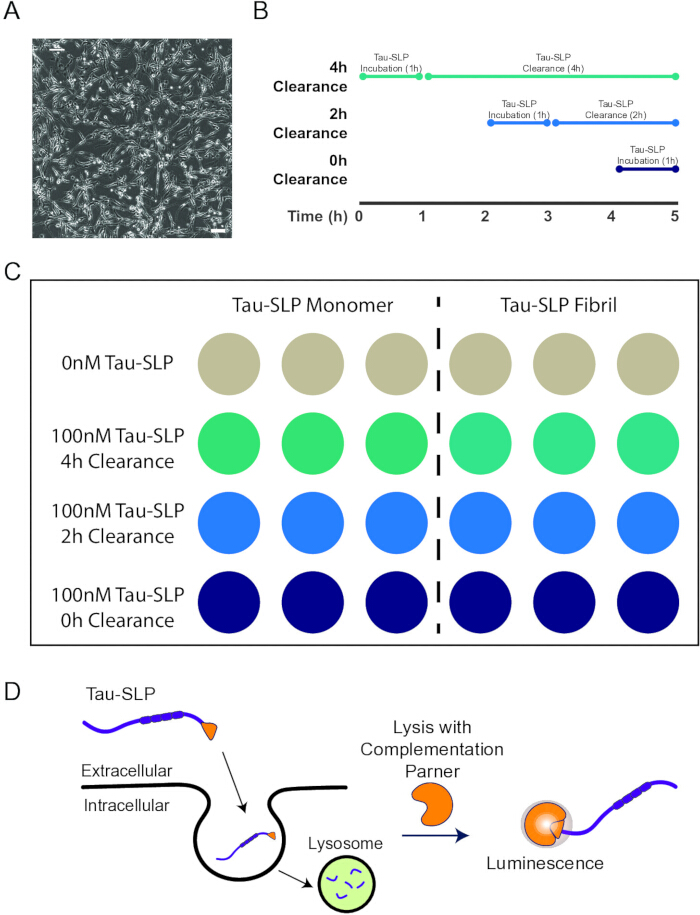
Figure 1: Representative experimental design of Tau-SLP clearance assay. (A) Representative brightfield image of iTF-Microglia utilized for this assay (scale bar = 100µm) (B) Human microglia (iTF-Microglia) are exposed to recombinant 2N4R-SLP in a staggered manner, enabling robust luminescence-based detection. Following a 1-hour incubation, extracellular tau is removed by media exchange and cells are washed with PBS. Cells are maintained in basal media during their respective clearance period (0 h, 2 h, 4 h) prior to harvest. At the endpoint, cells are lysed, and luminescence is measured. (C) Proposed plate design for measuring Tau-SLP clearance comparing monomeric to fibrillar degradation. (D) Tau-SLP protein is internalized into cells and routed for lysosomal degradation. Addition of LgBiT provides a quantitative readout of the amount of Tau-SLP left in the cell through complementation and production of luminescence. Please click here to view a larger version of this figure.
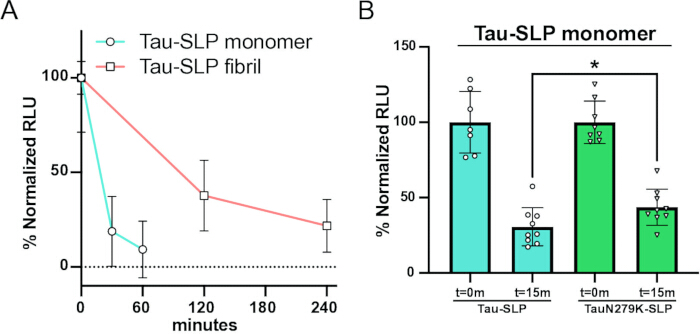
Figure 2: iTF-Microglia clear extracellular Tau-SLP. (A) iTF-Microglia were exposed to monomeric Tau-SLP (100nM) or fibrillar Tau-SLP (50nM), samples were normalized to respective initial uptake (t = 0; 100%) and to cells that were not exposed to Tau-SLP (0%) (mean ± SD; n = 9 from three independent differentiations). (B) iTF-Microglia were incubated with monomeric Tau-SLP or TauN279K-SLP (100nM), and samples were normalized to respective initial uptake (t = 0; 100%) and cells that were not exposed to Tau-SLP (0%) (mean ± SD; n ≥ 7 from three independent differentiations; ∗ p < 0.05 using unpaired Welch's t-test). Data reprinted from Shultz et al., 202524, and used under a Creative Commons Attribution (CC BY 4.0) license. Please click here to view a larger version of this figure.
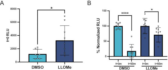
Figure 3: Lysosomal inhibition impairs Tau-SLP clearance. (A) Treatment with 1mM lysosomal inhibitor L-Leucyl-L-Leucine methyl ester (LLOMe) for 24 h increased initial Tau-SLP uptake (t = 0) in H4 neuroglioma cells (mean ± SD; n = 9 independent cultures; ∗ p < 0.05 using unpaired Welch’s t-test). (B) Treatment with LLOMe (1mM) for 24 h increased % normalized Tau-SLP RLU after a 60 min clearance time (t = 60min) compared to vehicle (DMSO). Luminescent signal is normalized to respective initial uptake levels (100%) and cells that were not exposed to Tau-SLP (0%) (mean ± SD; n = 9 independent cultures; ∗∗∗∗ p < 0.0001, ∗ p < 0.05 using unpaired Welch’s t-test). Data reprinted from Shultz et al., 202524, and used under a Creative Commons Attribution (CC BY 4.0) license. Please click here to view a larger version of this figure.
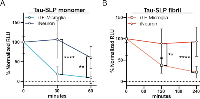
Figure 4: iTF-Microglia and iNeurons show differences in Tau-SLP clearance. (A) iTF-Microglia or iNeurons were incubated with monomeric Tau-SLP (100nM), samples were normalized to respective initial uptake (t=0; 100%) and cells that were not exposed to Tau-SLP (0%) (mean±SD; n ≥ 6 from two-three independent differentiations; ∗∗ p < 0.01, ∗∗∗∗ p < 0.0001 using a two-way ANOVA; time: F(1, 28) = 7.489, p = 0.0107; cell type: F(1, 28) = 46.61, p < 0.0001; interaction: F(1, 28) = 3.229, p = 0.0831; Sidak’s multiple comparisons test, p values indicated on figure, t=0 values are shown for reference but were excluded from statistical analysis). (B) iTF-Microglia or iNeurons were incubated with fibrillar Tau-SLP (50nM), samples were normalized to respective initial uptake (t=0; 100%) and cells that were not exposed to Tau-SLP (0%) (mean±SD; n ≥ 6 from two to three independent differentiations; ∗∗ p < 0.01, ∗∗∗∗ p < 0.0001 using a two-way ANOVA; time: F(1, 26) = 0.3888, p = 0.5384; cell type: F(1, 26) = 43.09, p < 0.0001; interaction: F(1, 26) = 1.209, p = 0.2816; Sidak’s multiple comparisons test, p values indicated on figure, t=0 values are shown for reference but were excluded from statistical analysis). Data reprinted from Shultz et al., 202524, and used under a Creative Commons Attribution (CC BY 4.0) license. Please click here to view a larger version of this figure.
Discussion
This assay provides a scalable and quantitative platform to measure extracellular Tau-SLP clearance by microglia with high sensitivity and temporal resolution. In contrast to traditional imaging-based approaches, which often require laborious sample processing and are limited in throughput and temporal sampling, this luminescence-based system enables direct, population-level quantification of tau handling in a microplate format. The resulting readout provides a quantitative measurement of Tau-SLP clearance following exposure to extracellular tau. Because measurements can be obtained repeatedly across multiple timepoints and with longer incubation periods, the assay enables a high-throughput, kinetically resolved analysis of tau uptake and clearance dynamics. Additionally, changes in luminescence over time are consistent with changes in intracellular Tau-SLP abundance, but do not distinguish between trafficking, degradation, or secretion without complementary approaches. Although Tau-SLP tagging did not significantly alter tau uptake or aggregation in our companion study24, exogenous tags may influence the behavior of other proteins. Therefore, assay findings should be complemented using orthogonal approaches and compared to untagged tau preparations where possible. An additional limitation is that this assay utilizes recombinant Tau-SLP species rather than endogenous tau or patient-derived pathological tau aggregates. While recombinant 2N4R tau provides a reproducible experimental substrate, it does not fully recapitulate the endogenous heterogeneity (i.e., post-translational modifications) of tau species isolated from human patient brains. Many protocols exist for in vitro phosphorylation of recombinant tau protein and could be easily incorporated into this workflow. This platform could also be adapted to study endogenous tau; this would require the use of genetic engineering to incorporate a SLP tag sequence at the tau (MAPT) locus.
Importantly, interpretation of signal reduction as degradation is contingent on the effective removal of extracellular and surface-bound tau, for example, through trypsinization or equivalent stripping steps. False negatives could occur if effective removal is not achieved. Apparent signal loss that would be interpreted as a false-positive of tau degradation is also possible. This could arise from cell detachment or death, secretion of Tau-SLP, or proteolytic cleavage that separates SLP from tau without degrading the protein. Accordingly, decreases in luminescence should be interpreted as reductions in intracellular Tau-SLP abundance rather than direct measurements of lysosomal degradation. To more definitively attribute signal changes to lysosomal degradation, orthogonal validation strategies are recommended. These may include pharmacological perturbation of lysosomal function, such as treatment with bafilomycin A1 or LLOMe (Figure 3), as well as pulse–chase experimental designs to resolve uptake versus degradation kinetics.
The flexibility of this assay makes it well-suited for interrogating mechanisms governing tau uptake, intracellular trafficking, and degradation, as well as for screening pharmacological or genetic modulators of tau clearance in human cellular systems. Although demonstrated here in iPSC-derived microglia, the platform is readily adaptable to other cell types (Figure 4), enabling broad application across models of tau biology and neurodegenerative disease, including Alzheimer’s disease and other primary and secondary tauopathies. Because this assay relies on the extracellular exposure of Tau-SLP and luminescence-based detection, it should be readily adaptable to astrocytes, oligodendrocytes, or commonly used cell lines, although optimization of uptake conditions may be necessary. Collectively, this approach provides a powerful tool for dissecting cellular pathways involved in proteostasis and advancing mechanistic understanding of neurodegenerative disease processes.
Disclosures
The authors have nothing to disclose.
Acknowledgements
This work was supported by grants from the National Institutes of Health (AG064116, AG077672)
Materials
| Name | Company | Catalog Number | Comments |
|---|---|---|---|
| Accutase | Thermo Scientific | A1110501 | |
| Conical Sterile Polypropylene Centrifuge Tubes | Fisher Scientific | 12-565-268 | |
| Corning Matrigel Growth Factor Reduced (GFR) Basement Membrane Matrix, Phenol Red-free, LDEV-free | Corning | 356231 | |
| DMEM/F-12, HEPES | Thermo Scientific | 11330057 | |
| Doxycycline | Clontech | 631311 | |
| DPBS | Thermo Scientific | 14-190-250 | |
| GlutaMAX | Thermo Scientific | 35050061 | |
| Graphing Software | GraphPad Prism (or similar) | n/a | |
| Human GM-CSF | Thermo Scientific | 300-03 | |
| KnockOut DMEM | Thermo Scientific | 10829018 | |
| Luminescent Plate Reader | SpectraMax i3x (or similar) - Molecular Devices | n/a | |
| Microcentrifuge Tubes | Fisher Scientific | 02-682-002 | |
| Microplate, 96 Well, PS, Half-area, White | Greiner | 675075 | |
| mTeSR Plus | STEMCELL Technologies | 100-0276 | |
| Nano-Glo HiBiT Lytic Detection System | Promega | N3030 | |
| Poly-L-Lysine hydrobromide | Fisher Scientific | ICN15017750 | |
| Recombinant Human IL-34 | Thermo Scientific | 200-34 | |
| Recombinant Human M-CSF | Thermo Scientific | 300-25 | |
| Recombinant Human TGF-β1 | Thermo Scientific | 100-21C | |
| Surface Treated Sterile Tissue Culture Plates | Fisher Scientific | FB012927 | |
| Tau-HiBiT protein | In house | n/a | Tau protein with a VSGWRLFKKIS peptide tag |
| Trypsin Inhibitor | Sigma Aldrich | T9128 | |
| Trypsin-EDTA | Thermo Scientific | 5-200-072 | |
| Y-27632 dihydrochloride | Bio-techne | 1254 |
References
- Vogel, J. W. et al. Spread of pathological tau proteins through communicating neurons in human Alzheimer’s disease. Nat Commun 11, 2612 (2020).
- Weber, A. J. et al. Tau seeds induce neurofibrillary tangle formation across brain regions via individual-specific connectivity. Neuron S0896627326001649 doi:10.1016/j.neuron.2026.03.001 (2026).
- Luo, W. et al. Microglial internalization and degradation of pathological tau is enhanced by an anti-tau monoclonal antibody. Sci Rep 5, 11161 (2015).
- Bolós, M. et al. Direct Evidence of Internalization of Tau by Microglia In Vitro and In Vivo. JAD 50, 77-87 (2016).
- Bolós, M. et al. Absence of CX3CR1 impairs the internalization of Tau by microglia. Mol Neurodegeneration 12, 59 (2017).
- Perea, J. R. et al. Extracellular Monomeric Tau Is Internalized by Astrocytes. Front. Neurosci. 13, 442 (2019).
- Fleeman, R. M., & Proctor, E. A. Astrocytic Propagation of Tau in the Context of Alzheimer’s Disease. Front. Cell. Neurosci. 15, 645233 (2021).
- Mothes, T. et al. Astrocytic uptake of neuronal corpses promotes cell-to-cell spreading of tau pathology. acta neuropathol commun 11, 97 (2023).
- Eltom, K., Mothes, T., Libard, S., Ingelsson, M., & Erlandsson, A. Astrocytic accumulation of tau fibrils isolated from Alzheimer’s disease brains induces inflammation, cell-to-cell propagation and neuronal impairment. acta neuropathol commun. 12, 34 (2024).
- Ferrer, I. et al. Involvement of Oligodendrocytes in Tau Seeding and Spreading in Tauopathies. Front. Aging Neurosci. 11, 112 (2019).
- Hopp, S. C. et al. The role of microglia in processing and spreading of bioactive tau seeds in Alzheimer’s disease. J Neuroinflammation 15, 269 (2018).
- Asai, H. et al. Depletion of microglia and inhibition of exosome synthesis halt tau propagation. Nat Neurosci 18, 1584-1593 (2015).
- Dickson, J. R. et al. Alzheimer disease-associated tau post-translational modification mimics impact tau propagation and uptake. Journal of Neuropathology & Experimental Neurology 84, 459-470 (2025).
- Weisová, P. et al. Therapeutic antibody targeting microtubule-binding domain prevents neuronal internalization of extracellular tau via masking neuron surface proteoglycans. acta neuropathol commun 7, 129 (2019).
- Shamir, D. B., Rosenqvist, N., Rasool, S., Pedersen, J. T., & Sigurdsson, E. M. Internalization of tau antibody and pathological tau protein detected with a flow cytometry multiplexing approach. Alzheimer’s & Dementia. 12, 1098-1107 (2016).
- Evans, L. D. et al. Extracellular Monomeric and Aggregated Tau Efficiently Enter Human Neurons through Overlapping but Distinct Pathways. Cell Reports 22, 3612-3624 (2018).
- Holmes, B. B. et al. Heparan sulfate proteoglycans mediate internalization and propagation of specific proteopathic seeds. Proc. Natl. Acad. Sci. U.S.A. 110, (2013).
- Boland, B. et al. Autophagy induction and autophagosome clearance in neurons: relationship to autophagic pathology in Alzheimer’s disease. J Neurosci 28, 6926-6937 (2008).
- Mandrekar, S. et al. Microglia Mediate the Clearance of Soluble Aβ through Fluid Phase Macropinocytosis. J. Neurosci. 29, 4252-4262 (2009).
- Oh-hashi, K., Furuta, E., Fujimura, K., & Hirata, Y. Application of a novel HiBiT peptide tag for monitoring ATF4 protein expression in Neuro2a cells. Biochemistry and Biophysics Reports. 12, 40-45 (2017).
- Schwinn, M. K. et al. CRISPR-Mediated Tagging of Endogenous Proteins with a Luminescent Peptide. ACS Chem. Biol. 13, 467-474 (2018).
- Boursier, M. E. et al. The luminescent HiBiT peptide enables selective quantitation of G protein–coupled receptor ligand engagement and internalization in living cells. Journal of Biological Chemistry 295, 5124-5135 (2020).
- Gibson, D. G. et al. Enzymatic assembly of DNA molecules up to several hundred kilobases. Nat Methods 6, 343-345 (2009).
- Shultz, A. et al. Extracellular tau clearance is governed by its aggregation state and independent of microglial activation by LPS and IFN-γ. Neurobiology of Disease 215, 107058 (2025).
- Rauch, J. N. et al. LRP1 is a master regulator of tau uptake and spread. Nature 580, 381-385 (2020).
- Rauch, J. N. et al. Tau Internalization is Regulated by 6-O Sulfation on Heparan Sulfate Proteoglycans (HSPGs). Sci Rep 8, 6382 (2018).
- Shultz, A., Mena, D., Roy, J. L., Nulman, J., & Rauch, J. N. Protocol for the removal of bound endotoxin from tau protein produced in Escherichia coli. STAR Protocols. 6, 104177 (2025).
- Smith, P. K. et al. Measurement of protein using bicinchoninic acid. Analytical Biochemistry 150, 76-85 (1985).
- Bradford, M. M. A rapid and sensitive method for the quantitation of microgram quantities of protein utilizing the principle of protein-dye binding. Analytical Biochemistry. 72, 248–254 (1976).
- Dräger, N. M. et al. A CRISPRi/a platform in human iPSC-derived microglia uncovers regulators of disease states. Nat Neurosci 25, 1149-1162 (2022).
- Elias, R. D. et al. Cathepsin C–Catalyzed Ligation Generates Intralysosomal Amyloid Fibrils from Dipeptide Esters. Preprint at https://doi.org/10.64898/2025.12.23.696283 (2025).
Reprints and Permissions
