Here we present a dissection protocol that preserves the pineal gland, which is often lost when the brain is removed from the cranial vault. This protocol ensures retention of the pineal gland in embryonic and early postnatal mouse brain preparations for downstream applications, including immunohistochemistry, in situ hybridization, and spatial transcriptomics.
Method Article
Preserving the Pineal Gland in Perinatal Mouse Brain Preparations
In This Article
Summary
Abstract
The pineal gland, a neuroendocrine structure that secretes melatonin, forms from an anlage at the dorsal midline in the embryonic forebrain. At embryonic day (E) 14.5 in the mouse, a pineal diverticulum is seen out-pouching from the roof of the diencephalon between the paired telencephalic vesicles. This diverticulum is in close contact with overlying cartilage, meningeal, and vascular tissue. It is easily lost when the cranial vault is opened to extract the brain at late embryonic/ early postnatal stages. Despite its important neuroendocrine role, the early development of the pineal gland and the signaling mechanisms that regulate this process remain poorly understood, partly due to the difficulty of preserving this structure intact during embryonic stages of mouse brain development.
This article presents an innovative dissection procedure that preserves the pineal gland while maintaining its structural integrity and its attachment to the roof of the diencephalon during brain isolation from embryonic stages through postnatal day (P) 7. The procedure is demonstrated using an E18.5 mouse brain and is therefore suitable for downstream applications requiring preservation of tissue architecture, including in situ hybridization, immunohistochemistry, and spatial transcriptomics. The protocol may also be used to isolate the developing pineal gland, thereby improving pineal-specific representation in transcriptomic and proteomic analyses, which is otherwise challenging because of its small size.
Introduction
The pineal gland is a small neuroendocrine structure located at the dorsal midline of the vertebrate brain and plays a central role in regulating circadian physiology through the rhythmic secretion of melatonin1,2. During mammalian embryogenesis, the pineal gland arises from the dorsal midline of the developing forebrain. In mice, pineal development begins when a specialized region of the dorsal diencephalic neuroepithelium forms the pineal anlage/pineal recess, which subsequently evaginates to form the pineal diverticulum at embryonic day (E) 14.53,4,5. This diverticulum is attached to the roof of the diencephalon and seen between the paired telencephalic vesicles. As development proceeds, this diverticulum becomes closely associated with overlying structures at the dorsal midline of the brain, in particular the superior sagittal sinus (SSS), and the transverse sinus (TS), venous channels running along the dorsal surface of the forebrain6. The developing pineal gland lies immediately beneath the intersection of the SSS and the TS, and its delicate attachment to the dorsal diencephalon is particularly vulnerable when the overlying vasculo-meningeal structures are removed to extract the brain from the cranial vault7. This makes the pineal gland particularly susceptible to damage, as it may be torn off when the cranial vault is opened for brain extraction. As a result, the pineal gland is frequently lost during routine dissections, which limits analysis of its structure and molecular composition in intact preparations.
Here, we describe a step-by-step protocol for dissecting the mouse brain while preserving the pineal gland and its attachment to the roof of the diencephalon. The method is applicable to embryonic stages from E16.5 up to early postnatal stages (P7), and is suitable for studies of pineal gland development using in situ hybridization, immunohistochemistry, and spatial transcriptomics. As a demonstration of downstream applications, in situ hybridization and immunohistochemistry for the pineal-enriched marker CRX are presented.
The key innovation in this method is that stress on midline structures is minimized by carefully removing physical attachments that would otherwise shear the pineal gland off when the brain is removed from the cranial vault. This is an improvement over the standard procedure.
This dissection method is limited to stages up to P7 because progressive ossification of the cranial vault during early postnatal development increases skull rigidity and strengthens its association with meningeal and vascular structures. While several studies have described pineal gland dissection procedures in adult rodents8, very few have focused on preserving the pineal gland during embryonic and early postnatal stages, when the gland is particularly fragile and susceptible to damage during brain isolation.
Protocol
All procedures are approved and conducted in accordance with the Institutional Animal Ethics Committee of the Tata Institute of Fundamental Research.
1. Material preparation
- Ensure all required instruments for microdissection (see the Table of Materials) are prepared before the procedure. Sterilize all instruments using 70% ethanol. For collecting preparations for downstream applications like spatial transcriptomic/in situ hybridization, wipe the instruments with RNase decontamination solution.
- Clean the dissection microscope stage with RNase decontamination solution and keep a bottle of ice-cold 1× PBS handy near the dissection bench.
- Keep the following instruments and plasticware ready before this procedure:
- For animal preparation, keep the following ready: 2 pairs of blunt forceps, 1 pair of surgical scissors, 2 petri dishes (10 cm), several petri dishes (6 cm; as needed, one for each head).
- For isolating the brain, keep the following ready: 1 pair of #5 forceps, 1 pair of #55 forceps, 1 pair of microdissection scissors, and collection tubes/dishes as suited for the downstream application.
- NOTE: If the downstream application involves in situ hybridization or spatial transcriptomics, clean the dissection instruments with RNase decontamination solution prior to use.
2. Animal preparation
- Follow steps (2.1.1– 2.1.6) for animals E14.5–P0
- Euthanize the pregnant dam in accordance with the Institutional Animal Ethics Committee guidelines. In the example shown, the dam was euthanized by cervical dislocation.
- Make a midline incision in the abdominal cavity of the pregnant dam to expose the uterus. Using blunt forceps, gently pull out the uterine horns and place them in a sterile 10 cm dish containing ice-cold 1× PBS.
- Carefully isolate individual fetuses by separating them from the surrounding amniotic sac and placenta.
- Transfer the fetuses into a fresh 10 cm petri dish containing ice-cold 1× PBS and maintain on ice until each one is taken for isolation of the brain. Transfer a single fetus to a 6 cm petri dish containing ice-cold PBS.
- For postnatal stages up to P7, euthanize the pup in accordance with the Institutional Animal Ethics Committee guidelines. Then proceed from step 2.1.6 onwards.
- Using a pair of surgical scissors, separate the head from the body such that the hindbrain is included in the head to protect the forebrain from inadvertent damage.
- Wash the head with cold PBS to remove the blood. Transfer to a fresh 6 cm petri dish containing ice-cold 1× PBS. Keep the head submerged in the PBS to allow blood and debris to be washed away during the subsequent dissection steps.
3. Dissection procedure to isolate the brain with the pineal gland intact
NOTE: All steps below are to be performed using a dissection microscope. Adjust the magnification to at least 2× (or as needed) to clearly visualize the brain and surrounding structures throughout the dissection. The entire process of dissection should be carried out in ice-cold PBS to minimize RNA and protein degradation. For steps 3.1–3.10, use the non-dominant hand to hold the head firmly and stabilize it throughout the procedure per the instructions in step 3.1. Steps 3.2–3.9 are to be performed with the non-dominant hand stabilizing the head, and the dominant hand for the fine motor skills, starting with a #5 forceps, then switching to #55 forceps or microdissection scissors as specified for each step.
- Use a #5 forceps in the non-dominant hand. Insert the two prongs into the eye sockets using a firm downward motion. Maintain downward pressure to pin the brain to the bottom of the petri dish until step 3.9 is complete.
NOTE: The brain rests above the eye sockets and is not affected by the insertion of the forceps into the eye sockets. Maintaining steady downward pressure stabilizes the brain throughout the dissection. - Using the dominant hand, pinch off and remove the skin covering the head with a #5 forceps.
NOTE: At this stage, the smooth chondrocranium is seen covering the brain. - Remove the portion of the chondrocranium that overlies the midbrain-hindbrain as follows:
- Use the #5 forceps to make two penetrating incisions in the chondrocranium on either side of the midbrain, just where it meets the telencephalon.
- Slide one prong of the #5 forceps tangentially under the chondrocranium along the imaginary tangential line separating the telencephalon from the midbrain. Grip firmly and tear open the chondrocranium.
- Continue to remove the chondrocranium such that the midbrain and hindbrain are exposed.
- Switch to #55 forceps. Insert one prong just under the lip of the caudal margin of the chondrocranium overlying the telencephalon. Gently separate the chondrocranium by detaching any connections with vascular and meningeal tissues.
- Using a microdissection scissor, cut along the dorsal telencephalic midline towards the olfactory bulb. Using a closed #55 forceps, gently clear any attachments of the chondrocranium to the brain. Separate the two halves of the chondrocranium covering the telencephalic hemispheres using the #5 forceps.
NOTE: At this stage, the SSS is seen as a dark red vein along the superior margin of the chondrocranium in the brain, and the TS is seen orthogonal to the SSS. - The pineal gland is just underneath the SSS. Make a cut at the rostral end of the SSS using a microdissection scissor.
- Using a #55 forceps, remove the dorsal portion of the chondrocranium completely from each side to better expose the pineal, SSS, and TS.
- Make two cuts in the TS on either side of the pineal, using a microdissection scissor. Using a #55 forceps, detach the lateral attachments of the TS.
- Using a closed #5 forceps, gently dislodge the rostral portion of the brain from the ventral cranial vault. Slide the closed forceps under the ventral side of the brain, dislodging it completely.
- Store the brain with the pineal gland intact as appropriate for further downstream applications.
4. Critical steps in the protocol
NOTE: The success of this dissection protocol depends on careful handling of the vascular attachments surrounding the pineal gland. The following points are critical for maintaining the pineal gland intact throughout the dissection procedure:
- Stabilize the head by inserting the prongs of a #5 forceps into both eye sockets and maintain firm downward pressure throughout steps 3.1–3.9. This provides stable support while avoiding direct pressure on the brain.
- During removal of the chondrocranium (step 3.3), avoid excessive pulling or peeling actions, as these may result in detachment of the pineal gland from the dorsal diencephalon.
- During clearance of meningeal and vascular attachments (step 3.4), and while opening the dorsal telencephalic chondrocranium (step 3.5), use only the tips of the #55 forceps. Deep penetration of the forceps may damage the pineal gland, which lies immediately beneath the caudal telencephalic roof.
- Complete the transection of the superior sagittal sinus (step 3.6) and bilateral transection of the transverse sinus (step 3.8) before extracting the brain. These cuts relieve tension within the dorsal venous network and prevent shearing of the pineal gland during brain removal.
- In step 3.9, gently dislodge the brain using closed forceps without allowing the dorsal midline to be disturbed. The brain should separate freely from the cranial vault once all meningeal and vascular attachments have been released.
Results
This dissection protocol enables consistent isolation of embryonic and early postnatal mouse brains from E16.5–P7 with the pineal gland preserved in its native anatomical position (Figure 1).
Stepwise removal of the chondrocranium, combined with careful clearing of meningeal and vascular attachments, prevents inadvertent shearing stress on the pineal gland. Moreover, controlled severing of the superior sagittal sinus (SSS) at its rostral end, along with bilateral cuts in the transverse sinus (TS), effectively releases tension in the dorsal vascular network. This protects the pineal gland from being sheared off during subsequent brain extraction. As a result, the pineal gland remains intact and attached to the diencephalic midline after isolation of the brain from the cranial vault (Figure 2A).
To test the integrity of the preserved pineal gland and its suitability for downstream applications, in situ hybridization for the pineal gland marker Crx, and immunohistochemistry for CRX protein were performed9,10(Figure 2B–D). This confirmed the identity and structural integrity of the pineal gland.
The dissection protocol was validated across multiple developmental stages, and the pineal gland was preserved intact in a large majority of trials: E16.5 (N = 10), E18.5 (N = 15), P0 (N = 10), and P7 (N = 6). This demonstrates that the method is robust and highly reproducible for preserving the pineal gland during embryonic and early postnatal brain isolation (Figure 2E).
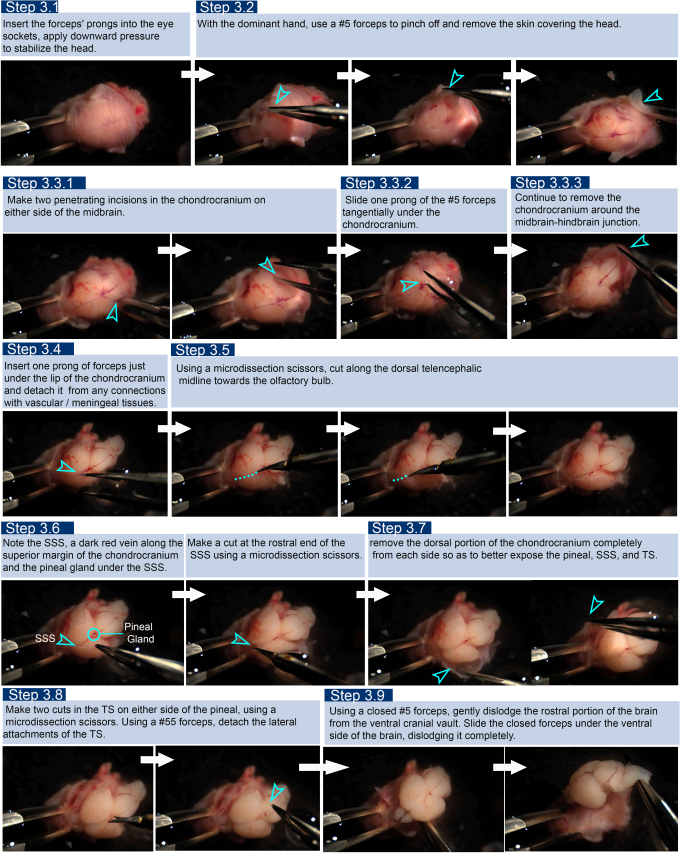
Figure 1: Stills from the video showing the step-by-step procedure for isolation of an E18.5 mouse brain, keeping the pineal gland intact. Please click here to view a larger version of this figure.
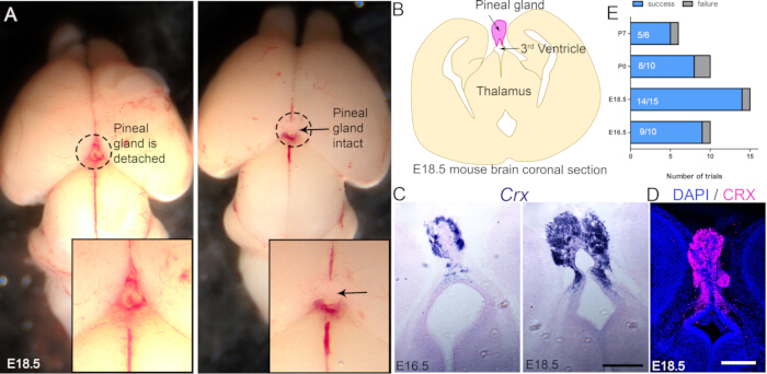
Figure 2: Representative results. (A) Representative examples of two E18.5 mouse brains in which the pineal gland is detached during the standard dissection procedure (left), compared with a brain where the pineal gland remains intact using the described method (right). (B) Schematic illustration of an E18.5 coronal section showing the pineal gland in pink. (C) In situ hybridization for Crx at E16.5 and E18.5, demonstrating pineal gland-specific expression. (D) Immunostaining for CRX at E18.5. (E) Bar graph showing the success (brain isolated with pineal intact) and failure (pineal damaged or lost) rates of the protocol at embryonic (E16.5 and E18.5) and postnatal (P0 and P7) developmental stages. Scale bars: 100 µm. N = 6 biological replicates for A, C, and D. Please click here to view a larger version of this figure.
Discussion
Standardized pineal gland dissection methods have been developed for the human fetal/infant brain, aiding forensic pathologists in understanding the cause of sudden infant death syndrome11. Previous protocols demonstrated removal of the pineal gland along with surface vasculature, meninges, and choroid plexus12; however, methods to reliably retain an intact pineal gland attached to the diencephalic roof during stages of its early development in mouse or other rodents have not been described.
Much of the classical anatomical and methodological literature describing pinealectomy and analysis has been developed using rat models13, where the gland is larger and easier to identify. In contrast, the mouse pineal gland is considerably smaller and more fragile, making it particularly susceptible to loss or damage during routine brain extraction procedures. Some studies have attempted to address this challenge by fixing the brain within the entire head. However, this introduces additional technical limitations during preparation of the tissue for sectioning. For example, during cryoprotection in 30% sucrose following fixation in 4% paraformaldehyde, the cranial vault may exert pressure on the underlying brain tissue, resulting in compression or distortion of dorsal forebrain structures. Such artifacts can compromise brain morphology and complicate analyses that rely on accurate anatomical organization. Furthermore, fixing the tissue may not be suitable for all applications. Therefore, methods that preserve the pineal gland and maintain its attachment to the roof of the diencephalon without compromising overall brain integrity are needed to facilitate developmental and molecular studies of the pineal gland in the mouse.
Reliable preservation of the pineal gland in intact brain preparations is important for several experimental applications. Histological and molecular approaches, such as immunohistochemistry and in situ hybridization, require preservation of tissue architecture to examine gene expression and cellular organization within the gland. More recently, emerging technologies such as spatial transcriptomics and other spatially resolved molecular profiling methods also rely on intact tissue morphology to map gene expression within specific anatomical contexts. To date, no spatial transcriptomic map of the embryonic mouse pineal gland has been reported. This may, in part, reflect the technical difficulty of preserving the intact pineal gland during conventional brain dissection. Therefore, methods that reliably maintain pineal gland integrity are likely to facilitate the application of emerging spatial transcriptomic approaches in pineal gland biology. In addition, because the pineal gland represents a very small fraction of total brain tissue, isolating the brain with the pineal gland intact permits a subsequent add-on step of harvesting the pineal gland specifically, and pooling such harvests would substantially improve enrichment of pineal-specific representation in bulk analysis in transcriptomic and proteomic studies.
It is necessary to stabilize the head well using a #5 forceps in the non-dominant hand. This needs to be maintained throughout the dissection process; therefore, the dominant hand must manage the changeover of instruments as needed. Tweak the illumination to better see the attachments of the pineal to the overlying vasculo-meningeal tissue that need to be removed.
Disclosures
All the authors declare no competing interests.
Acknowledgements
We thank Dr. Shital Suryavanshi and the animal house staff of the Tata Institute of Fundamental Research for their excellent support. We thank Dr. Sundar Naganathan for generously allowing the use of the Olympus MVX10 stereo macro zoom microscope in his lab for recording the video of the procedure. This work was funded by a grant from the Department of Atomic Energy (DAE), Government of India, Project Identification No. RTI4003 and RTI4015 (ST); ICMR-DHR SUG grant, GAP00303, ANRF PMECRG grant GAP00313 (AP).
Materials
| Name | Company | Catalog Number | Comments |
|---|---|---|---|
| Brightfield microscope | Olympus | IX73 | Any brightfield microscope with a color camera is suitable. |
| Confocal microscope | Olympus | FV3000 | Any confocal microscope or epifluoresence microscope is suitable depending on the resolution required |
| CRX antibody | Invitrogen | PA5-32182 | Immustaining protocol described in Parichha et al.9. |
| CRX probe | Custom made | Custom made | Insitu hybridization protocol Parichha et al.10. |
| Fine forceps # 5 | F.S.T | 11251-23 | May be substituted with similar tools from any company. |
| Fine forceps # 55 | F.S.T | 11255-20 | May be substituted with similar tools from any company. |
| PBS, pH 7.4 | Gibco | 10010023 | May be substituted with similar items from any company. |
| Petridish | Corning | CLS351029 | May be substituted with similar items from any company. |
| RNaseZap | Invitrogen | AM9782 | RNaseZap RNase Decontamination Solution is a surface decontamination solution that destroys RNases on contact |
| Spring scissors (microdissection scissors) | F.S.T | 15020-15 | May be substituted with similar tools from any company. |
| Stereozoom microscope | Nikon | SMZ745 | Any dissection microscope is suitable |
References
- Nakahara D, Nakamura M, Iigo M, Okamura H. Bimodal circadian secretion of melatonin from the pineal gland in a living CBA mouse. Proc Natl Acad Sci U S A. 2003;100(16):9584-9.
- Christ E, Pfeffer M, Korf HW, von Gall C. Pineal melatonin synthesis is altered in Period1-deficient mice. Neuroscience. 2010;171(2):398-406.
- Rath MF. Homeobox gene-encoded transcription factors in development and mature circadian function of the rodent pineal gland. J Pineal Res. 2024;76(3):e12950.
- Rath MF, Rohde K, Klein DC, Møller M. Homeobox genes in the rodent pineal gland: roles in development and phenotype maintenance. Neurochem Res. 2013;38(6):1100-12.
- Estivill-Torrús G, Vitalis T, Fernández-Llebrez P, Price DJ. The transcription factor Pax6 is required for development of the diencephalic dorsal midline secretory radial glia that form the subcommissural organ. Eur J Neurosci. 2001;14(6):1011-22.
- Dias M, et al. Transection of the superior sagittal sinus enables bilateral access to the rodent midline brain structures. eNeuro. 2021;8(4):ENEURO.0146-21.2021.
- Matsunaga MM, et al. Morphologic analysis of mice's pineal gland. J Morphol Sci. 2011;28(3):157-60.
- Boeckmann D. Morphological investigation of the deep pineal of the rat. Cell Tissue Res. 1980;210(2):283-94.
- Parichha A, et al. Constitutive activation of canonical Wnt signaling disrupts choroid plexus epithelial fate. Nat Commun. 2022;13:633.
- Parichha A, et al. Dentate gyrus morphogenesis is regulated by β-catenin function in hem-derived fimbrial glia. Development. 2022;149(21):dev200953.
- Sparks DL, Coyne CM, Sparks LM, Hunsaker JC 3rd. Recommended technique for brain removal to retain anatomic integrity of the pineal gland in order to determine its size in sudden infant death syndrome. J Forensic Sci. 1997;42(1):100-2.
- Bowyer JF, et al. A visual description of the dissection of the cerebral surface vasculature and associated meninges and the choroid plexus from rat brain. J Vis Exp. 2012;69:e4285.
- Calvo J, Boya J. Embryonic development of the rat pineal gland. Anat Rec. 1981;200(4):491-500.
Reprints and Permissions
