This protocol describes a rapid saliva pretreatment method for improving cortisol detection. A handheld filtration device is used to remove mucins and other interfering substances while preserving the target analyte. The pretreated saliva exhibits improved capillary flow, thereby enabling more consistent and reproducible assay performance.
Method Article
Handheld Device for Saliva Pretreatment to Improve Cortisol Detection
* These authors contributed equally
In This Article
Summary
Abstract
Salivary cortisol detection offers a noninvasive alternative to blood-based analysis. However, its practical implementation remains limited by the intrinsic viscosity of saliva and the presence of interfering components. In particular, mucins and salivary proteins impede capillary-driven flow and compromise the reliability of subsequent lateral flow assays (LFAs), thereby necessitating effective and accessible pretreatment strategies. Here, we present a handheld saliva pretreatment device, termed SaliFilter, for point-of-care cortisol detection. The device employs a filtration mechanism that selectively removes high-molecular-weight interfering components while allowing the passage of small molecules, including cortisol. Operated by simple manual pressure, it enables rapid sample processing (~2 min) without the need for external equipment, making it suitable for decentralized settings. The performance of the device was evaluated using turbidity measurements, protein quantification, and SDS-PAGE, which confirmed efficient removal of mucin and salivary proteins. Treated samples exhibited enhanced capillary flow, resulting in stable and reliable LFA operation. Quantitative analysis demonstrated improved signal reproducibility, and cortisol detection achieved a limit of detection of 1.47 ng mL⁻1 over clinically relevant concentration ranges. This protocol establishes an effective saliva pretreatment strategy that improves analytical sensitivity and reliability without compromising analyte integrity. The proposed approach provides a practical, portable solution for saliva-based diagnostics, with potential applicability for point-of-care testing and self-monitoring.
Introduction
Cortisol, a glucocorticoid hormone secreted by the adrenal cortex, is a key biomarker for assessing physiological stress1,2. It can be measured in multiple biological fluids, including serum, plasma, urine, interstitial fluid, and saliva, with blood and saliva being the most commonly used in clinical practice3. Although blood-based measurements provide reliable quantitative information, they require invasive venipuncture and trained personnel, which limits their suitability for frequent or longitudinal monitoring4,5,6. In contrast, salivary cortisol measurement offers a noninvasive and convenient alternative, enabling point-of-care and self-monitoring applications7,8. Notably, salivary cortisol levels correlate strongly with blood concentrations, supporting saliva as a reliable surrogate sample9. However, the intrinsic viscosity of saliva and the presence of interfering components significantly hinder analytical performance, necessitating effective pretreatment prior to analysis10,11,12.
Various pretreatment strategies have been explored to address these challenges. Centrifugation is widely used to remove large components such as mucins, proteins, and cellular debris. However, it requires bulky laboratory equipment and multiple processing steps, which limits its applicability in decentralized settings13,14. Dilution with buffer solutions can reduce viscosity and improve sample flow15. Nevertheless, it simultaneously lowers analyte concentration, thereby compromising analytical sensitivity. As a result, existing approaches remain insufficient for point-of-care applications, where rapid, equipment-free processing and preservation of target analytes are essential.
To overcome these limitations, we developed SaliFilter, a handheld device for saliva pretreatment tailored to point-of-care cortisol detection. The device employs a filtration mechanism that selectively removes high-molecular-weight interfering substances while permitting the passage of small molecules, including cortisol16. Operated manually, the device enables rapid, user-friendly processing without the need for external instrumentation. By eliminating interfering components, this approach improves capillary flow in lateral flow assays, thereby enhancing analytical reliability. This protocol describes the fabrication of the device and its application for rapid saliva pretreatment prior to cortisol detection using lateral flow assays.
Protocol
Commercially obtained de-identified pooled human saliva was used in this study. No human participants were recruited, and institutional ethics approval was not required.
1. Saliva sample preparation
- Instruct participants to refrain from eating or drinking for at least 30 min prior to sample collection.
- Prepare the saliva sample according to the supplier’s instructions.
NOTE: This study used commercially available pooled human saliva for protocol-level validation. - Gently mix the saliva sample before use to ensure homogeneity.
- Store the saliva samples at 4 °C and use them within the supplier's recommended storage period.
2. Preparation of the RBCM-coated membrane (Figure 1A)
- Isolate RBCM from whole blood according to a previously reported protocol16.
- Use commercially obtained human whole blood anticoagulated with K2EDTA as the RBCM source.
- Centrifuge the whole blood at 800 × g for 5 min and collect the red blood cell fraction after removing the plasma and buffy coat.
- Wash the isolated red blood cell pellet three times with ice-cold 1× phosphate-buffered saline (PBS) using gentle manual mixing.
- Resuspend the washed red blood cell pellet in ice-cold 0.25× PBS for 30 min to induce hemolysis.
- Separate free hemoglobin by centrifuging the lysed suspension at 20,000 × g for 30 min and collecting the RBCM pellet.
- Wash the collected RBCM pellet three additional times with 0.25× PBS and store the pale pink RBCM pellet at −80 °C until further use.
- Prepare a polyethersulfone (PES) membrane with a pore size of 30 nm and a diameter of 13 mm.
- Deposit 100 µL of 2% (w/v) RBCM solution onto one side of the PES membrane. Gently spread or dispense the solution dropwise until the entire membrane surface is fully wetted.
- Incubate the membrane at 50 °C for 30 min to ensure uniform coating and drying.
- Store the coated membrane at room temperature and use it within 15 days after preparation17.
NOTE: Detailed characterization of the RBCM-coated PES membrane has been described in our previous study16.
3. Fabrication of the SaliFilter device (Figure 1B)
- Place the RBCM-coated membrane into the designated filter holder.
- Assemble the device by securely connecting the inlet and outlet components.
- Attach a 1 mL syringe to the inlet of the device.
CAUTION: Ensure that all components are tightly sealed to prevent leakage during operation.
4. Saliva pretreatment
- Load 500 µL of saliva into the syringe connected to the SaliFilter device.
- Apply gentle manual pressure to pass the saliva sample through the membrane (~ 2 min).
- Collect the pretreated saliva from the outlet.
- Use the pretreated sample immediately or store it at 4 °C until further analysis.
5. SDS-PAGE analysis of salivary proteins
- Mix 15 µL of the sample with 5 µL of Laemmli sample buffer.
- Load the mixture into separate wells of an 8% SDS-PAGE gel.
- Perform electrophoresis at 150 V for 40 min.
- Stain the gel with Coomassie blue for 2 h.
- Destain the gel in distilled water overnight.
- Capture images of the gel using a smartphone or imaging system.
6. Cortisol detection using Lateral Flow Assay (LFA)
- Apply 150 µL of pretreated saliva to the sample pad of the LFA strip.
- Allow the sample to migrate along the strip for 10 min.
- Capture images of the test results using a smartphone or imaging system.
- Open the LFA image in ImageJ and use Image > Color > Split Channels to separate the RGB channels.
- Use the green channel image for test-line intensity analysis.
- Select a rectangular Region of Interest (ROI) of 100 × 10 pixels covering the test-line and apply the same ROI size to all strip images.
- Measure the mean intensity value of the test-line ROI. Select an adjacent blank membrane region with the same ROI size and measure the background intensity.
- Calculate the normalized test-line intensity as follows: normalized intensity = test-line intensity − background intensity.
7. Statistical analysis
- Present quantitative data as mean ± standard deviation (SD), unless otherwise stated. Define error bars as SD.
NOTE: In this study, all reported N values represent technical replicates performed using aliquots of commercially obtained pooled human saliva. - Perform linear regression analysis using the normalized test-line intensity values obtained from cortisol concentrations of 0–8 ng mL−1 and calculate the coefficient of determination (R2) from the fitted curve.
- Calculate the limit of detection (LOD) using the following equation: LOD = 3.3 × SE / |slope|, where SE is the standard error of the y-intercept and |slope| is the absolute value of the slope obtained from the linear regression equation. Use the absolute value of the slope because the competitive LFA produces a negative slope.
NOTE: In this study, no inferential statistical tests were performed, and no significance criteria were applied.
Results
Effective saliva pretreatment requires removing interfering components, including mucins and proteins, that compromise downstream analytical performance. The filtration capability of SaliFilter was first evaluated by comparing turbidity changes with those obtained using commercial saliva collection kits (Figure 2A). Untreated saliva exhibited high turbidity, whereas SaliFilter-treated samples appeared visibly clear, indicating efficient removal of mucins and suspended components. In contrast, samples processed using commercial kits showed only limited improvement in clarity.
Protein removal was quantitatively assessed by measuring absorbance at 280 nm, which demonstrated an approximate 69% reduction following filtration (Figure 2B). This result was further supported by a Bradford assay, which showed a marked decrease in color intensity in SaliFilter-treated samples (Figure 2C), corresponding to an approximately 80% reduction in total protein concentration (Figure 2D). Consistently, SDS-PAGE analysis revealed the absence of detectable protein bands in processed samples, confirming effective depletion of salivary proteins (Figure 2E).
The impact of pretreatment on capillary-driven flow was subsequently evaluated in the context of LFAs. A custom-made competitive LFA was used to assess the performance of pretreated saliva samples. Removal of mucins, which are known to impede fluid transport, restored capillary flow in the processed samples (Figure 3A). SDS-PAGE analysis further confirmed that mucins were retained upstream of the membrane and were absent from the processed samples (Figure 3B). As a result, only the processed samples produced well-defined test and control lines in the competitive LFA, whereas untreated and commercially processed samples failed to generate reliable signals (Figure 3C). Quantitative analysis of signal intensity corroborated these observations, demonstrating significantly improved reproducibility and signal stability (Figure 3D,E).
Finally, the assay's analytical performance was evaluated using cortisol-spiked saliva samples. In the original validation study17, ELISA-based recovery analysis confirmed that ~ 90% cortisol was recovered in the SaliFilter-treated filtrate, supporting the compatibility of the pretreatment step with downstream cortisol detection. As expected in a competitive LFA format, increasing cortisol concentration led to a progressive decrease in test-line intensity (Figure 4A). Quantitative analysis revealed a clear inverse relationship between cortisol concentration and signal intensity (Figure 4B), with a strong linear correlation at low concentrations (R2 ≈ 0.99). The limit of detection was approximately 1.47 ng mL⁻1, within clinically relevant ranges.
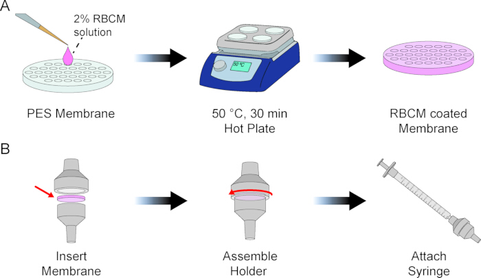
Figure 1: Fabrication and assembly of the SaliFilter. (A) Preparation of the RBCM-coated membrane. (B) Assembly of the handheld SaliFilter. Please click here to view a larger version of this figure.
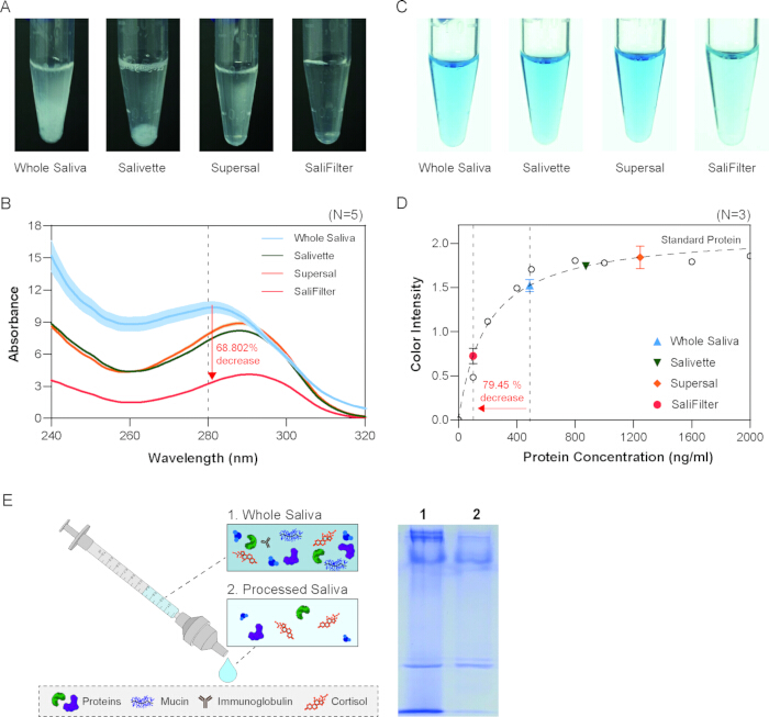
Figure 2: Evaluation of protein-removal performance. (A) Visual comparison of saliva turbidity before and after pretreatment. (B) Protein quantification based on absorbance at 280 nm. (C) Bradford assay showing colorimetric changes. (D) Quantitative protein analysis based on the Bradford assay. (E) SDS-PAGE analysis of salivary proteins before and after pretreatment. Reproduced with permission from Kim et al.17, Copyright © 2025 Elsevier. Please click here to view a larger version of this figure.
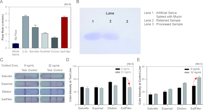
Figure 3: Improved capillary flow and lateral flow assay (LFA) performance following saliva pretreatment. (A) Comparison of capillary flow in untreated, commercially processed, and SaliFilter-treated saliva samples. (B) SDS-PAGE analysis demonstrating mucin retention. (C) Representative LFA results. Quantitative analysis of (D) test-line intensity and (E) control-line intensity. Reproduced with permission from Kim et al.17, Copyright © 2025 Elsevier. Please click here to view a larger version of this figure.
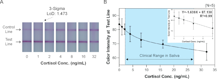
Figure 4: Quantitative cortisol detection using SaliFilter-treated saliva. (A) Representative LFA images obtained at varying cortisol concentrations. (B) Quantitative analysis of test-line intensity as a function of cortisol concentration. Reproduced with permission from Kim et al.17, Copyright © 2025 Elsevier. Please click here to view a larger version of this figure.
Discussion
Saliva-based diagnostics offer a noninvasive alternative to blood analysis. However, their broader adoption has been constrained by the intrinsic viscosity of saliva and the presence of interfering components such as mucins and proteins. These factors disrupt capillary-driven flow and compromise the performance of simple analytical platforms, including LFAs, thereby necessitating effective sample pretreatment.
In this study, we demonstrate that the proposed handheld filtration device, SaliFilter, effectively removes high-molecular-weight interfering substances while preserving small target analytes such as cortisol. The RBCM coating serves as a biologically derived interfacial layer on the PES membrane, contributing to the removal of high-molecular-weight salivary components such as mucins and proteins. By eliminating flow-disrupting components, the device enables direct integration of saliva samples into LFAs without the need for complex or instrument-dependent preprocessing. This finding highlights the critical role of pretreatment in bridging the gap between raw biological samples and simplified diagnostic platforms.
Conventional pretreatment strategies, such as centrifugation and dilution, are inherently limited in point-of-care settings. Centrifugation requires laboratory infrastructure and multiple processing steps, whereas dilution reduces analyte concentration and compromises analytical sensitivity. In contrast, the present approach enables rapid, equipment-free sample processing while maintaining analyte integrity, resulting in improved capillary flow and enhanced analytical reliability. A detailed comparison with conventional saliva pretreatment methods, including centrifugation-based approaches, has been provided in our previous study17. These features make the device particularly well-suited for decentralized and user-operated diagnostic applications.
The observed improvement in capillary flow directly translates into stable and reproducible signal generation in LFAs, underscoring the mechanistic link between sample properties and assay performance. By enabling quantitative cortisol detection within clinically relevant ranges, this work further supports the feasibility of saliva as a practical medium for real-time stress monitoring and related applications.
Despite these advantages, several limitations warrant further investigation. Although the RBCM-coated membranes were used within 15 days after preparation in this protocol, further studies are needed to systematically evaluate the long-term shelf-life of both the coated membrane and the fully assembled SaliFilter device under controlled storage conditions, including temperature and humidity variations. The device's performance should also be evaluated across a broader range of biomarkers with diverse physicochemical properties. The cortisol detection experiments were performed using commercially obtained pooled human saliva, which may not fully reflect inter-individual variability observed in clinical saliva samples. Therefore, future studies should validate the platform using clinical saliva samples with documented sample characteristics to further evaluate inter-individual variability, filtration efficiency, and clinical applicability.
Overall, the simplicity, portability, and effectiveness of this approach position it as a promising solution for saliva pretreatment in point-of-care diagnostics. Integration with user-friendly detection platforms may facilitate the development of accessible tools for continuous health monitoring and biomarker-based assessment in non-clinical settings.
Disclosures
The authors have no conflicts of interest to declare.
Acknowledgements
This research was supported by the Regional Innovation System & Education (RISE) program through the Gangwon RISE Center, funded by the Ministry of Education (MOE) and the Gangwon State (G.S.), Republic of Korea (2026-RISE-10-001). The present research has been conducted by the Excellent Researcher Support Project of Kwangwoon University in 2026
Materials
| Name | Company | Catalog Number | Comments |
|---|---|---|---|
| Artificial saliva | Solarbio, China | A7990 | |
| Bio-Rad Protein Assay Dye Reagent | Bio-Rad, USA | 5000006 | For bradford assay |
| Coomassie blue | Abcam, UK | ab119211 | For protein band visualization |
| Cortisol ELISA Kit | Abcam, UK | ab154996 | |
| Cortisol LFA | In-house | ||
| Hot plate | Daihan Scientific, Republic of Korea) | ||
| Hydrocortisone (Cortisol) | Sigma-Aldrich, USA | PHR1014 | |
| Laemmli sample buffer | GenDEPOT, USA | L1200-001 | For protein denaturation prior to SDS-PAGE |
| Membrane holder | GVS Filter Technology | 1220950 | 13mm diameter holder |
| Mini-PROTEAN® Tetra Cell for 0.75 mm Gels | Bio-Rad, USA | 1658000 | For gel electrophoresis |
| Mucin | Sigma-Aldrich, USA | M2378 | |
| NanoDrop 2000 spectrophotometer | Thermo Fisher Scientific, USA | For protein assay | |
| PES membranes | STERLITECH, USA | PES00313100 | 0.03 Micron, 13 mm PES membrane |
| Phosphate-buffered saline (PBS) | DUKSAN, Republic of Korea | LB004 | |
| Pooled Human Saliva | Innovative Research Inc., USA | IRHUSL5ML | |
| RBCM | In-house | ||
| Resolving Gel Buffer for PAGE | Bio-Rad, USA | 1610798 | For resolving gel preparation |
| Salivette Cortisol | SARSTEDT, Germany | 51.1534.500 | Saliva collection kit |
| Stacking Gel Buffer for PAGE | Bio-Rad, USA | 1610799 | For stacking gel preparation |
| Super•SAL™ | Oasis Diagnostics, USA | SSAL-601 | Saliva collection kit |
| Syringe | koreavaccine, Republic of Korea) | 1 mL volume | |
| TEMED | Bio-Rad, USA | 1610800 | For acrylamide polymerization |
Reprints and Permissions
