This article primarily outlines the standard procedure for bedside screening for retinopathy of prematurity (ROP), which helps detect ROP early, enabling timely treatment and reducing the risk of blindness.
A subscription to JoVE is required to view this content. Sign in or start your free trial.
Method Article
A Standardized Bedside Screening Protocol for Retinopathy of Prematurity
* These authors contributed equally
In This Article
Summary
Abstract
Retinopathy of prematurity (ROP) screening is a time-sensitive, multidisciplinary clinical process that enables early detection of this vision-threatening disease while minimizing examination-related physiologic stress. This article describes a structured bedside workflow for ROP screening, including eligibility assessment, pre-examination preparation, retinal examination, differential diagnosis of uncertain findings, documentation, follow-up planning, referral, and post-examination care. Infants are selected for screening according to gestational age and birth weight criteria, with additional consideration given to clinically unstable infants considered at risk by the neonatology team. Before examination, parent communication, cardiorespiratory monitoring, pharmacologic mydriasis, and comfort measures are performed according to the screening workflow. Retinal evaluation is performed primarily by binocular indirect ophthalmoscopy combined with scleral depression, while adjunctive image documentation is obtained when available and clinically indicated. Examination findings are recorded according to the International Classification of Retinopathy of Prematurity, Third Edition (ICROP3), including zone, stage, and plus disease status, to support consistent classification and longitudinal comparison. Follow-up intervals, senior review, and referral for treatment evaluation are then assigned according to predefined decision criteria. This workflow may serve as a practical operational framework for bedside ROP screening in units seeking more uniform documentation and follow-up planning.
Introduction
Retinopathy of prematurity (ROP) is one of the major preventable causes of childhood blindness worldwide and primarily affects preterm infants and those with low birth weight1. With advances in perinatal medicine and neonatal intensive care, increasing numbers of extremely preterm and extremely low-birth-weight infants now survive, and the burden of ROP therefore remains substantial, particularly in low- and middle-income countries2. Without timely screening and intervention, ROP may progress to tractional retinal detachment and severe irreversible visual loss, and it is also associated with long-term ocular sequelae such as refractive error, high myopia, strabismus, amblyopia, and late retinal complications3. Importantly, many severe ROP-related visual outcomes can be prevented or mitigated through standardized screening, early recognition, and timely treatment.
Current ROP management is centered on risk-based screening, standardized disease classification, timely intervention, and longitudinal follow-up4. Indirect ophthalmoscopy remains the clinical gold standard for screening, ICROP3 provides a standardized framework for disease documentation and staging, and laser photocoagulation and intravitreal anti-vascular endothelial growth factor therapy are the principal treatment options for severe ROP5,6. In clinical practice, however, the operational workflow used to deliver ROP screening varies across institutions. Some centers rely primarily on bedside indirect ophthalmoscopy performed according to local routines, whereas others use image-based documentation or telemedicine-assisted review when trained ophthalmologists or bedside resources are limited7,8. These approaches may differ in staffing requirements, documentation quality, workflow consistency, and capacity for longitudinal comparison, particularly when screening is performed across multiple providers or over extended follow-up periods9,10.
A structured bedside workflow may offer practical advantages over non-standardized bedside practice by clarifying team roles, improving consistency of documentation, supporting follow-up planning, and facilitating communication between ophthalmologists, neonatologists, and nursing staff9,10. Such a workflow may be particularly useful in neonatal wards and neonatal intensive care units that perform repeated bedside screening, maintain longitudinal follow-up, or aim to strengthen training and quality control11. At the same time, implementation depends on the availability of trained personnel, monitoring equipment, and access to adjunct image documentation when needed. The protocol may therefore be most applicable in centers with established neonatal care infrastructure and ophthalmic support, whereas adaptation may be necessary in settings with limited personnel, limited imaging access, or greater reliance on telemedicine-assisted screening. In addition, the characteristics of infants at risk for ROP may vary across healthcare systems and geographic regions. In low- and middle-income settings, ROP may occur in larger and more mature infants than those typically included in screening programs in highly resourced tertiary centers12. For this reason, screening criteria, team organization, and referral pathways may require local adaptation according to the patient population, available expertise, and institutional resources rather than strict reliance on a single operational model12.
Accordingly, this article presents a structured bedside ROP screening workflow applicable to neonatal wards and NICUs. The protocol focuses on screening eligibility assessment, pre-examination preparation, binocular indirect ophthalmoscopy combined with scleral depression, adjunctive image documentation when indicated, ICROP3-based documentation and grading, and decisions regarding follow-up and referral. Rather than proposing a new diagnostic standard, this article provides a practical operational framework intended to support more consistent implementation of bedside ROP screening and to facilitate training, documentation, and multidisciplinary coordination in routine clinical practice.
Access restricted. Please log in or start a trial to view this content.
Protocol
This protocol describes a structured bedside ROP screening workflow for neonatal wards and the neonatal intensive care unit (NICU). The study was approved by the Ethics Committee of the Children’s Hospital, Zhejiang University School of Medicine (ID: 2021-IRB-026). All examinations were performed according to routine clinical practice, and written informed consent was obtained from the parents or legal guardians. A standardized parental informed consent form used in this protocol is provided in Supplementary File 1.
1. Determining screening eligibility
- Identify infants eligible for screening. Review gestational age (GA), birth weight (BW), and neonatal records. Include infants with a GA of ≤30 weeks and/or a BW of ≤1500 g.
- Assess infants outside the routine thresholds. Review the systemic course of infants whose GA or BW exceeds the routine thresholds. Consult the neonatology and ophthalmology teams and determine whether screening is indicated.
- Schedule the first examination. Determine the timing of the first examination according to postmenstrual age (PMA) and postnatal age. Examine infants born at ≤27 weeks’ gestation at approximately 31 weeks’ PMA and examine infants born at >27 weeks’ gestation at approximately 4 weeks after birth13.
- Create the screening registry. Record the infant’s name, hospital identification number, GA, BW, PMA, general systemic condition, oxygen supplementation history, and results of prior ophthalmic examinations. Use the registry to arrange scheduling, follow-up, and interdisciplinary communication.
2. Preparation for bedside screening
- Prepare the team. Distribute workflow responsibilities among the ophthalmologist, neonatal team, and nursing staff before beginning bedside screening. Complete administrative preparation, pupillary dilation, and routine monitoring setup in advance when possible so that the bedside retinal examination can proceed efficiently once the ophthalmologist is present.
- Assemble the multidisciplinary team. Identify the ophthalmologist, the responsible neonatal team member, and the nursing staff involved in the screening workflow. Assign responsibilities for registry review, pupillary dilation, monitoring setup, bedside examination, documentation, and follow-up communication before the examination begins.
- Verify infant identity. Confirm the infant’s identity at the bedside using standard clinical identification procedures. Complete identity verification before medication administration and examination setup.
- Review systemic stability. Review heart rate, respiratory status, oxygen saturation, and overall systemic condition immediately before the examination. Reassess the plan if instability is present.
NOTE: For the purposes of this protocol, instability or clinically significant deterioration includes oxygen saturation <80%, heart rate <100 beats/min, apnea, gasping, or persistent increased work of breathing. - Prepare emergency support. Place resuscitation and cardiorespiratory support equipment at the bedside for infants with limited systemic reserve. Confirm immediate availability before starting the examination.
- Define bedside attendance according to task requirements. Ask nursing staff to complete preparatory steps that do not require the ophthalmologist at the bedside, including infant identification, routine monitoring setup, and scheduled pupil dilation. Request neonatal physician presence when systemic assessment, stabilization, or urgent medical support is required, rather than requiring continuous bedside attendance throughout the entire screening sequence.
- Prepare the instruments and materials
- Prepare and disinfect the examination instruments. Prepare the binocular indirect ophthalmoscope, condensing lens, infant eyelid speculum, scleral depressor, and related screening instruments. Clean and disinfect all reusable instruments before the examination and maintain aseptic handling during setup.
NOTE: Use disposable contact materials whenever possible. - Adjust the ophthalmoscope and condensing lens. Place the head-mounted indirect ophthalmoscope securely and adjust the interpupillary distance until a stable binocular view is obtained. Select the condensing lens used in the local screening protocol and reduce illumination to the lowest level that still permits reliable fundus visualization.
- Prepare adjunct imaging and monitoring equipment. Prepare wide-field retinal imaging for adjunctive documentation and assessment when it is available and clinically indicated14,15. Connect continuous monitoring devices for heart rate and oxygen saturation before the examination. The imaging device used for adjunct fundus documentation is shown in Figure 1.
NOTE: Perform adjunct imaging using a 130° pediatric wide-field lens and the manufacturer’s standard neonatal imaging mode. - Prepare the documentation forms. Place the standardized examination record form and report form at the bedside before starting the examination. Record findings during or immediately after the examination.
- Perform hand hygiene and infection-prevention procedures. Perform hand hygiene before patient contact and before instrument handling. Follow the institutional infection-prevention procedures throughout the bedside workflow.
- Prepare and disinfect the examination instruments. Prepare the binocular indirect ophthalmoscope, condensing lens, infant eyelid speculum, scleral depressor, and related screening instruments. Clean and disinfect all reusable instruments before the examination and maintain aseptic handling during setup.
- Prepare the infant. Complete infant preparation steps in advance whenever possible. Perform pupillary dilation, routine monitoring, registry confirmation, and bedside setup in parallel for more than one infant when the clinical workload and staffing pattern allow.
- Communicate with the parents or legal guardians. Explain the purpose, procedure, possible discomforts, and safety precautions of bedside ROP screening. Answer questions and complete written informed consent before pupil dilation.
- Review contraindications and medication history. Review contraindications, allergy history, and relevant systemic risk factors before administering ophthalmic medications. Confirm the absence of known adverse reactions to mydriatic agents or topical anesthetic agents.
- Monitor vital signs before dilation. Record heart rate, respiratory rate, and oxygen saturation before administering the mydriatic agent. Continue monitoring during pupil dilation and reassess the infant immediately before starting the ocular examination16.
CAUTION: Stop the workflow and reassess the infant if oxygen saturation falls below 80%, if heart rate falls below 100 beats/min, or if apnea, marked increased work of breathing, or other clinically significant deterioration occurs. - Administer mydriatic agents. Instill compound tropicamide (tropicamide 0.5% and phenylephrine 0.5%), 1 drop in each eye, before the examination. Allow 30–40 min for pupil dilation before beginning the ocular examination. Ask the nursing staff to administer the mydriatic agent according to the screening schedule. If pupil dilation remains inadequate after the waiting period, delay classification and reassess the infant according to the local neonatal ophthalmic workflow.
- Prepare ocular surface support. Instill proparacaine hydrochloride 0.5%, 1 drop in the eye, immediately before scleral depression-assisted examination. Wait 1 min for the topical anesthetic to take effect, then begin the examination. Apply lubricating drops or artificial tears when corneal dryness, unstable tear film, or reduced optical clarity is present.
- Provide comfort measures. Provide swaddling, non-nutritive sucking, and oral sucrose when appropriate17,18. Select the comfort strategy according to the infant’s clinical condition and the local neonatal care practice.
- Confirm readiness for examination. Confirm adequate pupil dilation before inserting the eyelid speculum and beginning indirect ophthalmoscopy. Define adequate dilation as a pupil diameter of at least 5 mm or as sufficient dilation to allow visualization of the posterior pole and the retinal periphery required for classification.
- Stabilize the infant. Ask the nursing staff to stabilize the infant’s body and head before the examination begins. Minimize unnecessary restraint and excessive stimulation.
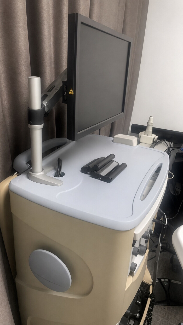
Figure 1: Imaging device used for adjunct fundus documentation in bedside ROP screening. Please click here to view a larger version of this figure.
3. Performing bedside retinal examination
- Position and monitor the infant.
- Position the infant. Place the infant in the supine position in the incubator or on a warming bed. Expose the eyes adequately and minimize unnecessary body movement.
- Start continuous physiologic monitoring. Monitor heart rate, respiratory status, and oxygen saturation continuously throughout the examination16. Pause the examination immediately if oxygen saturation falls below 80%, if heart rate falls below 100 beats/min, or if respiratory instability develops, including apnea, gasping, or persistent increased work of breathing.
- Open the eyelids and protect the ocular surface.
- Insert the eyelid speculum. Instill a topical anesthetic when necessary and insert an infant eyelid speculum gently. Expose the globe without applying excessive pressure to the eyelids.
- Maintain ocular surface clarity. Inspect the ocular surface before proceeding. Apply lubricating drops or artificial tears as needed to maintain corneal moisture and optical clarity.
- Examine the posterior pole and retinal vascularization.
- Examine the first eye. Examine the right eye first. Identify the optic disc and posterior pole and assess retinal vessel dilation, tortuosity, and pre-plus or plus vascular changes.
- Assess vascular termination and lesion extent. Identify the termination of retinal vessels and determine the extent and location of retinal lesions. Assign the retinal zone and note the findings relevant to stage classification.
- Examine the peripheral retina with scleral depression. Apply scleral depression gently and inspect the peripheral retina. Identify avascular retina, a demarcation line, a ridge, and extraretinal fibrovascular proliferation, and inspect the area near the ora serrata when needed.
- Examine the fellow eye. Repeat the same sequence in the left eye. Record posterior pole findings, vascular termination, lesion extent, and peripheral retinal findings separately for that eye.
- Acquire adjunct images when indicated.
- Acquire adjunct images. Acquire adjunct images when one or more of the following conditions are present: (1) posterior pole vascular changes require documentation; (2) zone or stage classification remains uncertain after bedside examination; (3) peripheral retinal findings require image confirmation; (4) secondary review or remote consultation is anticipated; (5) longitudinal comparison is required at follow-up; or (6) image documentation is required by the local screening workflow.
- Perform image acquisition after completion of the core bedside examination or during a brief pause when the infant remains clinically stable. Use the standard neonatal imaging mode of the device unless pupil size, media clarity, or infant tolerance requires adjustment.
NOTE: In routine neonatal bedside screening, no additional parameter adjustment was usually required, and image acquisition was generally performed using the manufacturer’s default neonatal settings unless pupil size, media clarity, or infant tolerance limited image quality.
- Perform image acquisition after completion of the core bedside examination or during a brief pause when the infant remains clinically stable. Use the standard neonatal imaging mode of the device unless pupil size, media clarity, or infant tolerance requires adjustment.
- Evaluate image adequacy before classification support. Confirm that the image shows the posterior pole or peripheral target region with sufficient focus, illumination, and field coverage before using it to support classification or documentation. Repeat image acquisition if vessel margins are blurred, the retinal region of interest is incompletely captured, motion artifact obscures interpretation, or illumination is insufficient for grading.
- Acquire adjunct images. Acquire adjunct images when one or more of the following conditions are present: (1) posterior pole vascular changes require documentation; (2) zone or stage classification remains uncertain after bedside examination; (3) peripheral retinal findings require image confirmation; (4) secondary review or remote consultation is anticipated; (5) longitudinal comparison is required at follow-up; or (6) image documentation is required by the local screening workflow.
- Resolve limited visualization and uncertain findings.
- Repeat the examination when visualization is limited. Repeat the examination if poor dilation, corneal dryness, media opacity, motion artifact, or infant intolerance limit visualization. Improve the ocular surface, reposition the infant, and repeat the view before assigning classification.
- Escalate uncertain findings. Proceed with classification only after confirming all of the following: (1) pupil dilation is adequate for visualization of the posterior pole and the retinal periphery required for staging; (2) retinal vessels and lesion borders are sufficiently visible for zone and stage assessment; and (3) plus or pre-plus vascular changes can be judged without major uncertainty.
NOTE: Delay final classification and request repeat examination, adjunct imaging, or senior review if any of these criteria are not met.
4. Documentation and classification of disease
- Record findings according to ICROP3.
- Record bilateral findings. Record retinal zone, stage, plus disease status, and aggressive posterior ROP (AP-ROP) status for each eye separately. Complete the standardized examination record form for both eyes (Supplementary Table 1).
- Record additional retinal features. Record clock-hour extent, vascular termination, avascular retina, demarcation line, ridge, extraretinal neovascularization, posterior pole vascular findings, and other relevant fundus findings when present.
NOTE: The examination form should allow bilateral recording of ICROP3 classification, posterior pole vascular findings, peripheral retinal findings, image acquisition status, and any uncertainty requiring secondary review.
- Label and archive image data.
- Label image files. Label each image file with the examination date, PMA, laterality, bed number, or hospital identification number, and screening type. Use a consistent naming format across all examinations.
- Archive image files. Store the labeled image files in the designated archive immediately after acquisition. Verify successful storage before moving to the next case.
- Perform secondary review when interpretation is uncertain.
- Send uncertain cases for secondary review. Send cases with poor image quality, borderline findings, or uncertain grading to a second ophthalmologist for review. Document the secondary review in the record.
- Escalate discrepant interpretations. Refer discrepant interpretations to a senior ophthalmologist for final adjudication. Update the final classification after adjudication.
- Generate the standardized report.
- Complete the report form. Summarize disease classification, the recommended follow-up interval, referral status for treatment evaluation, and the need for remote expert review in the standardized report form (Supplementary Table 2). Finalize the report promptly after the examination.
NOTE: The report form should summarize the final classification for each eye, the assigned follow-up interval, the need for senior review, referral for treatment evaluation, and whether adjunct images were archived and linked to the medical record. - Link the report to the medical record. Attach the final report to the infant’s clinical record. Confirm successful entry before ending the documentation workflow.
- Complete the report form. Summarize disease classification, the recommended follow-up interval, referral status for treatment evaluation, and the need for remote expert review in the standardized report form (Supplementary Table 2). Finalize the report promptly after the examination.
5. Planning the follow-up and referral
- Assign the follow-up interval
- Assign a follow-up interval according to disease severity. Assign follow-up within 1 week or less for posterior disease, zone I disease, stage 3 disease, pre-plus disease, plus disease, suspected aggressive ROP, or uncertain progression risk. Assign follow-up within 1–2 weeks for less severe zone II disease or immature vascularization without immediate treatment-warranted features.
- Assign follow-up within 2–3 weeks for mild or regressing disease without high-risk features. Apply the same interval assignment criteria consistently across all examinations to maintain uniform follow-up planning.
- Record the follow-up interval. Record the assigned interval in the standardized report form and communicate it to the neonatal care team before ending the screening workflow. Confirm the planned date or time window for the next examination at the time of documentation.
NOTE: Adjust the interval according to the infant’s systemic condition and the local screening guideline if earlier re-examination is clinically necessary.
- Assign a follow-up interval according to disease severity. Assign follow-up within 1 week or less for posterior disease, zone I disease, stage 3 disease, pre-plus disease, plus disease, suspected aggressive ROP, or uncertain progression risk. Assign follow-up within 1–2 weeks for less severe zone II disease or immature vascularization without immediate treatment-warranted features.
- Identify infants requiring short-interval re-examination.
- Flag high-risk infants. Flag infants for short-interval re-examination when one or more of the following findings are present: posterior immature vascularization, zone I disease, stage 3 ROP, pre-plus disease, suspected aggressive ROP, poor retinal visualization, or findings with uncertain progression risk.
- Prioritize re-examination scheduling. Schedule these infants for early re-examination and identify them clearly in the registry and report form. Communicate the short-interval plan directly to the neonatal care team and the responsible ophthalmology team member.
- Identify infants requiring immediate senior review.
- Escalate concerning findings immediately. Refer the case to a senior ophthalmologist immediately when the examination shows severe posterior vascular changes, suspected plus disease, possible aggressive posterior ROP, posterior zone I or posterior zone II involvement, poor-quality visualization that limits reliable grading, or persistent uncertainty in classification after repeat examination.
- Document the reason for escalation. Record the indication for senior review in the examination record and report form. Complete the final classification only after senior review or adjudication.
- Refer treatment-warranted disease.
- Transfer treatment-warranted cases to the treatment pathway. Identify treatment-warranted disease when one or more of the following findings are present: zone I ROP with plus disease at any stage, zone I stage 3 ROP without plus disease, or zone II stage 2 or 3 ROP with plus disease.
- Transfer the infant to the treatment pathway.
Transfer infants with treatment-warranted findings to the treatment pathway without delay. Initiate referral for treatment evaluation, document the referral decision in the medical record, and communicate the result directly to the neonatal care team and the infant’s guardians. - Use structured support for decision-making. Use the follow-up and referral decision matrix provided in Supplementary Table 3 to support interval assignment, escalation, and treatment referral. Apply the matrix together with bedside findings and the infant’s current systemic condition.
6. Performing post-examination care
- Monitor the infant after the examination.
- Continue post-examination observation. Observe the infant until heart rate, respiratory status, oxygen saturation, and overall tolerance remain stable. Continue bedside monitoring through the nursing team after the examination ends.
- Protect the ocular surface.
- Provide ocular surface care. Apply lubricating drops or artificial tears after the examination if ocular surface exposure or irritation is present. Reassess the eye if prolonged speculum use or repeated manipulation occurred.
- Clean and disinfect the instruments.
- Process the reusable instruments. Clean and disinfect all reusable instruments immediately after use according to institutional infection-control procedures. Complete instrument processing before moving to the next infant.
- Archive data and upload the report.
- Archive the image data. Archive all image data in the designated storage system. Confirm successful storage before ending the workflow.
- Upload the report. Upload the final standardized report to the medical record system. Verify successful upload before closing the case.
Access restricted. Please log in or start a trial to view this content.
Results
Approximately 25 eyes underwent bedside ROP screening per week. The examination completion rate was 100%, and the average examination duration was 2–3 min per eye. Standardized report generation was completed in all screened cases, and follow-up assignment was completed for all cases. Adverse events, including apnea and allergic reactions, occurred only rarely, indicating good overall tolerability of the bedside workflow.
Figure 2 summarizes the overall workflow o...
Access restricted. Please log in or start a trial to view this content.
Discussion
This article describes a structured bedside workflow for retinopathy of prematurity (ROP) screening in neonatal wards and neonatal intensive care units (NICUs). The protocol integrates screening eligibility assessment, pre-examination preparation, bedside retinal examination, adjunct image acquisition when indicated, ICROP3-based documentation, follow-up planning, referral, and post-examination care into a single operational sequence. Rather than establishing a new diagnostic standard, the protocol is intended to provide...
Access restricted. Please log in or start a trial to view this content.
Disclosures
The authors have no conflicts of interest to disclose.
Acknowledgements
This research was supported by the Scientific Research Fund of Zhejiang Provincial Education Department under Grant Y202454758. The authors would like to express gratitude to the people working on this standard screening procedure.
Access restricted. Please log in or start a trial to view this content.
Materials
| Name | Company | Catalog Number | Comments |
|---|---|---|---|
| 90D condensing lens | VOLK Optical Inc. | V503085 | used for bedside retinal examination with binocular indirect ophthalmoscopy |
| Binocular indirect ophthalmoscope (YZ25C) | Suzhou 66 Vision Technology Co., Ltd. | V150981 | bedside retinal examination |
| RetCam3 | Natus Medical Incorporated | AZ23001062-0301 | wide-field retinal imaging |
References
- Wood EH, et al. 80 years of vision: preventing blindness from retinopathy of prematurity. J Perinatol. 2021;41(6):1216-24.
- García H, et al. Global prevalence and severity of retinopathy of prematurity over the last four decades (1985-2021): a systematic review and meta-analysis. Arch Med Res. 2024;55(2):102967.
- Porteny JR, et al. A scoping review of adverse visual outcomes among preterm infants without versus those with retinopathy of prematurity. Surv Ophthalmol. 2026;71(3):992-6.
- Athikarisamy SE, Lam GC. Screening and surveillance for retinopathy of prematurity: a Wilson and Jungner framework approach. J Glob Health. 2023;13:03028.
- Chiang MF, Quinn GE, Fielder AR, Ostmo SR, Paul Chan RV, Berrocal A, et al. International classification of retinopathy of prematurity, third edition. Ophthalmology. 2021;128(10):e51-e68.
- Fierson WM. Screening examination of premature infants for retinopathy of prematurity. Pediatrics. 2013;131(1):189-95.
- Brady CJ, D'Amico S, Campbell JP. Telemedicine for retinopathy of prematurity. Telemed J E Health. 2020;26(4):556-64.
- Sekeroglu MA, Hekimoglu E, Sekeroglu HT, Arslan U. Alternative methods for the screening of retinopathy of prematurity: binocular indirect ophthalmoscopy versus wide-field digital retinal imaging. Eye (Lond). 2013;27(9):1053-7.
- Visser L, Singh R, Young M, Lewis H, McKerrow N. Guideline for the prevention, screening and treatment of retinopathy of prematurity (ROP). S Afr Med J. 2012;103(2):116-25.
- Padhi TR, et al. Outcome of real-time telescreening for retinopathy of prematurity using videoconferencing in a community setting in Eastern India. Indian J Ophthalmol. 2024;72(5):697-703.
- Song A, et al. Retinopathy of prematurity training and education: a systematic review. Surv Ophthalmol. 2026;71(1):248-61.
- Darlow BA, Gilbert C. Retinopathy of prematurity: a world update. Semin Perinatol. 2019;43(6):315-6.
- Yu Y, Tomlinson LA, Binenbaum G, Ying GS. Incidence, timing and risk factors of type 1 retinopathy of prematurity in a North American cohort. Br J Ophthalmol. 2021;105(12):1724-30.
- Roti EV, et al. Ultra-widefield optical coherence tomography beyond the ora serrata in retinopathy of prematurity. JAMA Ophthalmol. 2025;143(2):165-70.
- Chen L, Feng Y, Xu Z. Neonatal retinal hemorrhage and retinopathy of prematurity: a critical review of current practices and future directions. Ther Adv Ophthalmol. 2025;17:25158414251386295.
- Purohit R, et al. Physiological responses to retinopathy of prematurity screening: indirect ophthalmoscopy versus ultra-widefield retinal imaging. Pediatr Res. 2025;98(5):1801-8.
- Boyle EM, et al. Sucrose and non-nutritive sucking for the relief of pain in screening for retinopathy of prematurity: a randomized controlled trial. Arch Dis Child Fetal Neonatal Ed. 2006;91(3):F166-8.
- Dilli D, et al. Oral sucrose and non-nutritive sucking goes some way to reducing pain during retinopathy of prematurity eye examinations. Acta Paediatr. 2014;103(2):e76-9.
- Chatmethakul T, et al. Impact of telemedicine on retinopathy of prematurity screening at a level II neonatal intensive care unit in Southwest Oklahoma: a 6-year experience. Telemed J E Health. 2026;32(3):315-22.
- Nguyen TP, et al. Discrepancies in diagnosis of treatment-requiring retinopathy of prematurity. Ophthalmol Retina. 2024;8(1):88-91.
Access restricted. Please log in or start a trial to view this content.