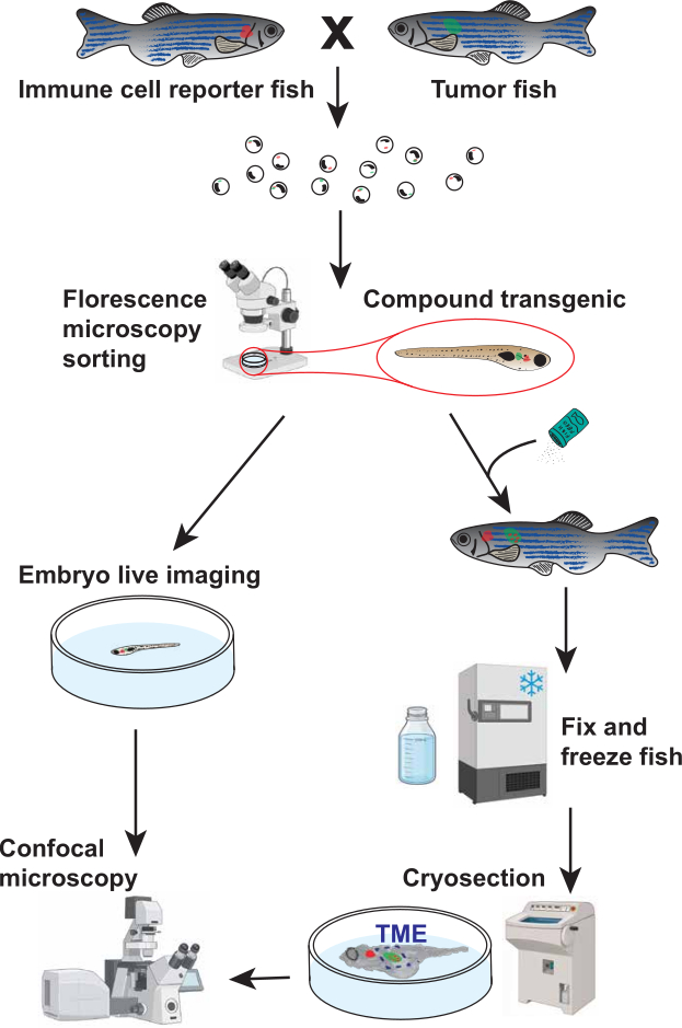
This protocol describes methods for generating compound transgenic zebrafish, imaging immune cells associated with tumors in larval and adult zebrafish, and quantifying immune cell frequency within the tumor microenvironment.
A subscription to JoVE is required to view this content. Sign in or start your free trial.
Method Article
Imaging and Quantification of Immune Cells in the Tumor Microenvironment Using a Zebrafish Model of Neuroblastoma
* These authors contributed equally
In This Article
Summary
Abstract
The recruitment of immune cells to the tumor microenvironment (TME) is an important component of immune regulation and tumor progression. Studying the spatial and temporal relationships between immune and tumor cells in vivo can provide insight into interactions within the TME. However, visualizing and quantifying these interactions in intact organisms remains challenging using conventional imaging and histological approaches. Zebrafish provide a valuable model for cancer research owing to their genetic tractability, rapid development, and optical transparency during early developmental stages, enabling visualization of fluorescently labeled cells in vivo. Here, we present a protocol to generate, identify, image, and quantify immune and tumor cells that are differentially labeled with fluorescent markers in a zebrafish model of neuroblastoma. The protocol includes breeding and screening of compound transgenic zebrafish, live imaging of larvae, preparation and imaging of adult tumor-bearing fish, and quantification of immune cells surrounding and within tumor regions. Using transgenic reporter lines, CD4+ immune cells and MYCN-driven neuroblastoma cells can be visualized and analyzed at different developmental stages. The workflow enables assessment of immune cell localization relative to tumors in both larval and adult fish and can be used to compare immune cell abundance across disease stages. This protocol provides a reproducible approach for imaging and quantifying immune cells within the TME in vivo using zebrafish and may facilitate studies of immune–tumor interactions in other cancer models.
Introduction
The tumor microenvironment (TME) is an ecosystem composed of tumor, immune, fibroblast, and vascular cells1. The TME plays an essential role in cancer progression, therapeutic response, and anti-tumor immunity. Within the TME, immune cells are key regulators of tumor fate, either participating in anti-tumor responses or inducing immunosuppression2. Understanding the recruitment, distribution, and function of immune cells within the TME is critical for elucidating the mechanisms by which tumors evade immune surveillance. However, conventional approaches to studying the TME, such as endpoint histology, flow cytometry, and ....
Access restricted. Please log in or start a trial to view this content.
Protocol
All animal procedures were approved by the Institutional Animal Care and Use Committee (IACUC) at Boston University Chobanian & Avedisian School of Medicine under protocol PROTO202100001. The overall workflow for generation of compound transgenic zebrafish, larval imaging, adult tissue preparation, and image analysis is summarized in Figure 1.
Access restricted. Please log in or start a trial to view this content.
Results
The overall experimental workflow is shown in Figure 1. Adult Tg(cd4-1:mCherry) zebrafish were crossed with Tg(dβh:MYCN;dβh:EGFP) zebrafish to generate compound transgenic offspring in which CD4+ cells and dβh-expressing cells were differentially labeled with mCherry and EGFP fluorescence, respectively. Embryos were screened for the appropriate fluorescent markers and subsequently analyzed by either live confocal imaging during larval developmen.......
Access restricted. Please log in or start a trial to view this content.
Discussion
The protocol described here utilizes zebrafish as an in vivo model to visualize and quantify immune cells within the TME. Several factors are critical for successful implementation of this approach. First, appropriate selection and validation of compound transgenic zebrafish lines are essential. Larvae should be screened for the expected fluorescent expression patterns and, if required, confirmed by genotyping. Immune-reporter and oncogene expression should be verified prior to experimentation to ensure accurate.......
Access restricted. Please log in or start a trial to view this content.
Disclosures
The authors declare no competing interests.
Acknowledgements
We acknowledge support from the National Institutes of Health (NIH; 1UL1TR001430, CA215059, and NS140967), the American Cancer Society (RSG-17-204-01-TBG), the National Science Foundation (1911253), and Alex’s Lemonade Stand Foundation to H.F.; and from a Warren Alpert Distinguished Scholars Fellowship to X.Q. Y.W. was supported by the Boston University Undergraduate Research Opportunities Program (UROP) and the St. Baldrick’s Summer Fellowship Program. M.V. was supported by the St. Baldrick’s Summer Fellowship Program. The content of this article is solely the responsibility of the authors and does not necessarily represent the official views of the....
Access restricted. Please log in or start a trial to view this content.
Materials
| Name | Company | Catalog Number | Comments |
|---|---|---|---|
| 200 µL Gel Loading Tip | MilliporeSigma | 1022-0600 | Used for larval manipulation |
| Artificial Plants | The Hidden Reef | 62874200000000000 | Optional breeding enrichment |
| Bleach | Fisher Scientific | 50371500 | Used for embryo decontamination |
| Cell Counter Plugin | National Institutes of Health (NIH) | N/A | Fiji plugin used for cell quantification |
| Computer and Monitor | HP | 2TB68A8#ABA | Used for image acquisition and analysis |
| Confocal Microscope | Zeiss | LSM 710 | Used for larval and adult imaging |
| Cryomold, Plastic | Epredia | 22-050-160 | Used for OCT embedding |
| Cryostat | Thermo Fisher Scientific | 95-664-0EC70 | Used for cryosectioning |
| Dry Ice | Fisher Scientific | N/A | Used for sample transport |
| Fiji Software | National Institutes of Health (NIH) | Version 1.54p | Image analysis software |
| Fine-Mesh Net | Carolina Biological | 651345 | Used for embryo collection |
| Fish Incubator | VWR | 35960-056 | Maintained at 28.5°C |
| Forceps | Fisher Scientific | 10-316B | Used for sample handling |
| Glass-Bottom Dish | IBIDI | 81218-200 | Used for imaging |
| Low-Melting Agarose | Prolab Supply | 9012-36-6 | Used for larval and adult mounting |
| Mating Tank (main tank, divider, grated section) | Carolina Biological | 161937 | Used for zebrafish breeding |
| Nutating Rocker | Midsci | R2D-30 | Used for fixation during overnight incubation |
| OCT Compound | Tissue-Tek | 4583 | Embedding medium for cryosectioning |
| Paraformaldehyde (PFA) | Sigma Aldrich | 158127-500G | Fixative |
| Petri Dish, Plastic | MilliporeSigma | BR452000 | Used for embryo and larval handling |
| Phosphate-Buffered Saline (PBS) | Gibco | 14080-055 | Used for washing and fixation procedures |
| Plastic Transfer Pipette | Fisher Scientific | 50-998-100 | Used for embryo and larval transfer |
| Razor Blade | Fisher Scientific | 12-640 | Used during sample preparation |
| Conductivity Meter | YSI | EC300ACC-04 | Used to verify rotifer water salinity |
| Rotifers | The Hidden Reef | 116151603 | Used for larval rearing |
| Sodium Thiosulfate | Thermo Fisher Scientific | AAA1762936 | Used for bleach neutralization |
| Sucrose | Fisher Scientific | S25590A | Cryoprotection reagent |
| Tricaine Methanesulfonate | Western Chemical | NC0872873 | Anesthetic; sold by Fisher Scientific |
| Tweezers | Fisher Scientific | 12-000-122 | Used for sample handling |
| Upright Fluorescence Microscope | Leica | M165 | Used for fluorescence screening |
References
- Anderson NM, Simon MC. The tumor microenvironment. Curr Biol. 2020;30(16):R921-R925.
- Kumagai S, Momoi Y, Nishikawa H. Immunogenomic cancer evolution: A framework to understand cancer immunosuppression. Sci Immunol. 2025;10(105):eabo5570.
- Keren L, et al. A structured tumor-immune microenvironment in triple negative breast cancer revealed by multiplexed ion beam imaging. Cell. 2018;174(6):1373-1387.e19.
- Gerner MY, et al. Histo-cytometry: A method for highly multiplex quantitative tissue imaging analysis applied to dendritic cell subset microanatomy in lymph nodes. Immunity. 2012;37(2):364-376.
- Pittet MJ, Weissleder R. Intravital imaging. Cell. ....
Access restricted. Please log in or start a trial to view this content.
Reprints and Permissions
This article has been published
Video Coming Soon