Here, we present a robust method to physically immobilize liquid–liquid phase-separated condensates within a porous low-melting agarose hydrogel matrix without disrupting their internal liquid dynamics. The approach minimizes droplet sedimentation, coalescence, and surface-wetting artifacts, enabling reproducible imaging and quantitative biophysical analysis over periods of hours.
Method Article
Stabilizing Protein and Protein—RNA Condensates in Low-Melting Agarose Hydrogels for Quantitative Biophysical Measurements
In This Article
Summary
Abstract
Biomolecular condensates formed by liquid–liquid phase separation are increasingly recognized as fundamental organizational units of the cell, with roles ranging from RNA processing to stress granule assembly. In vitro reconstitution of such condensates using purified proteins and RNA provides model systems, yet liquid droplets coarsen, sediment, and spread on surfaces over time, introducing significant variability into quantitative measurements over experimental timeframes. Embedding these condensates within a sparse, optically transparent, and chemically inert hydrogel—here prepared from low-melting agarose (LMA) at 0.3–1.0% w/v—stabilizes droplets in three dimensions, enabling the use of methodologies with longer measurement times or experimental setups with repeated measurements. This paper presents a detailed, step-by-step protocol for LMA stock preparation, droplet formation, and embedding, and provides examples of specific downstream biophysical readouts. We also show that specific droplet properties, such as molecular diffusion, are preserved after LMA embedding in the representative systems tested. Since the method is compatible with confocal, widefield, or super-resolution microscopy, fluorescence recovery after photobleaching, or nuclear magnetic resonance spectroscopy, among others, it can support quantitative characterization of biomolecular condensates relevant in health and disease.
Introduction
Liquid–liquid phase separation (LLPS) drives the formation of membraneless organelles and biomolecular condensates in cells, including stress granules, P-bodies, and the nucleolus1,2,3,4,5. These condensates concentrate specific proteins and nucleic acids while maintaining rapid exchange with the surrounding cytoplasm or nucleoplasm – a hallmark of liquid-like behavior1,2,3,4,5. Dysregulation of LLPS has been linked to various diseases, such as neurodegenerative disorders3,6,7 and is also implicated in the replication of several viruses, like SARS-CoV-28,9,10,11,12.
In vitro reconstitution of condensates using purified proteins and RNA under defined buffer conditions has become a cornerstone approach to study LLPS mechanisms3,4. However, droplets in a test tube or on a glass surface are susceptible to three major artifacts: (i) gravitational sedimentation, causing droplets to accumulate at the bottom; (ii) coalescence, where droplets fuse over time into fewer, larger structures; and (iii) surface wetting, where droplets spread and partially adsorb onto the coverslip. These phenomena reduce the reproducibility of quantitative measurements and make it difficult to track individual droplets in their physiologically relevant dimensions13. These phenomena further limit the use of methods that require extended measurement times of minutes to hours (e.g., nuclear magnetic resonance [NMR] spectroscopy or super-resolution microscopy techniques such as STORM) and prohibit repeated measurements over extended timescales.
Physical immobilization within a hydrogel network addresses all three artifacts simultaneously. Agarose forms a physically crosslinked gel at temperatures below 34–38 °C. At concentrations of 0.3–1.0% w/v, the mesh size of agarose is large enough to encapsulate intact micrometer-scale droplets while remaining sufficiently porous not to impede intra-droplet molecular dynamics13. Additionally, the porosity of the agarose matrix permits diffusion of macromolecules and small molecules in and out of the embedded droplets. For particularly temperature-sensitive systems involving proteins with folded domains or following an upper/lower critical solution temperature (UCST/LCST)-type phase separation, low-melting agarose (LMA) with a gelling point of 26–30 °C can be used instead. Alternative anti-wetting approaches, such as surface passivation, can reduce condensate adhesion and thus wetting, but often require specialized preparation and do not prevent droplet sedimentation or coalescence. In contrast, agarose embedding provides a straightforward, broadly accessible method for spatially immobilizing condensates. Importantly, agarose has been validated to not perturb the conformational state of fused in sarcoma (FUS) and other RNA-binding proteins upon droplet formation up to agarose concentrations of 1.0% w/v13. This protocol standardizes the agarose embedding approach for general use with many phase-separating protein or protein-RNA systems of interest, providing step-by-step guidance for reagent preparation and sample preparation.
Protocol
1. Preparation of agarose stock
IMPORTANT: Prepare the agarose stock in the same buffer used to make the phase-separated samples (hereafter called “droplet buffer”) to prevent buffer mismatch14. Example: The nucleocapsid sample is prepared in buffer A (25 mM HEPES, 50 mM NaCl, pH 7.2), which should also be used to prepare the agarose stock.
NOTE: Final experimental conditions, including but not limited to buffer composition, protein concentration, the use of additives, and final sample volumes, depend heavily on the specific biological system under investigation and the specific downstream application. Likewise, acquisition parameters, instrument setup, and data analysis depend strongly on the sample and the specific experiment. For example, the preparation of the SARS-CoV-2 nucleocapsid with poly(A) is described in Figure 1A (confocal fluorescence microscopy).
- Weigh the appropriate amount of LMA or regular agarose into a 1.5 mL tube containing droplet buffer. Example: For a 2% w/v stock, dissolve 20 mg of agarose in 1 mL of buffer A.
NOTE: Final agarose stock concentration should be 1–2% w/v, depending on the desired final agarose concentration in the sample. Exceeding 2% w/v is not recommended as it becomes increasingly difficult to pipette. Cut the narrow end of the pipette tip to make pipetting easier, especially at high agarose percentages. Consult the discussion section to determine if LMA or regular agarose is required for the samples. - Heat the agarose stock to 80–90 °C in a heating block or water bath and fully dissolve the agarose by vortexing (takes approx. 10 min).
CAUTION: Molten agarose is hot. Handle with care. - Equilibrate the agarose stock to 37 °C (LMA) or 55 °C (regular agarose) in the heating block or water bath. Verify the solution remains clear and liquid at this temperature. Verify the temperature of the agarose stock with a thermometer if needed.
PAUSE POINT: Molten agarose stocks can be kept warm for 30–60 min.
Heating the agarose in a water bath is strongly recommended to ensure more homogeneous heating and maintain the target temperature with sufficient precision. This is especially important when working with LMA, as small deviations in temperature can cause premature gelation. - After sample preparation (section 2), store leftover agarose stocks at +4 °C. Follow steps 1.2 and 1.3 above to re-melt and re-equilibrate the agarose stock before use.
NOTE: We recommend keeping the agarose stock for a maximum of 2 weeks at +4 °C and subjecting it to no more than 2–3 solidify/melt cycles. Use good manufacturing practices to prevent contamination. Discard stocks earlier if they fail to re-melt or re-solidify, or if visible contamination occurs.
2. Sample preparation
- Prewarm all materials that come into direct contact with the agarose (e.g., pipette tips, tubes, microscopy slides) to approximately 37 °C by placing them on the heating block or in an incubator for 2–3 min.
- Prepare the agarose stock as outlined in Section 1.
- Prepare droplet buffer, the phase-separating components (e.g., protein or RNA stocks), and any other additives (e.g., crowding agents, dyes) for the samples according to the standard protocol. Equilibrate to room temperature (RT). Example: Buffer A (25 mM HEPES, 50 mM NaCl, pH 7.2), nucleocapsid protein stock (60 µM, in buffer A), poly(A) stock (30 µM, in buffer A).
NOTE: For fluorescence recovery after photobleaching (FRAP), confocal fluorescence microscopy, or stochastic optical reconstruction microscopy (STORM), add a small fraction of fluorescently labeled protein or RNA to the protein or RNA stocks, respectively (recommended: <5% labeled fraction). - Calculate the volumes of droplet buffer, agarose stock, the phase-separating components (e.g., protein or RNA of interest), and any supplementary additives (e.g., crowding agents) needed for the sample. Example: 6 µL of buffer A, 5 µL of 2% agarose stock (final 0.5%), 5 µL of nucleocapsid protein (final 15 µM), 4 µL of poly(A) (final 6 µM). Final volume 20 µL.
NOTE: While highly dependent on the dish, tube, plate, or other vessel used for the experiment, commonly used volumes are 150 µL for a 3 mm NMR tube, 20 µL for microscopy or FRAP experiments in a 6-channel microscopy slide, and 20–30 µL for STORM experiments in 18-well microscopy slides (see the Table of Materials).- As starting values, use final 0.3–0.5% w/v agarose for standard biophysical assays, and 0.8–1.0% w/v for long-term stabilization (e.g., aging experiments).
NOTE: For long-term stabilization and storage of samples, measures should be taken to prevent sample dehydration (e.g., sealing the sample container, storing in a humidified chamber, and adding an evaporation barrier) and to ensure that buffer composition and pH remain stable during prolonged incubations. - For NMR samples, add final 5% v/v D2O to the sample for locking.
NOTE: The concentration of all other components and the total sample volume depend heavily on the specific application and experimental setup.
- As starting values, use final 0.3–0.5% w/v agarose for standard biophysical assays, and 0.8–1.0% w/v for long-term stabilization (e.g., aging experiments).
- Add the corresponding volumes of buffer, phase-separating components, agarose stock, and additives into a prewarmed 1.5 mL tube.
NOTE: We recommend adding the buffer and agarose first, followed by any supplementary additives. We further recommend adding the phase-separating components last to trigger condensate formation. However, the optimal mixing order depends on the system and should be determined in advance. This is especially relevant for systems in which stoichiometry and the order of addition can strongly influence droplet size or compartmentalization. Example: The mixing order for the nucleocapsid sample is first Buffer A, second agarose stock, third nucleocapsid protein, and last poly(A), using the volumes noted in Step 2.4. In this sample, LLPS is triggered by the addition of poly(A). For systems in which LLPS produces visible turbidity, turbidity can serve as a qualitative QC indicator; however, onset time and visibility are system-dependent, and lack of immediate turbidity does not by itself indicate protocol failure.- Keep the tube in the heating block or water bath while pipetting up and down a few times. Avoid introducing air bubbles.
NOTE: While the agarose is still liquid, droplets can fuse. Thus, varying the pre-incubation time on the heating block or in a water bath prior to initiating gelation can be a useful tool for investigating droplets of different sizes. Characterizing droplet growth kinetics in the absence of agarose prior to selecting the pre-incubation time interval allows investigators to reproducibly target a specific droplet size range.
- Keep the tube in the heating block or water bath while pipetting up and down a few times. Avoid introducing air bubbles.
- For microscopy / FRAP samples:
- Use a normal pipette to quickly transfer the sample into/onto the microscopy slide of choice (e.g., channel microscopy slide with coverslip bottom).
- Allow gelation at RT for 5–10 min (see note in discussion section).
NOTE: Do not move the microscopy slide during this period. Protect fluorescent samples from light. - Measure the sample according to the standard protocol. Example: spinning disk confocal microscope, 100x 1.45 plan apo oil objective, excitation wavelength 561 nm for Cy3 dye, acquisition 100 ms at 30% laser power (Cy3) and 50 ms at 6% light intensity (brightfield). For FRAP: Bleach region 1/10th of droplet diameter placed in the middle of the droplet, 25 prebleach frames (acquisition with no delay), 60 post-bleach frames (taken in 1 s intervals), laser pulse for bleaching 15 ms at 70% laser power. Analyze the FRAP recovery by full-scale normalization in open-access software; report kinetic fitting parameters only if fitting is performed.
- For STORM samples:
- Use a normal pipette to quickly transfer the sample into the STORM slide of choice (e.g., 18-well chamber slide with coverslip bottom). Dispense the sample as a drop.
- Allow gelation at RT for 5–10 min (see note in discussion section).
NOTE: Do not move the microscopy slide during this period. Protect fluorescent samples from light. - Fill the entire well with STORM buffer. Seal the well according to the standard protocol.
- Equilibrate the sample for at least 1–2 h prior to measurement to allow the STORM buffer to diffuse into the agarose hydrogel.
NOTE: Successful diffusion of the STORM buffer into the sample can be easily verified if the fluorescent dyes blink during STORM acquisition. Depending on the sample specifications, longer equilibration times might be needed. - Measure the sample according to the standard protocol used. Example: Ti2 inverted microscope, SR apochromat TIRF 100x 1.49 oil objective, highly inclined thin illumination. Acquisition of 20,000 frames per sample, field of view 64 x 64 pixels, exposure time 10 ms with a 125 mW 647 nm laser for Cy5 dye, and using a 20 mW 405 nm laser to assist the transition from the long-lived dark state into the ground state. Images were processed using maximum likelihood estimation with multi-emitter fitting. Drift correction was applied, and only data points with localization precision of 25 nm or better were included in the final visualization. Final images are produced using the average-shifted-histograms method at 20x magnification (1 pixel = 8 nm in the final image).
- For NMR samples:
- Use a long, thin glass pipette (prewarmed) to rapidly transfer the sample into the NMR tube. Dispense the sample at the bottom of the tube.
- Allow gelation at RT for 5–10 min (see note in discussion section).
NOTE: Do not shake the NMR tube during this period. - Measure the sample according to the standard protocol used. Example: Field strength of NMR 750 MHz with PATXI room temperature probe, 3 mm tube, acquisition at 25 °C, standard pulse sequence (stebpgp1s19) for diffusion (DOSY) experiments, parameters: 4,096 points with 32 scans, gradient strength 2–95%, diffusion time 0.08 s, gradient pulse 12 ms, relaxation delay (d1) 5 s.
Results
The protocol was applied to condensates formed by the FUS N-terminal domain (FUS NTD) or by the SARS-CoV-2 nucleocapsid (N) protein with poly(A) or s2m RNA as representative examples of both protein-only and protein-RNA droplets. To assess whether agarose detectably affects condensate formation under the tested conditions, we formed droplets with N protein and poly(A) in the presence and absence of 0.5% w/v LMA. Since LMA solidifies after approximately 5 min at RT, we recorded fluorescence microscopy images at this time point and observed that droplets were roughly the same size in both cases (Figure 1A,B). To demonstrate the preventive effect of agarose on droplet wetting and fusion, we recorded another set of images 60 min after sample preparation. While the agarose-embedded droplets did not change in size, condensates in the absence of LMA showed a drastic increase in size due to fusion and surface wetting (Figure 1A,B). While we embedded the droplets relatively early after nucleation, droplets of different sizes can be embedded by varying the pre-embedding incubation time. Despite embedding the droplets in agarose hydrogel, the measured condensate dynamics were not detectably altered. FRAP recovery and, therefore, apparent droplet diffusion were highly similar with and without LMA in the sample (Figure 1C). Furthermore, previous diffusion-ordered NMR spectroscopy (DOSY) experiments with FUS NTD condensates have shown that diffusion rates were not detectably influenced by agarose concentration, at least over the range 0.25–1% w/v (Figure 1D) (adapted from Emmanouilidis et al.13). These results demonstrate that an agarose hydrogel is suitable for trapping phase-separated condensates in space and time, enabling repeated measurements over extended time frames.
While both microscopy and FRAP measurements are fairly quick to record (seconds to minutes), some experiments require several minutes (e.g., STORM, 1D 1H NMR) or even hours (e.g., 2D [15N 1H] HSQC NMR) to acquire. For such experiments, it is crucial that droplets remain stationary throughout acquisition and do not sediment over time. Here, we show that we stabilized N protein + s2m RNA droplets in LMA for long enough to acquire STORM images (Figure 1E). Furthermore, NMR spectra of FUS NTD droplets in the presence and absence of agarose had comparable line widths, suggesting no detectable interaction between agarose hydrogels and the protein under these conditions (Figure 1F) (adapted from Emmanouilidis et al.13). In contrast, spectra of agarose-embedded condensates showed higher signal intensity, likely because non-stabilized droplets sedimented in the NMR tube during acquisition and were thus no longer available for measurement (Figure 1F) (adapted from Emmanouilidis et al.13). In conclusion, stabilization of condensates in agarose hydrogels not only enables time-course experiments but also allows the use of methodologies with extended acquisition times, while preserving the measured droplet properties in the representative systems tested.
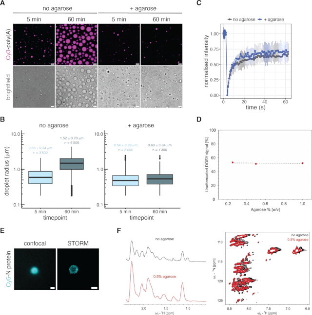
Figure 1: Condensate stabilization by agarose embedding without detectably perturbing measured droplet properties. (A) Fluorescence microscopy of 15 µM nucleocapsid protein with 6 µM Cy3-poly(A) (5% labeled) in buffer A (25 mM HEPES, 50 mM NaCl, pH 7.2) in the presence (+ agarose) or absence (no agarose) of 0.5% w/v LMA. Images were acquired 5 min or 60 min after mixing / agarose embedding. Images are representative of 7 images acquired in n = 2 independent experiments. Scale bar = 5 µm. (B) Quantification of the droplet radii of the condensates shown in (A). Data are displayed as boxplots (box = interquartile range, center line = median, whiskers = range of remaining data, point = outliers). Average radius ± standard deviation (sd) and number of analyzed droplets in each condition are shown directly on the graph. Data follow a normal distribution. Individual droplets were not treated as independent biological replicates for statistical inference. No statistical test was performed; distributions are shown descriptively. (C) FRAP of the droplets shown in (A) at time point 5 min. Photobleaching was performed with the 561 nm laser (Cy3). Data show the normalized intensity (mean ± sd of n = 3 independent replicates per condition), which is obtained by normalizing the bleached signal to the pre-bleach values, an unbleached area of the same sample, and the background fluorescence. (D) Unattenuated DOSY signal of 200 µM FUS NTD protein in buffer B (30 mM HEPES, 200 mM KCl, 600 mM urea, pH 7.3) in the presence of 0.25, 0.5, or 1.0% w/v regular agarose. N = 1. (E) Confocal fluorescence microscopy and STORM images of 15 µM Cy5-nucleocapsid protein (5% labeled) with 6 µM viral s2m RNA in buffer A containing 0.5% w/v LMA. Representative of n = 3 independent experiments. Scale bar = 1 µm in both images. (F) 1D 1H NMR spectra (left) and 2D [15N 1H] HSQC NMR spectra (right) of 200 µM FUS NTD in buffer B in the presence or absence of 0.5% w/v regular agarose. N = 1. Panels (D) and (F) are reproduced from Emmanouilidis et al.13 Please click here to view a larger version of this figure.
Discussion
The success of agarose-embedding of biomolecular condensates relies on several factors. First, agarose must be maintained at temperatures that are high enough to prevent premature gelation, and at the same time low enough not to perturb biomolecular structures or interactions. Low-melting agarose has a gelling point of 26–30 °C and a melting point of 60–65 °C, while regular agarose has a gelling point of 34–38 °C and a melting point of 90–95 °C. Second, rapid mixing and transfer into the final reaction vessel (e.g., NMR tube) are critical, as small deviations in timing or temperature can lead to uneven embedding and thus compromised reproducibility.
Despite these critical points, the method can be readily adapted to different experimental needs. For phase-separating systems with intrinsically disordered proteins or systems that are not prone to heat denaturation, regular agarose can be used instead of LMA to save cost. When preparing samples containing proteins with folded domains, or any other temperature-sensitive (bio-)molecule prone to heat denaturation, LMA should be used to prevent denaturation. A heat-only/no-agarose control sample can be prepared to separate the effects of agarose embedding from the effects of transient sample warming. To reduce the risk of those temperature-dependent denaturation effects, low-melting agarose is particularly suitable for temperature-sensitive samples, while agarose concentration can be adjusted to modulate mesh size and the degree of mechanical confinement. For highly sensitive diffusion measurements, such as the recently developed NMR method called LLPS REDIFINE15,16, the lowest agarose concentration that prevents sedimentation is usually preferable, typically 0.3–0.5% w/v. For maturation experiments requiring weeks to months of stability, 0.8–1% w/v is preferable. These parameters should be optimized to ensure effective stabilization without detectably altering condensate morphology or dynamics and should always be adapted to the specific system. This validation requires comparison of samples with and without agarose embedding (and otherwise identical conditions) or comparison of different agarose concentrations (to exclude concentration-dependent mesh effects), for example, using microscopy to assess droplet morphology, FRAP to assess diffusion, or any other method suitable to assess droplet properties important to the investigator.
Gelation time is another important consideration. The time it takes for agarose to solidify depends on several factors, such as the type of buffer, pH, final agarose concentration, starting temperature, and whether low-melting or regular agarose is used. Typically, the agarose solidifies within 5–10 min. The gelation of the agarose stock can be used as an indication for the gelation of the sample. Similarly, varying the pre-incubation times prior to agarose embedding enables reproducible characterization of different droplet sizes. Buffer choice is also critical14. It is vital that the agarose stock is prepared in the same buffer used to prepare the samples to prevent buffer mismatch. The buffer should be one in which the system is known to phase separate.
Temperature sensitivity should be considered for each system. Many phase-separating systems exhibit thermo-reversible phase behavior, known as UCST/LCST-type LLPS. Therefore, brief exposure to slightly elevated temperatures during agarose mixing should be validated to ensure that it does not perturb phase behavior or the measured droplet properties. If issues arise, they can typically be addressed by refining temperature conditions, minimizing handling time, or adjusting agarose concentration. For example, although the LMA stock is ideally heated to 37 °C, LMA stocks will still remain liquid at 33–35 °C.
Troubleshooting is important because different failure modes may arise depending on the sample and experimental setup. If droplets dissolve upon mixing with agarose, this may be caused by LMA being too warm or by buffer mismatch. In this case, the LMA stock should be maintained exactly at or a few degrees below the target temperature, and prepared in an identical buffer to the protein/RNA of interest. If premature gelation occurs in the pipette tip, it may be due to the LMA being too cold or the tips not being pre-warmed. To avoid this, tips and tubes should be prewarmed to 37 °C, the operator should work quickly, and the heating block should be kept at the target temperature. If wetting, coalescence, or sedimentation occurs over time, it may be due to the gel being too sparse or to uneven gelation. In this case, the LMA concentration can be increased, and even gelation at room temperature should be ensured. If the agarose solution contains visible particles, it may be due to incomplete agarose dissolution. In this case, the agarose should be heated until fully transparent and mixed thoroughly before use (e.g., vortexed). If air bubbles are introduced during mixing or pipetting, these may interfere with imaging and lead to uneven condensate distribution. To minimize bubble formation, solutions should be mixed gently by slowly pipetting up and down. If heterogeneous gelation or uneven condensate distribution is observed, it may be due to insufficient mixing or local temperature gradients during gelation. In this case, samples should be kept on the heating block (or water bath) until all components are added, mixed immediately but gently, and allowed to solidify under uniform temperature conditions. Steep temperature gradients should be avoided; solidification at room temperature is recommended (and solidification in the fridge should be avoided). In long-term experiments, sample evaporation may alter solute concentrations and condensate properties. To minimize evaporation, samples should be sealed appropriately and maintained in a humidity-controlled environment whenever possible.
In contrast to methods such as paraformaldehyde fixation, agarose embedding does not chemically fix samples. Therefore, condensates remain dynamic, which is advantageous for functional studies but limits long-term storage. For extended experimental time frames, precautions must be taken to prevent evaporation and buffer changes, such as pH shifts. Additionally, while agarose is generally low-interacting in these assays, potential indirect effects on diffusion or the local environment should be considered. If agarose–biomolecule interactions are suspected, such as non-specific binding of the protein to agarose, alternative hydrogel matrices such as photo-crosslinked polyethylene glycol diacrylate (PEG-DA), with matching mesh size and porosity, can be considered. The absence of interactions should be validated by measuring FRAP in the presence and absence of the matrix.
In addition to chemical fixation, surface passivation strategies such as BSA-, PEG-, or surfactant-coated surfaces and supported lipid bilayers are commonly used to reduce condensate adhesion and wetting. While these approaches effectively minimize surface interactions, they do not prevent condensate movement, sedimentation, or fusion. Agarose embedding, therefore, complements these methods by providing physical immobilization while maintaining condensate dynamics, making it particularly useful for prolonged experiments, three-dimensional imaging, and quantitative measurements that require stable sample positioning. The choice of method should therefore be guided by the experimental objective.
Overall, agarose hydrogels provide a simple yet effective means to stabilize condensates in three dimensions. This prevents droplet wetting and fusion without detectably perturbing the measured intrinsic properties in the representative systems tested, enabling time-course experiments and techniques requiring long acquisition times, such as NMR spectroscopy15. While this method might not be suitable for all phase-separating systems for a number of reasons, the method presented here is applicable to the study of several biomolecular condensates and can be particularly useful for investigating maturation processes relevant to neurodegenerative disease mechanisms17.
Disclosures
The authors have no conflicts of interest to declare.
Acknowledgements
This work was supported by Swiss National Science Foundation (SNSF) grants 310030-215555 (F.H.-T.A.), 4078P0_198253 (F.H.-T.A.), CRSII5_205922 (F.H.-T.A.), 205321_204920 (F.H.-T.A.), NCCR RNA & Disease grant 51NF40-182880 (F.H.-T.A.), and EMBO Long-Term Fellowship LTF-388-2018 (L.E.).
Materials
| Name | Company | Catalog Number | Comments |
|---|---|---|---|
| 1.5 mL tubes | any brand is OK | N/A | |
| 18-well chamber slides | ibidi | 81816 | for STORM |
| agarose, low gelling temperature | Sigma-Aldrich | A9414 | molecular biology grade |
| agarose, ultra pure | Invitrogen | 16500500 | |
| channel slides | ibidi | 80606 | for microscopy |
| confocal microscope | Nikon | N/A | |
| Cy5-dye, NHS chemistry | Jena Biosciences | FP-201-CY5 | for labeling protein |
| D2O (deuterium oxide) | Sigma-Aldrich | 151882 | |
| easyFRAP | Koulouras et al. 2018 | N/A | https://pubmed.ncbi.nlm.nih.gov/29901776/ |
| Everspark 2.0 dSTORM buffer | idylle | KMO|ETE+ | |
| FUS NTD protein | N/A | N/A | expressed recombinantly in our lab, as described in ref 13 (Emmanouilidis et al. Nat Chem Biol 2021) |
| heating block or water bath | any brand is OK | N/A | any make or model will do |
| HEPES | Sigma-Aldrich | H3375 | |
| image analysis software | Fiji | N/A | https://fiji.sc/ |
| KCl | Sigma-Aldrich | P5405 | |
| NaCl | Sigma-Aldrich | S9888 | |
| NMR spectrometer | Bruker | N/A | 750 MHz, PATXI room-temperature probe, see ref 13 for more details (Emmanouilodis et al. Nat Chem Biol 2021) |
| NMR tube | Bruker | N/A | 3 mm, glass |
| P10, P20, P200 and P1000 pipettes | any brand is OK | N/A | |
| pCp-Cy3-dye | Jena Biosciences | NU-1706-CY3 | for labelling RNA |
| poly(A) RNA | N/A | N/A | in-house in vitro transcribed in our lab, length approx 50 nucleotides, as described in Kathe et al. Mol Biol Cell 2024 |
| s2m RNA | N/A | N/A | in-house in vitro transcribed in our lab, length 45 nucleotides, sequence: 5'-GGUUCACCGAGGCCACGCGGAGU ACGAUCGAGUGUACAGUGAACC-3', as described in Kathe et al. Mol Biol Cell 2024 |
| SARS-CoV-2 nucleocapsid protein | N/A | N/A | expressed recombinantly in our lab, as described in Kathe et al. Mol Biol Cell 2024 |
| software for plotting (e.g. R) | R-Project | Version 4.2.1 | https://www.r-project.org/ |
| STORM microscope | Nikon N-STORM | N/A | |
| ThunderSTORM plugin | Ovesny et al. 2014 | N/A | https://pmc.ncbi.nlm.nih.gov/articles/PMC4207427/ |
| tips for the used pipettes | any brand is OK | N/A | |
| TopSpin | Bruker | Version 3.2 and 4.1 | |
| urea | Sigma-Aldrich | U5128 |
References
- Alberti S. Phase separation in biology. Curr Biol. 2017;27(20):R1097-102.
- Rabouille C, Alberti S. Cell adaptation upon stress: the emerging role of membrane-less compartments. Curr Opin Cell Biol. 2017;47:34-42.
- Boeynaems S, et al. Protein phase separation: a new phase in cell biology. Trends Cell Biol. 2018;28(6):420-35.
- Hyman AA, Weber CA, Jülicher F. Liquid-liquid phase separation in biology. Annu Rev Cell Dev Biol. 2014;30:39-58.
- Banani SF, Lee HO, Hyman AA, Rosen MK. Biomolecular condensates: organizers of cellular biochemistry. Nat Rev Mol Cell Biol. 2017;18(5):285-98.
- Patel A, et al. A liquid-to-solid phase transition of the ALS protein FUS accelerated by disease mutation. Cell. 2015;162(5):1066-77.
- Shen Y, et al. The liquid-to-solid transition of FUS is promoted by the condensate surface. Proc Natl Acad Sci U S A. 2023;120(33):e2301366120.
- Brocca S, Grandori R, Longhi S, Uversky V. Liquid-liquid phase separation by intrinsically disordered protein regions of viruses: roles in viral life cycle and control of virus-host interactions. Int J Mol Sci. 2020;21(23):9045.
- Scoca V, Di Nunzio F. Membraneless organelles restructured and built by pandemic viruses: HIV-1 and SARS-CoV-2. J Mol Cell Biol. 2021;13(4):259-68.
- Su J, Wilson M, Samuel C, Ma D. Formation and function of liquid-like viral factories in negative-sense single-stranded RNA virus infections. Viruses. 2021;13(1):126.
- Etibor TA, Yamauchi Y, Amorim MJ. Liquid biomolecular condensates and viral lifecycles: review and perspectives. Viruses. 2021;13(3):366.
- Guseva S, et al. Measles virus nucleo- and phosphoproteins form liquid-like phase-separated compartments that promote nucleocapsid assembly. Sci Adv. 2020;6(14):eaaz7095.
- Emmanouilidis L, et al. NMR and EPR reveal a compaction of the RNA-binding protein FUS upon droplet formation. Nat Chem Biol. 2021;17(5):608-14.
- Kathe NC, Novakovic M, Allain FHT. Buffer choice and pH strongly influence phase separation of SARS-CoV-2 nucleocapsid with RNA. Mol Biol Cell. 2024;35(5):ar73.
- Novakovic M, Schmoll J, Emmanouilidis L, Allain FHT. Using solution NMR to characterize biomolecular condensates under biphasic conditions. J Vis Exp. 2026;(230):e70530.
- Novakovic M, et al. LLPS REDIFINE allows the biophysical characterization of multicomponent condensates without tags or labels. Nat Commun. 2025;16(1):4628.
- Emmanouilidis L, et al. A solid beta-sheet structure is formed at the surface of FUS droplets during aging. Nat Chem Biol. 2024;20(8):1044-52.
Reprints and Permissions
