Advances in deep brain stimulation (DBS) technology now allow electrophysiological signals to be recorded from implanted electrodes in addition to providing therapeutic stimulation. Long-term intracranial DBS recordings can characterize the neural effects of pharmacological agents, such as sleep-promoting medications, providing a unique opportunity to understand their circuit-based mechanisms of action.
Method Article
Leveraging Longitudinal DBS Recordings to Probe Pharmacological Mechanisms: Investigating the Role of Orexin Antagonists on Extended Amygdala Activity
In This Article
Summary
Abstract
Advances in deep brain stimulation (DBS) technology now allow electrophysiological signals to be recorded from implanted electrodes in addition to providing stimulation. These intracranial recordings provide a rare opportunity to measure brain activity in human subjects over time in relation to symptoms and naturalistic behaviors. Objective biomarkers can be used to identify the circuit-level mechanisms of action of treatment interventions, such as pharmacological agents, in human subjects. Here, the authors describe methods for conducting long-term electrophysiological recordings in naturalistic settings during pharmacological interventions to characterize the neural effects of psychotropic medications. The authors present an illustrative case of a patient receiving DBS for their severe, refractory OCD with co-morbid insomnia in which beta activity was being longitudinally monitored from the bed nucleus of the stria terminalis (BNST). The authors find that dose reductions of a sleep-promoting orexin antagonist for insomnia, taken at night, significantly elevated the BNST beta biomarker of arousal overnight. This case study demonstrates a role for orexin signaling in regulating arousal-related BNST activity and highlights the utility of DBS recordings for identifying circuit-based mechanisms of action of pharmacologic agents.
Introduction
Dysregulated brain electrophysiological activity lies at the core of the pathophysiology of many neuropsychiatric disorders. Pharmacological agents for treating these conditions must therefore act by modifying the brain’s endogenous electrophysiological activity to change symptoms and behavior. However, the methods that authors have for monitoring brain electrical activity remain limited. Currently, functional imaging is the primary modality for characterizing the neural effects of pharmacological agents in human subjects. While functional imaging offers the benefits of being non-invasive and spatially comprehensive, it often utilizes metabolic activity as a proxy measure for electrophysiological activity and is limited in its temporal sampling, capturing only a brief snapshot of brain activity at specific instances in time. For this reason, methods capable of directly monitoring brain electrical activity across the timescales of naturalistic symptoms and behaviors would greatly aid in the ability to understand the mechanisms of pharmacological agents.
Deep brain stimulation (DBS) is a targeted circuit-based treatment that is often used to treat refractory neuropsychiatric disorders, such as movement disorders, epilepsy, and obsessive-compulsive disorder (OCD)1,2. DBS involves electrical stimulation delivered through electrodes implanted in deep brain structures to modulate dysregulated neural circuits. Advances in DBS technology now allow local field potential (LFP) signals to be recorded from electrodes in addition to providing stimulation, allowing for the longitudinal measurement of brain activity using chronic intracranial recordings1,2,3,4. These electrophysiological signals can be used to identify objective biomarkers of disease-specific symptoms and therapeutic responses5,6,7. Currently, these clinical biomarkers have been used to develop adaptive DBS, which adjusts stimulation parameters in response to detected shifts in pathological brain activity3,6,8. These electrophysiological recordings can also be utilized to identify neural correlates of cognitive and behavioral states over the course of days to months6,8,9.
In addition to characterizing the impact of neuromodulatory interventions, intracranial recordings provide a rare opportunity to continuously characterize electrophysiological changes in activity in response to pharmacological agents in naturalistic settings. For example, it has been observed that dopamine replacement medications in Parkinson’s disease can suppress pathological cortico-basal ganglia beta oscillations, which are associated with improvements in motor symptoms10. Similar approaches can be utilized to characterize the circuit-based mechanisms of action of medications in other neuropsychiatric conditions, such as sleep disorders8,11.
Here, the authors describe a protocol for acquiring longitudinal intracranial electrophysiology data using the Medtronic Percept BrainSense recording system, in parallel with clinical symptom measures and wearable sensor-based behavior measures under naturalistic conditions. Currently, commercially available DBS devices can continuously record brain activity within a frequency-limited band for several weeks. This protocol assumes that the spectral frequency of the biomarker of interest is already known through prior investigational efforts. In this case study, researchers chronically record electrophysiological activity related to circadian sleep-wake activity and the effects of the orexin antagonist lemborexant. However, the methods described here are general and can be applied widely across other symptom/behavioral domains and classes of pharmacological agents.
Protocol
This protocol has been reviewed and approved by the University of California, San Francisco (UCSF). The data presented here are from an individual with severe, treatment-refractory OCD and co-morbid insomnia undergoing deep brain stimulation (DBS) for OCD using an implantable pulse generator (IPG) approved under the Humanitarian Device Exemption. Written informed consent was obtained from the participant for a UCSF Institutional Review Board (IRB)-approved protocol (IRB: 20-31239 ) permitting the collection of chronic intracranial neural recordings, wearable physiological data, and self-report behavioral measures. This individual’s insomnia was being treated with the sleep-promoting medication lemborexant. Lemborexant is a dual inhibitor of the orexin-1 and orexin-2 receptors. Here, the authors characterize the effects of decreasing lemborexant from 10 mg to 5 mg on intracranial beta signals recorded within the bed nucleus of the stria terminalis (BNST) as this participant was being weaned off the medication following a period of improvement in his insomnia.
1. Device setup for longitudinal intracranial electrophysiological recordings
- Access the BrainSense programming feature within the clinician programmer to set up the recording configuration. This can be accessed from the Stimulation settings under the BrainSense tab. Click the ‘Setup’ button and then the ‘Run Signal Test’ icon.
- On the ‘Sensing Setup- Electrode Configuration’ screen, select the bipolar recording contacts. Recordings can only be acquired with monopolar stimulation, and recording contacts must flank the monopolar stimulation contacts.
- Specify the center frequency that you want to record from.
NOTE: The BrainSense timeline recordings will longitudinally collect the power over 10-minute intervals within a predefined 5 Hz frequency band centered at the specified frequency. The IPG can store timeline data for about a month. Then click ‘Save and Exit’ and end the session.
2. Log of medication schedule and dose adjustments
- Document the medications that a participant is taking, including the name of the medication that they are taking, dose, frequency, time, and date of administration.
- Create a structured online database that allows a user to record the time when they are taking their medications. Record this information throughout the DBS recording period to enable temporal alignment of pharmacological events with the neural data.
- For every change to an existing medication, the researcher logs the type of change (initiation, dose increase, dose decrease, or discontinuation), the date of the change, the new dose and frequency, and the clinical rationale.
3. Acquiring online self-report behavioral measures
- Create an online database to log self-reported changes in symptoms.
NOTE: For many neuropsychiatric conditions, repeated self-reported measures of symptom change can be acquired in parallel with intracranial recording data following a medication adjustment. These more densely collected self-report measures can complement clinician-administered measures collected at fixed intervals. - Ask the participant to complete online questionnaires of self-reported symptoms in parallel with intracranial recording data following a medication adjustment. These measures should be short to reduce the burden on study participants.
4. Collection of wearable sensor data
- Provide the participant with a wearable sensor that can be used to passively collect behavioral data.
NOTE: Many sensors collect actigraphy data using accelerometers, heart rate using pulse oximetry, or autonomic measures using skin conductance, and stream these data to a personal phone or computer via a device-specific application, which can be accessed by the study team. - Collect wearable sensor data in parallel with intracranial recording measures. It is important to ensure that these wearable data can be synchronized with the DBS recording data in a timely manner.
5. Data integration and analysis aligned to medication events
- Following each scheduled visit, download ambulatory timeline data from the Medtronic Percept IPG. To do this, go to the ‘Reports’ screen of the DBS programming software on the clinician programmer.
- Select ‘Export Json Session Data’ to generate the JSON data containing the 10-minute average power timeline data over several weeks.
- Convert these files from their native JSON format and extract into MATLAB-compatible .mat structures for offline processing.
- Convert timestamps from coordinated universal time (UTC) to local time to ensure accurate alignment with local medication administration times and wearable sleep data.
- Sort individual recording files chronologically, then concatenate into a single continuous LFP power timeline, screen to remove missing data epochs prior to analysis.
- Check the quality of the LFP neural data by looking for any sudden shifts in the collected timeline data that can be attributed to changes in stimulation settings or impedance.
NOTE: Many regions of the brain exhibit regular circadian changes in neural activity, as reflected in the timeline data, which can be used to assess recording stability over time.
6. Epoch definition relative to medication change events
- Align the LFP timeline data relative to the medication adjustments to identify the neural effects of pharmacological changes.
- Factor in pharmacokinetic and pharmacodynamic considerations to ensure that there is an adequate amount of time for the drug to both build up to steady state and have an effect on neural circuits.
- Perform statistical analysis by comparing the LFP power timeline distribution before and after medication adjustments.
- To develop neural biomarkers of symptoms and behavior, identify periods of overlap between the LFP timeline data and the acquired self-report surveys and wearable sensor data.
- Correlate the LFP timeline data from these periods with self-report and wearable sensor data to develop neural biomarkers.
- Align the self-report and wearable data relative to the change in medication. Determine if there are any changes in the acquired self-report surveys and wearable sensor data in response to the changes in medication.
7. Circadian and time-of-day analysis
- Statistically model the variation in the collected LFP timeline data.
NOTE: Many regions of the brain have a circadian pattern of activity. Also, medications are taken at a specific time of day. For this reason, the authors recommend aligning the electrophysiology data by time of day or time to determine whether activity changes before and after the medication change. For example, a sinusoidal linear regression model can be used to characterize circadian variation in the neural data and capture any changes due to medication changes. A non-parametric permutation test examining daily average power, by permuting labels for days before and after medication changes, can also be used to assess the significance of observed differences in LFP activity. Alternatively, a nonparametric Wilcoxon rank-sum test controlling for time of day can be used to identify specific time points during the day when neural activity changes with medication changes.
Results
Chronic intracranial LFP recordings were obtained from the right BNST in a participant with treatment-refractory OCD implanted with bilateral DBS leads via the Medtronic Percept IPG (Figure 1). This individual also had insomnia that was being treated with the medication lemborexant taken in the evenings to promote sleep. Based upon the prior studies, the recording contact used for all subsequent analyses captured striato-limbic beta-band activity (23.44 ± 2.5Hz) within the right BNST because of its strong circadian and sleep-wake dependence8,11 (Figure 2). The participant was also provided with a wearable ring that allowed the researchers to monitor his sleep/wake status, and he completed an online Pittsburgh Sleep Quality Index (PSQI) questionnaire, capturing his daytime self-reported arousal level (Figure 2). As the authors previously described, intracranial BNST beta activity demonstrates circadian variation with beta being higher during the day and lower during the night, consistent with the participant’s sleep-wake cycle11.
To characterize the pharmacologically driven shifts in intracranial BNST beta power, they looked for changes in circadian beta activity immediately before and after the reduction in orexin antagonist dose (Figure 3). Researchers statistically analyzed the 30-day period prior to and after the dose reduction. Following the dose reduction, there was a significant increase in beta power taking into account the circadian variation (p = 1.76 × 10⁻²⁵, t tests: H0:βmed_red = 0) and in daily average beta power (p = 0.003, permutation test) (Figure 4). During the 30-day period of time before and after the dose reduction, there was no significant change in the average weekly PSQI (PSQIpre = 4 ± 1.1, PSQIpost = 4 ± 0.25; ±sem, n = 4).
Given its strong circadian dependence, the authors wanted to determine if there was a specific time of day when the intracranial BNST beta power was elevated following the dose reduction of the orexin antagonist. In general, they found that intracranial BNST beta power was significantly elevated when the participant was asleep overnight and during an afternoon period around 2–3 pm when he would take a nap following the dose reduction (Figure 5, p < 0.05, Wilcoxon rank-sum test). However, the beta was not significantly different during the mornings and other periods of wakefulness during the day.
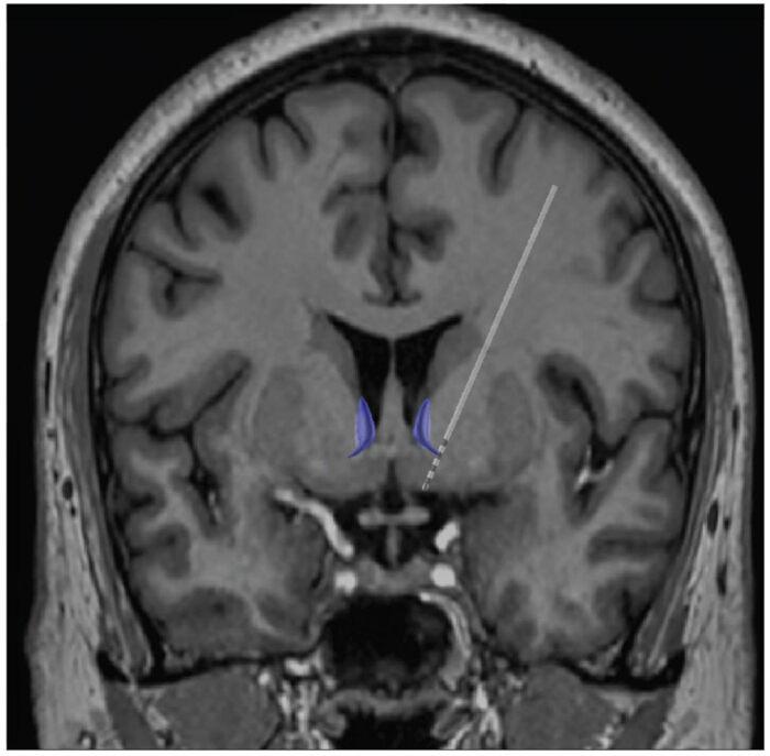
Figure 1: Localization of DBS recording lead. The recording electrode localization was positioned adjacent to the BNST (purple). Please click here to view a larger version of this figure.
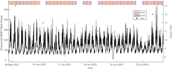
Figure 2: Longitudinal striato-limbic beta power aligned to self-report surveys and wearable sensor monitoring. Continuous DBS recordings were acquired over several days. Participant logged their sleep quality using the Pittsburgh Sleep Quality Index (PSQI) (dark blue dots). Sleep-wake state was assessed using data from a wearable ring (red- awake, blue- sleep). Please click here to view a larger version of this figure.
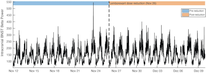
Figure 3: Striato-limbic beta power before and after lemborexant dose reduction. Continuous recording of striato-limbic beta power within the right bed nucleus of the stria terminalis (BNST) centered on the lemborexant dose reduction (dashed vertical line). Please click here to view a larger version of this figure.
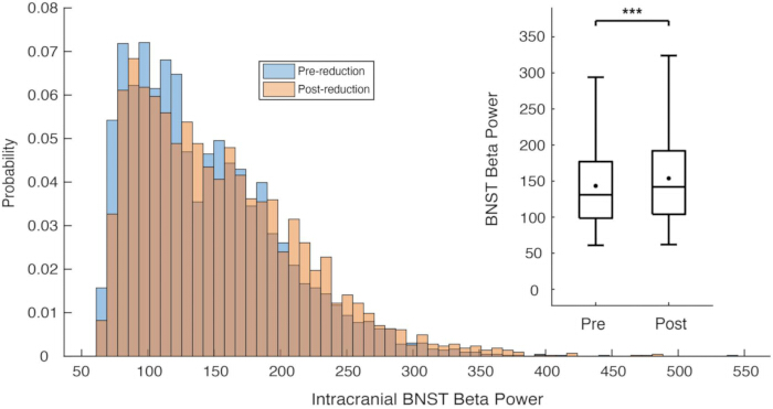
Figure 4: Increase in Striato-limbic beta power before and after lemborexant dose reduction. Probability distributions of intracranial BNST beta power comparing 30 days before (blue) and 30 days after (orange) the medication reduction. Box-and-whisker plots comparing BNST beta power across the full 30-day pre- and post-reduction windows. Filled circles denote group means. (*** p < 0.001, t-test: H0: βmed_red= 0). Please click here to view a larger version of this figure.
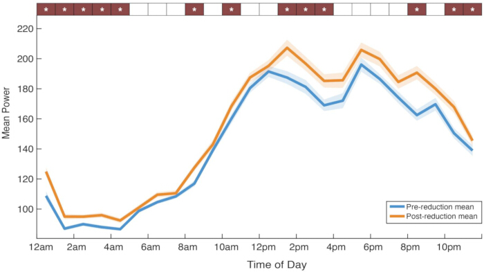
Figure 5: Increase in striato-limbic beta power during sleep, but not wake. Mean intracranial BNST beta power across time of day computed in 1-hour bins across the 60-day window, 30 days for pre-reduction (blue) and 30 days post-reduction (orange) epochs. Shaded bands represent ±SEM. (*p < 0.05, Wilcoxon rank-sum tests with False Discovery Rate correction). Please click here to view a larger version of this figure.
Discussion
DBS is an established therapy for Parkinson’s disease, essential tremor, dystonia, epilepsy, and refractory neuropsychiatric disorders, such as OCD12,13. The development of sensing technologies now enables chronic intracranial recordings in human subjects undergoing DBS treatment, providing a rare opportunity to characterize the circuit-based mechanisms of action of pharmacological agents. Here, the authors describe a workflow for collecting chronic intracranial recordings, self-report measures, and wearable sensors in the context of medication changes to understand the neural mechanisms of action of pharmacological agents. Because many regions exhibit natural diurnal variation, the stability of circadian activity over time can be used to troubleshoot and verify that DBS recordings capture biologically relevant signals. Large drifts in circadian activity or dramatic outliers may be related to artifact infiltration or slow changes in recording impedance over time. Researchers also describe methods for synchronizing and aligning these data sets to perform analyses that identify changes in neural activity before and after medication changes. The ability to directly and longitudinally monitor electrophysiological neural data from a limited number of implanted brain regions complements other methods, such as functional imaging, which is spatially comprehensive but offers more limited temporal sampling.
To illustrate the method, the authors present data from an individual with severe, treatment-refractory OCD and co-morbid insomnia undergoing DBS with the Medtronic Percept implantable pulse generator (IPG) and bilateral lead system. This individual’s insomnia was being treated with the sleep-promoting medication lemborexant, which blocks the wake-promoting neuropeptide orexin from acting on the orexin-1 and 2 receptors. The data were opportunistically collected in the context of medication adjustments primarily conducted as part of normal clinical care. Following a reduction in the subject’s nightly lemborexant dose, the authors found a significant increase in circadian arousal-associated BNST beta activity at night, but not in the morning or most of the day. Researchers previously found evidence that BNST beta activity is causally associated with circadian arousal, increasing prior to wakefulness and decreasing during the night prior to sleep8,11. These data are consistent with a circuit model in which orexin antagonists administered at night can suppress wake-promoting BNST beta activity14.
This case study has limitations. Because these data were acquired opportunistically in the context of a patient’s regular clinical care, it was not possible to perform randomized controlled studies where the investigators and the participant were blinded. It is possible that the shifts in BNST beta activity could also be related to other factors, such as changes in symptom state over time that happen to coincide with the change in medication dose, drift, or seasonal changes in sleep patterns, or other factors. Also, this case study remains an N = 1 investigation. It remains to be seen whether these results can be replicated in a larger cohort of subjects. Lastly, the authors primarily relied on participants to accurately self-report changes in the schedule of their self-administered medications to minimize study burden and barriers to participation in this naturalistic study. However, when there are concerns about medication adherence and timing, a more stringent study design with observed medication administration can be implemented. If concerns related to medication bioavailability exist in addition to adherence, regular blood draws may be warranted to measure medication levels and active metabolites while concurrent neural recordings are collected.
The case described here focuses on the use of sleep medications to treat insomnia in naturalistic clinical settings. However, the methods described here can be applied to characterize the electrophysiological effects of a wide range of pharmacological agents across different neuropsychiatric disorders. For example, numerous studies have characterized the effects of dopaminergic medications in Parkinson’s Disease, identifying suppression of synchronized beta activity within basal ganglia circuitry as a potential mechanism of therapeutic action for this class of pharmacological agents. Moreover, the methods for collecting intracranial DBS recordings, wearable sensor measures, and symptom assessments could be expanded to include additional modalities of longitudinal behavioral tracking in naturalistic settings. Given that dysregulated brain activity underlies the pathophysiology of many neuropsychiatric disorders, the ability to directly monitor brain electrical activity with longitudinal DBS recordings over symptom-relevant timescales is likely to open a window into the network mechanisms of action of pharmacological agents.
Disclosures
All authors declare that they have no conflicts of interest.
Acknowledgements
This work was supported by the Foundation for OCD Research (A.M.L.), One Mind (A.M.L.), National Institutes of Mental Health (NIMH) K23MH125018 (A.M.L.), NIMH R21MH130914 (A.M.L), and NARSAD Young Investigator grant from the Brain & Behavior Research Foundation (A.M.L.).
Materials
References
- Gilron R, et al. Long-term wireless streaming of neural recordings for circuit discovery and adaptive stimulation in individuals with Parkinson's disease. Nat Biotechnol. 2021;39(9):1078-85.
- Starr PA. Totally Implantable Bidirectional Neural Prostheses: A Flexible Platform for Innovation in Neuromodulation. Front Neurosci. 2018;12:619.
- Oehrn CR, et al. Chronic adaptive deep brain stimulation versus conventional stimulation in Parkinson's disease: a blinded randomized feasibility trial. Nat Med. 2024.
- Swann NC, et al. Chronic multisite brain recordings from a totally implantable bidirectional neural interface: experience in 5 patients with Parkinson's disease. J Neurosurg. 2018;128(2):605-16.
- de Hemptinne C, et al. Therapeutic deep-brain stimulation reduces cortical phase-amplitude coupling in Parkinson's disease. Nat Neurosci. 2015;18(5):779-86.
- Scangos KW, et al. Closed-loop neuromodulation in an individual with treatment-resistant depression. Nat Med. 2021;27(10):1696-700.
- Frank AC, et al. Identification of a personalized intracranial biomarker of depression and response to DBS therapy. Brain Stimul. 2021;14(4):1002-4.
- Guillermo N, et al. Sleep-aware adaptive deep brain stimulation to mitigate insomnia in OCD. Brain Stimul. 2026;19(1):103009.
- de Hemptinne C, et al. Prefrontal Physiomarkers of Anxiety and Depression in Parkinson's Disease. Front Neurosci. 2021;15:748165.
- Iskhakova L, et al. Modulation of dopamine tone induces frequency shifts in cortico-basal ganglia beta oscillations. Nat Commun. 2021;12(1):7026.
- Fan JM, et al. Intracranial beta activity is a biomarker of circadian and stimulation-induced arousal in obsessive-compulsive disorder. Brain Stimul. 2024;17(1):29-31.
- Krauss JK, et al. Technology of deep brain stimulation: current status and future directions. Nat Rev Neurol. 2021;17(2):75-87.
- Sheth SA, Mayberg HS. Deep Brain Stimulation for Obsessive-Compulsive Disorder and Depression. Annu Rev Neurosci. 2023;46:341-58.
- Giardino WJ, Pomrenze MB. Extended Amygdala Neuropeptide Circuitry of Emotional Arousal: Waking Up on the Wrong Side of the Bed Nuclei of Stria Terminalis. Front Behav Neurosci. 2021;15:613025.
Reprints and Permissions
This article has been published
Video Coming Soon