This narrative review examines the bidirectional relationships between anxiety and depression and coronary artery disease, acute coronary syndrome, and heart failure. It summarizes neuroendocrine dysfunction, inflammation, mitochondrial impairment, gut microbiota alterations, heart-brain signaling, multimodal management strategies, and future research directions.
Review Article
Research Progress on the Potential Mechanisms and Multimodal Management of Psychocardiological Disease
* These authors contributed equally
In This Article
Summary
Abstract
Cardiovascular disease frequently coexists with anxiety and depression, creating a complex bidirectional relationship between mental and cardiovascular health. Anxiety and depression may increase cardiovascular risk, impair treatment adherence and self-management, and contribute to poorer long-term outcomes. Conversely, acute cardiovascular events, persistent physical symptoms, and functional limitations may trigger or exacerbate psychological distress. This narrative review focuses on coronary artery disease, acute coronary syndrome, and heart failure, and summarizes the epidemiological associations, clinical implications, potential mechanisms, and management strategies related to anxiety and depression. The mechanisms discussed include neuroendocrine and autonomic dysregulation, inflammation, mitochondrial dysfunction, gut microbiota alterations, and heart–brain communication. The review also examines integrated approaches involving cardiovascular treatment, psychological interventions, exercise rehabilitation, pharmacotherapy, social support, and digital management. Future research should standardize diagnostic criteria and outcome measures, clarify key mechanistic pathways, develop clinically meaningful phenotypic classifications, identify useful biomarkers, and evaluate comprehensive interventions in multicenter studies. These efforts may strengthen early detection, risk stratification, individualized treatment, and long-term management for patients with coexisting cardiovascular and mental health conditions.
Introduction
Cardiovascular diseases and mental health disorders, particularly anxiety and depression, are major contributors to the global disease burden, and their coexistence further complicates clinical management. Psychocardiological disease broadly refers to a clinical condition in which cardiovascular disease or cardiovascular-related symptoms coexist with mental health problems, with each condition influencing the onset, progression, and prognosis of the other1. Anxiety and depression are common among patients with coronary artery disease, including acute coronary syndrome and chronic coronary artery disease, as well as among those with heart failure. The coexistence of cardiovascular disease and these mental health disorders may impair treatment adherence and quality of life while increasing the risks of rehospitalization, adverse cardiovascular events, and mortality2.
Accumulating evidence supports a bidirectional association between cardiovascular disease and depression3. Chronic psychological stress and negative emotional states may contribute to the development and progression of cardiovascular disease through activation of the hypothalamic–pituitary–adrenal (HPA) axis, autonomic dysfunction, immune and inflammatory responses, and unhealthy behaviors4. Conversely, cardiovascular events, persistent physical symptoms, and functional limitations may aggravate anxiety and depression.
Although research in this field has expanded in recent years, substantial heterogeneity remains across studies in disease categories, psychological assessment instruments, diagnostic criteria, and outcome measures, and some findings remain inconsistent. Clinical practice is also limited by inadequate recognition of mental health problems, poorly defined referral pathways, and insufficient interdisciplinary collaboration. A comprehensive synthesis of the available evidence and a clearer understanding of the bidirectional relationship between mental and cardiovascular health may facilitate early detection, standardized assessment, integrated management, and future research.
Review and Perspective
This study was conducted as a narrative review. A structured literature search was performed in PubMed, Web of Science, Embase, and the China National Knowledge Infrastructure (CNKI), with a search cutoff date of April 30, 2026. The search focused on the epidemiology, underlying mechanisms, screening, and treatment of coronary artery disease, heart failure, anxiety, depression, and psychological stress. Eligible publications included Chinese-language and English-language guidelines, expert consensus statements, systematic reviews, meta-analyses, clinical trials, observational studies, and relevant preclinical studies. Duplicate records, studies unrelated to the review, reports with insufficient information, articles without accessible full texts, and studies that did not support the main topics of this review were excluded.
After duplicate records were removed, titles and abstracts were screened, followed by full-text assessment. Priority was given to high-quality clinical evidence, while landmark studies were retained and supplemented with relevant high-quality publications from the preceding 3–5 years. The included evidence was classified as clinical, animal, cellular, or theoretical, with preclinical studies used primarily to support potential mechanisms. Evidence relating to interventions was narratively synthesized according to study design, consistency of findings, and potential for clinical translation to assess and compare the maturity of the available evidence.
1. Epidemiology
Anxiety and depression are common comorbidities among patients with cardiovascular disease. Among patients with heart disease, the reported prevalence rates of depressive symptoms, anxiety symptoms, and stress are 31.3%, 32.9%, and 57.7%, respectively5. The overall prevalence of depression among patients with cardiovascular disease is approximately 19%6. Among patients with heart failure, the prevalence of depression of any severity is estimated to be 41.9%7. However, the prevalence of anxiety and depressive symptoms varies substantially among hospitalized patients with heart failure8. Major depression affects approximately 19.8% of patients who survive an acute myocardial infarction9. Patients with myocardial infarction with nonobstructive coronary arteries (MINOCA) or Takotsubo syndrome also frequently report impaired mental health and reduced quality of life after an acute event. However, the available evidence is limited by a high risk of bias10.
Mental disorders and cardiovascular disease have a bidirectional relationship. Large prospective cohort studies in the general population have shown that depression is associated with increased risks of all-cause and cardiovascular mortality11. Among patients with myocardial infarction, studies have found that major depression after hospital discharge is associated with an increased risk of short-term cardiac mortality12. Mental stress-induced myocardial ischemia has been documented in patients with stable coronary artery disease13 and is associated with a higher risk of subsequent cardiovascular events14. Social isolation and living alone are also associated with an increased risk of all-cause mortality among patients with cardiovascular disease15. Mendelian randomization studies further suggest that depression may contribute to the risk of certain cardiovascular outcomes16, supporting early psychological screening and risk-stratified management.
2. Pathophysiological mechanisms
Psychocardiological disease may result from the combined effects of neuroendocrine and autonomic dysregulation, inflammation, mitochondrial dysfunction, gut microbiota dysbiosis, and altered communication between the heart and brain.
1. Neuroendocrine and autonomic regulatory axis
Negative emotional states, such as anxiety and depression, can activate the paraventricular nucleus of the hypothalamus and associated cortical and limbic circuits. This activation promotes the release of corticotropin-releasing hormone (CRH), arginine vasopressin, adrenocorticotropic hormone (ACTH), and cortisol. It also increases sympathetic activity and reduces parasympathetic regulation17, thereby affecting the sinoatrial node, myocardium, and coronary vasculature.
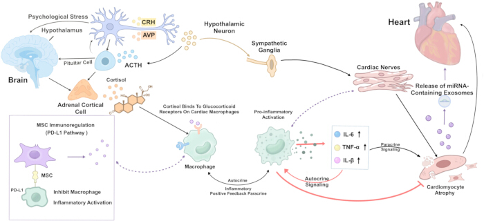
Figure 1: Psychological stress affects the heart through the hypothalamic–pituitary–adrenal axis and the autonomic nervous system. Psychological stress activates the paraventricular nucleus and stimulates the hypothalamic–pituitary–adrenal axis, leading to the release of corticotropin-releasing hormone, arginine vasopressin, adrenocorticotropic hormone, and cortisol. Cortisol acts via glucocorticoid receptors in cardiac macrophages, influencing inflammatory signaling, macrophage polarization, cardiomyocyte injury, and cardiac remodeling. In parallel, increased sympathetic activity affects the sinoatrial node, myocardium, and coronary arteries. The figure also shows regulatory interactions involving cardiomyocyte-derived exosomes, mesenchymal stem cells, and the PD-1/PD-L1 pathway. Abbreviations: PVN, paraventricular nucleus; CRH, corticotropin-releasing hormone; AVP, arginine vasopressin; ACTH, adrenocorticotropic hormone; GR, glucocorticoid receptor; IL-6, interleukin 6; TNF-α, tumor necrosis factor alpha; IL-1β, interleukin 1 beta; miRNA, microRNA; MSC, mesenchymal stem cell; PD-L1, programmed death-ligand 1; HPA, hypothalamic–pituitary–adrenal axis. Please click here to view a larger version of this figure.
Glucocorticoids regulate inflammatory responses and tissue repair through glucocorticoid receptors expressed in macrophages. In experimental models of myocardial infarction, the loss of glucocorticoid receptors in myeloid cells impairs the functional differentiation of monocyte-derived macrophages and is associated with abnormal scar formation, angiogenesis, and ventricular remodeling18. Dysregulated glucocorticoid receptor signaling may therefore alter inflammation and tissue repair after myocardial injury by disrupting macrophage function.
Following myocardial injury, the expression of inflammatory cytokines, including tumor necrosis factor alpha (TNF-α), interleukin 1 beta (IL-1β), and interleukin 6 (IL-6), increases in noncardiomyocytes such as macrophages. These changes are associated with collagen deposition and ventricular remodeling, suggesting that local inflammation and paracrine signaling contribute to the regulation of the myocardial microenvironment19. Injured cardiomyocytes can also release exosomes containing specific microRNAs. Among them, exosomal miR-146a-5p regulates macrophage polarization and the expression of inflammation-related genes, providing preclinical evidence of bidirectional communication between cardiomyocytes and immune cells20.
Preclinical evidence suggests that programmed death-ligand 1 (PD-L1) carried by apoptotic bodies derived from mesenchymal stem cells can interact with programmed cell death protein 1 (PD-1) on macrophages. This interaction induces metabolic reprogramming and promotes a shift from a proinflammatory to an anti-inflammatory phenotype. However, these findings were obtained from a model of acute lung injury, and their relevance to psychological stress-related cardiac injury requires further validation21. In a model of stress-induced cardiomyopathy, PD-1/PD-L1 signaling limited myocardial inflammation. Blocking this pathway prolonged the inflammatory response and delayed recovery of left ventricular structure and function22.
2. Immune-inflammatory and neuroinflammatory axis
Inflammatory activation is an important pathological feature shared by psychocardiological diseases. Patients with cardiovascular disease and depressive symptoms may have elevated inflammatory markers, including C-reactive protein (CRP), IL-6, and TNF-α23. Higher levels of IL-6, TNF-α, and composite inflammatory indices have been associated with the onset of depression, although findings regarding the longitudinal association between CRP and depression remain inconsistent24. Inflammatory mediators may affect the integrity and function of the blood-brain barrier, as well as brain regions involved in emotional regulation25. Neuroinflammation associated with cardiovascular disease may contribute to bidirectional communication between the heart and brain26. Preclinical studies also implicate a proinflammatory microglial phenotype in stress-related depressive-like behaviors. However, direct evidence for this mechanism in human psychocardiological disease remains limited.
The Toll-like receptor 4 (TLR4)/nuclear factor kappa B (NF-κB)/NOD-like receptor family pyrin domain containing 3 (NLRP3) inflammasome cascade represents a potential pathway linking innate immune activation, inflammatory amplification, and vascular endothelial injury.
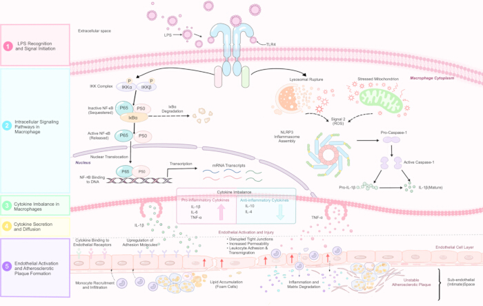
Figure 2: Lipopolysaccharide/Toll-like receptor 4-mediated inflammatory amplification and atherosclerosis. Lipopolysaccharide activates Toll-like receptor 4 on macrophages, triggering nuclear factor kappa B signaling and mitochondrial reactive oxygen species-dependent activation of the NOD-like receptor family pyrin domain containing 3 inflammasome. These pathways promote the production and maturation of proinflammatory cytokines, including interleukin-1 beta, interleukin-6, and tumor necrosis factor-alpha. Sustained inflammatory signaling contributes to endothelial activation, monocyte recruitment, foam cell formation, lipid accumulation, plaque development, and plaque instability. Abbreviations: LPS, lipopolysaccharide; TLR4, Toll-like receptor 4; NF-κB, nuclear factor kappa B; ROS, reactive oxygen species; NLRP3, NOD-like receptor family pyrin domain containing 3; IL-1β, interleukin 1 beta; IL-6, interleukin 6; TNF-α, tumor necrosis factor alpha; caspase-1, cysteine-aspartic protease 1. Please click here to view a larger version of this figure.
Cellular stress signals, including potassium efflux, mitochondrial reactive oxygen species, and lysosomal damage, can promote NLRP3 inflammasome assembly and caspase-1 activation. These processes drive the maturation and release of IL-1β and IL-1827. Increased expression of NLRP3 and caspase-1 in peripheral blood mononuclear cells from patients with major depressive disorder provides preliminary clinical evidence for the involvement of this pathway28. However, therapies targeting the NLRP3 inflammasome remain at an early stage of development29.
Mitochondrial DNA (mtDNA) released from damaged mitochondria can act as a damage-associated molecular pattern. Cytosolic mtDNA is recognized by cyclic GMP-AMP synthase (cGAS), which catalyzes the production of cyclic GMP-AMP (cGAMP) and subsequently activates the stimulator of interferon genes (STING). Activated STING promotes the expression of type I interferons and proinflammatory cytokines through TANK-binding kinase 1 (TBK1), interferon regulatory factor 3 (IRF3), and NF-κB signaling30. Crosstalk between the cGAS-STING and NLRP3 pathways may connect mitochondrial damage with innate immune activation and inflammatory amplification. Inflammation may also alter neural excitability and synaptic plasticity through the kynurenine pathway31.
3. Mitochondrial dysfunction and metabolic regulation
Mitochondrial dysfunction may provide a mechanistic link among oxidative stress, inflammation, myocardial injury, and emotional disorders. Psychological stress has been associated with alterations in mitochondrial energy metabolism, redox homeostasis, and structural integrity32. Chronic or excessive stress can impair oxidative phosphorylation, reduce adenosine triphosphate (ATP) production, and increase the accumulation of reactive oxygen species (ROS). These changes may disrupt mitochondrial membrane potential, calcium homeostasis, apoptosis, and cellular energy supply, thereby impairing both myocardial contractility and neuronal function.
Mitochondrial quality control involves biogenesis, fusion and fission, and mitophagy. Peroxisome proliferator-activated receptor gamma coactivator 1 alpha (PGC-1α)-dependent mitochondrial biogenesis and mitochondrial dynamics regulated by mitofusin 1 (MFN1), mitofusin 2 (MFN2), optic atrophy 1 (OPA1), and dynamin-related protein 1 (DRP1) jointly maintain mitochondrial network integrity and respiratory efficiency. PTEN-induced kinase 1 (PINK1)/Parkin-mediated mitophagy is also involved in mitochondrial homeostasis and tissue injury in experimental models of cardiovascular disease33. Under conditions of stress and inflammation, disruption of these processes can lead to excessive mitochondrial fragmentation, impaired function, and increased ROS production.
PINK1/Parkin-mediated mitophagy enables the recognition and removal of damaged mitochondria. When mitochondrial membrane potential declines, Parkin is selectively recruited to damaged mitochondria and promotes their autophagic degradation34. In vitro studies further indicate that phosphorylated ubiquitin generated through PINK1 activity recruits autophagy receptors, including optineurin (OPTN) and nuclear dot protein 52 (NDP52), thereby initiating mitophagy35.
When mitochondrial quality control is impaired, damaged mitochondria cannot be efficiently removed, resulting in the persistent accumulation of ROS, mtDNA, and other damage-associated molecules. Figure 3 illustrates the sequence linking defective mitochondrial quality control, danger signal release, and innate immune activation. These damage-associated signals can act as upstream activators of the cGAS-STING and NLRP3 pathways, thereby amplifying innate immune and inflammatory responses.
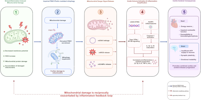
Figure 3: Inflammatory amplification involving mitochondrial quality-control dysfunction, innate immune activation, and cardiac and cerebral injury. Stress and inflammation can impair mitochondrial quality control and PINK1/Parkin-mediated mitophagy, leading to the accumulation of damaged mitochondria. These mitochondria release mitochondrial DNA, mitochondrial reactive oxygen species, and mitochondrial damage-associated molecular patterns, which activate the cGAS-STING and NLRP3 pathways. The resulting inflammatory response promotes type I interferon and nuclear factor kappa B signaling, caspase-1 activation, and cytokine release. Sustained inflammation may further aggravate mitochondrial injury and contribute to impaired cardiac function, arrhythmia susceptibility, reduced neuronal energy supply, altered synaptic plasticity, and impaired emotional regulation. Abbreviations: ROS, reactive oxygen species; PINK1, PTEN-induced kinase 1; Parkin, Parkin RBR E3 ubiquitin protein ligase; LC3, microtubule-associated protein 1 light chain 3; mtDNA, mitochondrial DNA; mtROS, mitochondrial reactive oxygen species; mtDAMPs, mitochondrial damage-associated molecular patterns; cGAS-STING, cyclic GMP-AMP synthase–stimulator of interferon genes; IFNs, interferons; NF-κB, nuclear factor kappa B; NLRP3, NOD-like receptor family pyrin domain containing 3; IL-1β, interleukin 1 beta; IL-18, interleukin 18; IL-6, interleukin 6; TNF-α, tumor necrosis factor alpha; ATP, adenosine triphosphate. Please click here to view a larger version of this figure.
4. Gut microbiota–gut–heart–brain axis
The gut microbiota contributes to both the gut–heart axis36 and the microbiota–gut–brain axis37 through immune, metabolic, neural, and intestinal barrier-related pathways. Microbial metabolites exert a wide range of biological effects. A human study showed that the gut microbiota metabolizes dietary phosphatidylcholine into trimethylamine, which is subsequently converted to trimethylamine N-oxide (TMAO), and that elevated circulating TMAO levels are associated with cardiovascular risk38. Among patients with heart failure, higher TMAO levels are also associated with poorer clinical outcomes39. Evidence from integrated human and animal studies indicates that dietary L-carnitine can be metabolized by the gut microbiota to generate TMAO and promote atherosclerosis40. TMAO may also increase platelet reactivity and thrombotic potential41. Other microbial metabolites, including short-chain fatty acids and tryptophan-derived metabolites, may contribute to immune regulation, intestinal barrier integrity, and neural function. However, their clinical significance remains unclear.
Metagenomic studies in patients with atherosclerotic cardiovascular disease have shown that alterations in the functional profile of the gut microbiota are associated with disease phenotypes42. Population-based studies have also identified associations between the functions of specific neuroactive gut microorganisms, quality of life, and depression43. Transplantation of gut microbiota from patients with depression into germ-free mice can induce depressive-like behaviors and metabolic alterations44. Similarly, another cross-species transplantation study found that depression-associated gut microbiota induced neurobehavioral changes in recipient rats45.
5. Structural and functional alterations in the heart and brain and cross-organ communication
At the organ level, the mechanisms described above may lead to structural and functional abnormalities in both the heart and brain. Among patients with heart failure, alterations in brain networks have been associated with lower left ventricular ejection fraction, cognitive impairment, and more severe depressive symptoms46. In the brain-to-heart direction, psychological stress and impaired central regulation may induce autonomic and neuroendocrine changes. The central autonomic network, sympathovagal balance, and broader brain–heart axis provide a physiological framework through which negative emotional states may influence heart rate, electrophysiological stability, and coronary vascular tone47.
Preclinical evidence suggests that extracellular vesicles released by cardiac cells may carry microRNAs, proteins, and inflammatory mediators involved in intercellular communication. Cardiac fibroblast-derived exosomes have been shown to alter cardiomyocyte phenotype and promote cardiomyocyte hypertrophy48. However, these findings primarily demonstrate local intercellular communication within the heart and do not prove that exosomes mediate long-distance signaling between the heart and brain in humans. Current clinical evidence provides stronger support for interactions among cardiac function, cerebral perfusion, and autonomic regulation. Evidence for long-range heart–brain communication mediated by extracellular vesicles remains mainly preclinical and theoretical.
3. Treatment strategies
Psychocardiological disease requires patient-centered, integrated management encompassing screening, comprehensive assessment49, intervention, and follow-up. The proposed framework incorporates cardiovascular treatment, psychological interventions, exercise rehabilitation, social support, and digital management, with screening, comprehensive assessment, tiered interventions, and long-term follow-up (Figure 4).
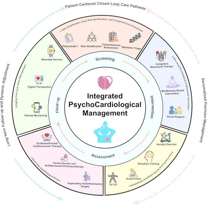
Figure 4: Integrated management framework for psychocardiological disease. The framework illustrates a patient-centered pathway that begins with psychological screening and comprehensive cardiovascular and psychosocial assessment. Patients are then stratified according to clinical risk and directed to appropriate interventions, including cardiovascular treatment, psychological therapy, exercise rehabilitation, social support, pharmacological treatment, and digital management. Long-term follow-up, reassessment, and treatment adjustment are incorporated throughout the pathway. Please click here to view a larger version of this figure.
1. Risk stratification, early identification, and comprehensive assessment
- Psychological screening and comprehensive assessment are essential components of psychocardiological care. They aim to identify anxiety, depression, suicide risk, and psychosocial factors that may affect treatment adherence and to guide risk-stratified referral. The Patient Health Questionnaire-9 (PHQ-9) can be used to assess the presence and severity of depressive symptoms50. The Generalized Anxiety Disorder-7 (GAD-7) is commonly used to screen for generalized anxiety symptoms and evaluate their severity51. The Hospital Anxiety and Depression Scale (HADS) is suitable for the initial assessment of anxiety and depressive symptoms in patients with physical illness52.
Patients with severe symptoms, marked functional impairment, or suicide risk should receive professional psychological evaluation. A comprehensive assessment should include cardiovascular disease severity, cardiac function, arrhythmia risk, metabolic and sleep disorders, concomitant medications, psychological symptoms, social support, and patient preferences. Mechanism-related biomarkers are not yet sufficiently validated for routine risk stratification. Management intensity should correspond to the patient’s level of risk within a pathway combining screening, diagnosis, referral, treatment, and follow-up.
2. Nonpharmacological, psychological, and behavioral interventions
- Structured psychotherapy may be considered for patients with persistent anxiety, depression, or specific psychological disorders. Meta-analyses suggest that psychosocial interventions can reduce anxiety and depressive symptoms among patients with heart failure, although their effects on quality of life, hospitalization, and mortality remain limited53. In the ENRICHD trial, the intervention improved depression and perceived social support but did not increase event-free survival54. Stress management may reduce psychological distress, but its effects on cardiovascular events require further confirmation55.
In patients with heart failure, cognitive behavioral therapy can improve depression and quality of life, but has not improved self-care behaviors or clinical outcomes56. Telephone-based collaborative care has been shown to improve depression and mental health-related quality of life after coronary artery bypass grafting (CABG)57. Hybrid collaborative care may also improve mood, although reductions in rehospitalization and mortality have not been demonstrated58. These findings indicate that improvements in psychological symptoms should not be regarded as evidence of cardiovascular protection.
3. Exercise, physical rehabilitation, and traditional Chinese medicine-based rehabilitation
- Exercise-based cardiac rehabilitation is a standard component of care for eligible patients with coronary heart disease and selected patients with stable heart failure. Meta-analyses indicate that exercise-based cardiac rehabilitation reduces the risks of cardiovascular death, myocardial infarction, and hospitalization among patients with coronary heart disease while also improving health-related quality of life59. The UPBEAT randomized trial showed that both aerobic exercise and sertraline reduced depressive symptoms in patients with coronary heart disease60.
A 2022 meta-analysis suggested that tai chi, when added to standard care, may improve depressive symptoms, exercise capacity, and quality of life in patients with chronic heart failure, with the certainty of evidence ranging from very low to moderate61. Acupuncture may also alleviate depressive symptoms in patients with cardiovascular disease and depression. Nevertheless, the evidence remains uncertain because of limited quality and substantial heterogeneity62.
These interventions should complement, rather than replace, standard cardiovascular and psychological treatments. Exercise prescriptions should be individualized according to cardiovascular risk, cardiac function, exercise capacity, frailty, and psychological status.
4. Pharmacological and targeted biological interventions
- Guideline-directed cardiovascular therapy
- Guideline-directed cardiovascular pharmacotherapy is a standard component of care. Patients with chronic coronary artery disease should receive antiplatelet therapy, lipid-lowering therapy, anti-ischemic treatment, and other secondary prevention measures63. Patients with heart failure should receive appropriate guideline-directed pharmacotherapy based on left ventricular ejection fraction, symptom severity, and comorbidities64.
- Antidepressant therapy
Antidepressants may be prescribed to patients with moderate-to-severe depressive or anxiety disorders following specialist evaluation, with the aim of alleviating psychological symptoms and improving functional impairment. The SADHART trial showed that sertraline had a favorable overall cardiovascular safety profile in patients with recent myocardial infarction or unstable angina and major depression. Its antidepressant effects were more pronounced in patients with recurrent or more severe depression65. The CREATE trial demonstrated that citalopram improved depressive symptoms in patients with coronary heart disease and depression66. The REMIT trial suggested that escitalopram may reduce the occurrence of mental stress-induced myocardial ischemia67.
Antidepressant therapy should generally be initiated at a low dose, with regular monitoring of therapeutic response, adherence, and cardiovascular safety. For patients with cardiovascular disease and depression, treatment efficacy and both cardiovascular and psychiatric safety should be evaluated throughout therapy68. Particular attention should be paid to QTc prolongation, orthostatic hypotension, hyponatremia, bleeding risk, and potential interactions with antiplatelet agents, anticoagulants, and antiarrhythmic drugs. Tricyclic antidepressants are generally not considered first-line treatment for patients with cardiovascular disease because of their potential to cause cardiac conduction abnormalities, anticholinergic adverse effects, and arrhythmias.- Anti-inflammatory and mechanism-targeted therapies
Anti-inflammatory and mitochondria-targeted interventions remain under investigation. The CANTOS trial showed that canakinumab reduced the risk of certain recurrent cardiovascular events but increased the risk of fatal infection69. However, psychological outcomes were not evaluated, and the findings therefore do not support the use of canakinumab specifically for patients with psychocardiological disease. Therapeutic strategies targeting cGAS-STING and NLRP3 pathways also remain in their early stages, with inadequate validation in clinical populations.
- Anti-inflammatory and mechanism-targeted therapies
5. Digital and Longitudinal Management
Wearable devices, digital therapeutics, and remote monitoring can supplement standard medical care and extend management beyond clinical settings. Wearable devices can continuously monitor heart rate, electrocardiographic activity, physical activity, and sleep70. Therapist-supported internet-based cognitive behavioral therapy (ICBT) has been shown to improve depressive symptoms and quality of life, although its effects on anxiety remain inconsistent71. Improvements in depression may persist for 6–12 months72. Digital cardiac rehabilitation may improve selected measures of physical activity and functional capacity, but evidence regarding long-term clinical outcomes remains limited73. Smartphone-based cardiac rehabilitation has also been associated with improvements in exercise capacity, health behaviors, quality of life, and anxiety after percutaneous coronary intervention (PCI)74. The eIMPACT intervention reduced depressive symptoms but did not improve several cardiovascular risk biomarkers75.
Digital tools should be integrated into established clinical referral and treatment pathways, with adequate validation of devices and algorithms, protection of data privacy, reduction of the digital divide, and improved accessibility across diverse patient populations.
6. Evidence-based and clinical positioning of interventions
To facilitate clinical comparison of management strategies, this review provides a narrative synthesis of interventions according to study design, consistency of findings, and potential for clinical translation (Supplementary Table 1). The strength of evidence is categorized as substantial, limited, or preliminary. The clinical role of each intervention is classified as standard management or treatment, a potential adjunctive intervention, an exploratory intervention, or an intervention not currently recommended as a priority. These classifications are intended only to describe the maturity, consistency, and scope of the available evidence rather than formal levels of evidence or classes of recommendation used in clinical practice guidelines.
Conclusions
Psychocardiological disease can be understood as a clinical condition in which cardiovascular disease and psychological disorders, particularly anxiety and depression, interact bidirectionally. Evidence from human studies supports associations among psychological distress, autonomic dysfunction, systemic inflammation, metabolic disturbances, and adverse cardiovascular outcomes. By contrast, specific molecular mechanisms, including the NLRP3 inflammasome, cGAS-STING signaling, PINK1/Parkin-mediated mitophagy, and the gut–heart–brain axis, are supported mainly by preclinical and observational studies. These mechanisms therefore cannot yet be regarded as established causal pathways in humans.
Clinical care should follow an integrated pathway linking screening, assessment, intervention, referral, and follow-up. Guideline-directed cardiovascular therapy, structured psychotherapy when clinically indicated, antidepressant treatment after appropriate evaluation, and exercise-based cardiac rehabilitation constitute the main components of management. Psychoeducation, social support, traditional rehabilitation approaches, and digital interventions may provide additional support. Cardiovascular therapies primarily improve cardiovascular outcomes, whereas psychotherapy and antidepressants mainly target psychological symptoms and associated functional impairment. Exercise-based cardiac rehabilitation may offer broader benefits by improving functional capacity, psychological well-being, and selected cardiovascular outcomes. Adjunctive interventions should complement rather than replace established cardiovascular and psychological treatments.
Anti-inflammatory and other mechanism-targeted therapies remain investigational. IL-1β inhibition has shown cardiovascular benefits in selected patients with residual inflammatory risk, but these findings cannot be directly extrapolated to patients with psychocardiological disease. Evidence supporting NLRP3-targeted interventions remains insufficient for routine clinical use. Strategies targeting cGAS-STING signaling or PINK1/Parkin-mediated mitophagy remain largely confined to preclinical and early translational research.
Future studies should standardize disease definitions, diagnostic criteria, and outcome measures. Prospective multicenter cohorts and mechanism-driven, stratified trials are needed, with psychological and cardiovascular outcomes assessed separately. Research should progress from validation of mechanisms in humans to biomarker qualification, safety assessment, and confirmation of clinical efficacy. Such efforts may facilitate the translation of mechanistic findings into practical and individualized approaches to psychocardiological care.
Supplementary Table 1: Evidence-based and clinical positioning of key interventions in psychocardiology. The table summarizes major intervention categories used in psychocardiological care, including psychological screening, psychotherapy, collaborative care, exercise rehabilitation, traditional rehabilitation approaches, antidepressant treatment, anti-inflammatory and mechanism-targeted therapies, digital interventions, and remote monitoring. For each intervention, it presents the main evidence and outcomes, supporting references, the evidence's maturity, and the proposed clinical role. Evidence is categorized as substantial, limited, or preliminary, and interventions are positioned as standard treatment, adjunctive care, exploratory therapy, or not currently recommended for routine use. These categories represent a narrative synthesis of the available evidence and do not constitute formal guideline recommendations. Please click here to download this file.
Disclosures
The authors declare no competing interests. This review is based exclusively on previously published studies and did not involve new human-participant research.
Acknowledgements
The authors thank the Teaching and Research Section of Psychology at Anhui University of Chinese Medicine and Miao Qing of Guangzhou University of Chinese Medicine for their guidance and support. This work was supported by the 2025 Key Humanities and Social Sciences Project for Higher Education Institutions of the Anhui Provincial Department of Education, “Coupling Mechanisms and Optimization Pathways for Cultivating a Sense of Community for the Chinese Nation through Cultural and Museum Research and Study from the Perspective of Cultural Embedding: An Empirical Study Based on Anhui–Xinjiang Practices” (Project No. 2025AHGXSK30457), and the Basic Research Operating Expenses Program for Higher Education Institutions in the Xinjiang Uygur Autonomous Region, “Underlying Mechanisms and Improvisational Strategies for Fostering a Sense of Community for the Chinese Nation through Cultural Heritage Research and Study Tours from a Cultural Embedding Perspective” (Project No. XJEDU2026P141).
References
- Rozanski A, et al. The epidemiology, pathophysiology, and management of psychosocial risk factors in cardiac practice: the emerging field of behavioral cardiology. J Am Coll Cardiol. 2005;45(5):637–651. doi:10.1016/j.jacc.2004.12.005.
- Levine GN, et al. Psychological health, well-being, and the mind-heart-body connection: a scientific statement from the American Heart Association. Circulation. 2021;143(10):e763–e783. doi:10.1161/CIR.0000000000000947.
- Wu J, et al. Cardiovascular disease and depression: a bidirectional relationship and its clinical implications. Front Psychiatry. 2026;16:1675680. doi:10.3389/fpsyt.2025.1675680.
- Vaccarino V, Bremner JD. Stress and cardiovascular disease: an update. Nat Rev Cardiol. 2024;21(9):603–616. doi:10.1038/s41569-024-01024-y.
- Karami N, et al. Global prevalence of depression, anxiety, and stress in cardiac patients: a systematic review and meta-analysis. J Affect Disord. 2023;324:175-189. doi:10.1016/j.jad.2022.12.055.
- Rafiei S, et al. Depression prevalence in cardiovascular disease: global systematic review and meta-analysis. BMJ Support Palliat Care. 2023;13(3):281-289. doi:10.1136/spcare-2022-003884.
- Moradi M, et al. Global prevalence of depression among heart failure patients: a systematic review and meta-analysis. Curr Probl Cardiol. 2022;47(6):100848. doi:10.1016/j.cpcardiol.2021.100848.
- Hamatani Y, et al. Prevalence, temporal change, and determinants of anxiety and depression in hospitalized patients with heart failure. J Card Fail. 2022;28(2):181-190. doi:10.1016/j.cardfail.2021.07.024.
- Thombs BD, et al. Prevalence of depression in survivors of acute myocardial infarction. J Gen Intern Med. 2006;21(1):30–38. doi:10.1111/j.1525-1497.2005.00269.x.
- Leissner P, et al. Mental health status and quality-of-life after an acute myocardial infarction with non-obstructive coronary arteries or takotsubo syndrome: a systematic review. Eur J Prev Cardiol. 2025:zwaf101. doi:10.1093/eurjpc/zwaf101.
- Meng R, et al. Association of depression with all-cause and cardiovascular disease mortality among adults in China. JAMA Netw Open. 2020;3(2):e1921043. doi:10.1001/jamanetworkopen.2019.21043.
- Frasure-Smith N, Lespérance F, Talajic M. Depression following myocardial infarction: impact on 6-month survival. JAMA. 1993;270(15):1819–1825. doi:10.1001/jama.1993.03510150053029.
- Jiang W, et al. Prevalence and clinical characteristics of mental stress-induced myocardial ischemia in patients with coronary heart disease. J Am Coll Cardiol. 2013;61(7):714-722. doi:10.1016/j.jacc.2012.11.037.
- Vaccarino V, et al. Association of mental stress-induced myocardial ischemia with cardiovascular events in patients with coronary heart disease. JAMA. 2021;326(18):1818–1828. doi:10.1001/jama.2021.17649.
- Long RM, et al. Loneliness, social isolation, and living alone associations with mortality risk in individuals living with cardiovascular disease: a systematic review, meta-analysis, and meta-regression. Psychosom Med. 2023;85(1):8-17. doi:10.1097/PSY.0000000000001151.
- Lu Y, et al. Genetic liability to depression and risk of coronary artery disease, myocardial infarction, and other cardiovascular outcomes. J Am Heart Assoc. 2021;10(1):e017986. doi:10.1161/JAHA.120.017986.
- Helman TJ, Headrick JP, Stapelberg NJC, Braidy N. The sex-dependent response to psychosocial stress and ischaemic heart disease. Front Cardiovasc Med. 2023;10:1072042. doi:10.3389/fcvm.2023.1072042.
- Galuppo P, et al. The glucocorticoid receptor in monocyte-derived macrophages is critical for cardiac infarct repair and remodeling. FASEB J. 2017;31(11):5122–5132. doi:10.1096/fj.201700317R.
- Yue P, Massie BM, Simpson PC, Long CS. Cytokine expression increases in nonmyocytes from rats with postinfarction heart failure. Am J Physiol Heart Circ Physiol. 1998;275(1):H250-H258. doi:10.1152/ajpheart.1998.275.1.H250.
- Chen C, et al. Role of cardiomyocyte-derived exosomal microRNA-146a-5p in macrophage polarization and activation. Dis Markers. 2022;2022:2948578. doi:10.1155/2022/2948578.
- Jiang T, et al. Apoptotic bodies inhibit inflammation by PDL1-PD1-mediated macrophage metabolic reprogramming. Cell Prolif. 2024;57(1):e13531. doi:10.1111/cpr.13531.
- Hayashi T, et al. The programmed death-1 signaling axis modulates inflammation and LV structure/function in a stress-induced cardiomyopathy model. JACC Basic Transl Sci. 2022;7(11):1120–1139. doi:10.1016/j.jacbts.2022.05.006.
- Varghese TP, et al. Interplay of inflammatory biomarkers in heart disease patients with depressive symptoms: an update. Curr Probl Cardiol. 2024;49(3):102352. doi:10.1016/j.cpcardiol.2023.102352.
- Shi R, et al. Inflammatory markers and incident depression: evidence in a population-based prospective study. Psychoneuroendocrinology. 2022;142:105806. doi:10.1016/j.psyneuen.2022.105806.
- Medina-Rodriguez EM, Beurel E. Blood brain barrier and inflammation in depression. Neurobiol Dis. 2022;175:105926. doi:10.1016/j.nbd.2022.105926.
- Hu JR, Abdullah A, Nanna MG, Soufer R. The brain-heart axis: neuroinflammatory interactions in cardiovascular disease. Curr Cardiol Rep. 2023;25(12):1745-1758. doi:10.1007/s11886-023-01990-8.
- Xu J, Nunez G. The NLRP3 inflammasome: activation and regulation. Trends Biochem Sci. 2023;48(4):331–344. doi:10.1016/j.tibs.2022.10.002.
- Alcocer-Gómez E, et al. NLRP3 inflammasome is activated in mononuclear blood cells from patients with major depressive disorder. Brain Behav Immun. 2014;36:111-117. doi:10.1016/j.bbi.2013.10.017.
- Xia CY, et al. The NLRP3 inflammasome in depression: potential mechanisms and therapies. Pharmacol Res. 2023;187:106625. doi:10.1016/j.phrs.2022.106625.
- Kim J, Kim HS, Chung JH. Molecular mechanisms of mitochondrial DNA release and activation of the cGAS-STING pathway. Exp Mol Med. 2023;55(3):510–519. doi:10.1038/s12276-023-00965-7.
- Badawy AA-B, Dawood S, Bano S. Kynurenine pathway of tryptophan metabolism in pathophysiology and therapy of major depressive disorder. World J Psychiatry. 2023;13(4):141-148. doi:10.5498/wjp.v13.i4.141.
- Picard M, McEwen BS. Psychological stress and mitochondria: a systematic review. Psychosom Med. 2018;80(2):141–153. doi:10.1097/PSY.0000000000000545.
- Wu Y, et al. PINK1/Parkin-mediated mitophagy in cardiovascular disease: from pathogenesis to novel therapy. Int J Cardiol. 2022;361:61–69. doi:10.1016/j.ijcard.2022.05.025.
- Narendra D, Tanaka A, Suen DF, Youle RJ. Parkin is recruited selectively to impaired mitochondria and promotes their autophagy. J Cell Biol. 2008;183(5):795–803. doi:10.1083/jcb.200809125.
- Lazarou M, et al. The ubiquitin kinase PINK1 recruits autophagy receptors to induce mitophagy. Nature. 2015;524(7565):309–314. doi:10.1038/nature14893.
- Bui TVA, et al. The gut-heart axis: updated review for the roles of microbiome in cardiovascular health. Korean Circ J. 2023;53(8):499–518. doi:10.4070/kcj.2023.0048.
- Yang H, et al. A narrative review of relationship between gut microbiota and neuropsychiatric disorders: mechanisms and clinical application of probiotics and prebiotics. Ann Palliat Med. 2021;10(2):2304–2313. doi:10.21037/apm-20-1365.
- Tang WHW, et al. Intestinal microbial metabolism of phosphatidylcholine and cardiovascular risk. N Engl J Med. 2013;368(17):1575–1584. doi:10.1056/NEJMoa1109400.
- Tang WHW, et al. Prognostic value of elevated levels of intestinal microbe-generated metabolite trimethylamine-N-oxide in patients with heart failure: refining the gut hypothesis. J Am Coll Cardiol. 2014;64(18):1908-1914. doi:10.1016/j.jacc.2014.02.617.
- Koeth RA, et al. Intestinal microbiota metabolism of L-carnitine, a nutrient in red meat, promotes atherosclerosis. Nat Med. 2013;19(5):576–585. doi:10.1038/nm.3145.
- Zhu W, et al. Gut microbial metabolite TMAO enhances platelet hyperreactivity and thrombosis risk. Cell. 2016;165(1):111–124. doi:10.1016/j.cell.2016.02.011.
- Jie Z, et al. The gut microbiome in atherosclerotic cardiovascular disease. Nat Commun. 2017;8(1):845. doi:10.1038/s41467-017-00900-1.
- Valles-Colomer M, et al. The neuroactive potential of the human gut microbiota in quality of life and depression. Nat Microbiol. 2019;4(4):623–632. doi:10.1038/s41564-018-0337-x.
- Zheng P, et al. Gut microbiome remodeling induces depressive-like behaviors through a pathway mediated by the host's metabolism. Mol Psychiatry. 2016;21(6):786-796. doi:10.1038/mp.2016.44.
- Kelly JR, et al. Transferring the blues: depression-associated gut microbiota induces neurobehavioural changes in the rat. J Psychiatr Res. 2016;82:109–118. doi:10.1016/j.jpsychires.2016.07.019.
- Gao Q, et al. Mapping brain synergy dysfunction in heart failure patients with reduced and mildly reduced ejection fraction using multimodal neuroimaging: functional and molecular insights. Eur J Radiol. 2026;203:113018. doi:10.1016/j.ejrad.2026.113018.
- Valenza G. Depression as a cardiovascular disorder: central-autonomic network, brain-heart axis, and vagal perspectives of low mood. Front Netw Physiol. 2023;3:1125495. doi:10.3389/fnetp.2023.1125495.
- Bang C, et al. Cardiac fibroblast-derived microRNA passenger strand-enriched exosomes mediate cardiomyocyte hypertrophy. J Clin Invest. 2014;124(5):2136–2146. doi:10.1172/JCI70577.
- Bueno H, et al. 2025 ESC Clinical Consensus Statement on mental health and cardiovascular disease: developed under the auspices of the ESC Clinical Practice Guidelines Committee. Eur Heart J. 2025;46(41):4156–4225. doi:10.1093/eurheartj/ehaf191.
- Kroenke K, Spitzer RL, Williams JBW. The PHQ-9: validity of a brief depression severity measure. J Gen Intern Med. 2001;16(9):606–613. doi:10.1046/j.1525-1497.2001.016009606.x.
- Spitzer RL, Kroenke K, Williams JBW, Löwe B. A brief measure for assessing generalized anxiety disorder: the GAD-7. Arch Intern Med. 2006;166(10):1092–1097. doi:10.1001/archinte.166.10.1092.
- Zigmond AS, Snaith RP. The hospital anxiety and depression scale. Acta Psychiatr Scand. 1983;67(6):361–370. doi:10.1111/j.1600-0447.1983.tb09716.x.
- Chernoff RA, et al. Psychosocial interventions for patients with heart failure and their impact on depression, anxiety, quality of life, morbidity, and mortality: a systematic review and meta-analysis. Psychosom Med. 2022;84(5):560-580. doi:10.1097/PSY.0000000000001073.
- Berkman LF, et al. Effects of treating depression and low perceived social support on clinical events after myocardial infarction: the Enhancing Recovery in Coronary Heart Disease Patients (ENRICHD) randomized trial. JAMA. 2003;289(23):3106-3116. doi:10.1001/jama.289.23.3106.
- Blumenthal JA, et al. Enhancing cardiac rehabilitation with stress management training: a randomized, clinical efficacy trial. Circulation. 2016;133(14):1341–1350. doi:10.1161/CIRCULATIONAHA.115.018926.
- Freedland KE, et al. Treatment of depression and inadequate self-care in patients with heart failure: one-year outcomes of a randomized controlled trial. Gen Hosp Psychiatry. 2023;84:82-88. doi:10.1016/j.genhosppsych.2023.06.001.
- Rollman BL, et al. Telephone-delivered collaborative care for treating post-CABG depression: a randomized controlled trial. JAMA. 2009;302(19):2095–2103. doi:10.1001/jama.2009.1670.
- Rollman BL, et al. Efficacy of blended collaborative care for patients with heart failure and comorbid depression: a randomized clinical trial. JAMA Intern Med. 2021;181(10):1369-1380. doi:10.1001/jamainternmed.2021.4978.
- Dibben G, et al. Exercise-based cardiac rehabilitation for coronary heart disease: a meta-analysis. Eur Heart J. 2023;44(6):452–469. doi:10.1093/eurheartj/ehac747.
- Blumenthal JA, et al. Exercise and pharmacological treatment of depressive symptoms in patients with coronary heart disease: results from the UPBEAT study. J Am Coll Cardiol. 2012;60(12):1053-1063. doi:10.1016/j.jacc.2012.04.040.
- Hui J, et al. Effects of Tai Chi on health status in adults with chronic heart failure: a systematic review and meta-analysis. Front Cardiovasc Med. 2022;9:953657. doi:10.3389/fcvm.2022.953657.
- Lu L, et al. Acupuncture in treating cardiovascular disease complicated with depression: a systematic review and meta-analysis. Front Psychiatry. 2022;13:1051324. doi:10.3389/fpsyt.2022.1051324.
- Virani SS, et al. 2023 AHA/ACC/ACCP/ASPC/NLA/PCNA guideline for the management of patients with chronic coronary disease. Circulation. 2023;148(9):e9-e119. doi:10.1161/CIR.0000000000001168.
- Heidenreich PA, et al. 2022 AHA/ACC/HFSA guideline for the management of heart failure. Circulation. 2022;145(18):e895-e1032. doi:10.1161/CIR.0000000000001063.
- Glassman AH, et al. Sertraline treatment of major depression in patients with acute myocardial infarction or unstable angina. JAMA. 2002;288(6):701-709. doi:10.1001/jama.288.6.701.
- Lespérance F, et al. Effects of citalopram and interpersonal psychotherapy on depression in patients with coronary artery disease: the Canadian Cardiac Randomized Evaluation of Antidepressant and Psychotherapy Efficacy (CREATE) trial. JAMA. 2007;297(4):367-379. doi:10.1001/jama.297.4.367.
- Jiang W, et al. Effect of escitalopram on mental stress-induced myocardial ischemia: results of the REMIT trial. JAMA. 2013;309(20):2139-2149. doi:10.1001/jama.2013.5566.
- Lichtman JH, et al. Depression and coronary heart disease: recommendations for screening, referral, and treatment: a science advisory from the American Heart Association. Circulation. 2008;118(17):1768–1775. doi:10.1161/CIRCULATIONAHA.108.190769.
- Ridker PM, et al. Antiinflammatory therapy with canakinumab for atherosclerotic disease. N Engl J Med. 2017;377(12):1119–1131. doi:10.1056/NEJMoa1707914.
- Spatz ES, Ginsburg GS, Rumsfeld JS, Turakhia MP. Wearable digital health technologies for monitoring in cardiovascular medicine. N Engl J Med. 2024;390(4):346-356. doi:10.1056/NEJMra2301903.
- Kwek SQ, et al. Effectiveness of therapist-supported internet-based cognitive behavioural therapy interventions on depression, anxiety and quality of life among patients with cardiovascular disease: a systematic review and meta-analysis. Eur J Cardiovasc Nurs. 2025;24(6):860-870. doi:10.1093/eurjcn/zvaf084.
- Westas M, et al. Effects of internet-delivered cognitive behavioural therapy adapted for patients with cardiovascular disease and depression: a long-term follow-up of a randomized controlled trial at 6 and 12 months posttreatment. Eur J Cardiovasc Nurs. 2022;21(6):559-567. doi:10.1093/eurjcn/zvab131.
- Heimer M, et al. eHealth for maintenance cardiovascular rehabilitation: a systematic review and meta-analysis. Eur J Prev Cardiol. 2023;30(15):1634-1651. doi:10.1093/eurjpc/zwad145.
- Lao SSW, Chair SY, Wang Q, Leong MLT. The feasibility and effects of smartphone-based application on cardiac rehabilitation for patients after percutaneous coronary intervention: a randomized controlled trial. J Cardiovasc Nurs. 2024;39(1):88-101. doi:10.1097/JCN.0000000000000993.
- Stewart JC, et al. Effect of modernized collaborative care for depression on depressive symptoms and cardiovascular disease risk biomarkers: eIMPACT randomized controlled trial. Brain Behav Immun. 2023;112:18-28. doi:10.1016/j.bbi.2023.05.007.