This protocol describes the standardized placement of bronchial blockers performed directly in the lateral decubitus position during video-assisted thoracic surgery. The goal is to improve device stability, reduce repositioning-related displacement, minimize the need for fiberoptic bronchoscopy, and optimize procedural efficiency and perioperative airway management during one-lung ventilation.
Research Article
Fiberoptic Bronchoscopy-Guided Bronchial Blocker Placement in Lateral Versus Supine Positions in Video-Assisted Thoracic Surgery: A Comparative Study
In This Article
Summary
Abstract
Bronchial blockers (BBs) are widely used for one-lung ventilation during video-assisted thoracic surgery (VATS), but malposition remains a common cause of repeated fiberoptic bronchoscopy (FOB) examinations and procedural inefficiency. Whether BB placement performed directly in the lateral decubitus position improves device stability and perioperative outcomes remains unclear. Clinical data from adult patients undergoing elective unilateral VATS with BB-assisted one-lung ventilation were analyzed to compare lateral-position BB placement with the conventional supine approach followed by repositioning. Patients were categorized according to the position used during BB deployment. BB malposition occurred less frequently in the lateral group than in the supine group (12.5% vs 30.8%; relative risk [RR] 0.41, 95% confidence interval [CI]: 0.23–0.72; p = 0.001). Lateral placement was associated with shorter BB placement time (3.8 ± 0.9 vs 5.6 ± 1.2 min; p < 0.001), fewer FOB examinations (1.3 ± 0.3 vs 2.1 ± 0.5; p < 0.001), and a shorter anesthesia-to-incision interval (18.4 ± 3.5 vs 24.7 ± 4.2 min; p < 0.001). Intraoperative oxygenation was improved (arterial partial pressure of oxygen [PaO2]: 186.5 ± 22.4 vs 158.3 ± 25.1 mmHg; p < 0.001), accompanied by lower peak airway pressures (23.4 ± 3.1 vs 26.8 ± 3.6 cmH2O; p < 0.001) and fewer hypoxemic events. Postoperative sore throat, hoarseness, hypoxemia, and airway mucosal injury occurred less frequently in the lateral group (all p < 0.05). In conclusion, BB placement in the lateral decubitus position was associated with greater device stability, improved procedural efficiency, and fewer airway-related complications. Aligning BB deployment with the final surgical position may provide a practical strategy to optimize lung isolation during minimally invasive thoracic surgery.
Introduction
One-lung ventilation (OLV) is a cornerstone of modern thoracic anesthesia, enabling optimal surgical exposure while minimizing trauma to the operative lung during video-assisted thoracic surgery (VATS)1. Achieving reliable lung isolation, however, remains technically demanding and clinically consequential. Malpositioning of airway isolation devices can compromise oxygenation, increase airway pressures, prolong operative time, and precipitate adverse events such as hypoxemia, atelectasis, or airway injury2,3,4. Although double-lumen tubes (DLTs) have long been considered the standard for lung isolation, bronchial blockers (BBs) have gained widespread acceptance as an alternative because they can be deployed through conventional single-lumen endotracheal tubes, accommodate difficult airways, and facilitate postoperative ventilation without tube exchange5,6,7. BBs are particularly attractive in VATS, where minimally invasive surgical approaches and enhanced recovery pathways prioritize safety, efficiency, and reduced airway trauma8. Nevertheless, despite their increasing use, BB placement remains prone to malposition, repeated fiberoptic bronchoscopy (FOB) adjustments, and time-consuming repositioning, all of which can diminish the anticipated advantages of BBs and compromise perioperative outcomes9,10,11.
Patient positioning is a critical yet underexplored determinant of airway device stability. Conventionally, BBs are inserted with the patient in the supine position, followed by repositioning to the lateral decubitus position required for thoracic surgery. This sequence introduces mechanical and anatomical perturbations that can displace airway devices. Changes in head and neck alignment, gravitational shifts of the carina and main bronchi, mediastinal movement, and alterations in cuff-to-airway contact forces during lateral positioning collectively predispose to device migration12,13,14. Indeed, a substantial proportion of lung isolation failures occur during or after the transition from supine to lateral positioning, necessitating repeat FOB and intraoperative adjustments15,16,17. While these phenomena have been well documented for DLTs, analogous vulnerabilities likely apply to BBs, which rely on precise endobronchial positioning and stable cuff apposition for effective lung isolation. In addition to technical inefficiency, repeated manipulations may increase the risk of airway mucosal injury, desaturation episodes during bronchoscopy, and contamination of the operative field, thereby undermining both patient safety and surgical workflow18,19,20,21.
Recent work in thoracic anesthesia has begun to challenge the dogma that airway devices must be placed in the supine position. For DLTs, intubation performed directly in the lateral position has been shown to reduce device malposition, decrease the need for repeated bronchoscopy, and improve perioperative stability compared with conventional supine intubation22,23. These findings provide mechanistic plausibility that maintaining the patient in the surgically required position during airway device placement may minimize subsequent displacement by avoiding post-placement repositioning. However, extrapolating these results to BBs is not straightforward. Unlike DLTs, BBs are coaxially deployed through single-lumen endotracheal tubes, and their functional performance depends on the interaction among blocker-tip geometry, bronchial anatomy, and dynamic respiratory mechanics. BBs are also more susceptible to subtle shifts in bronchial orientation and to changes in cuff-to-wall contact during lung deflation and surgical manipulation24,25,26. Despite the widespread clinical adoption of BBs, there is a striking paucity of evidence addressing whether the patient’s position at the time of BB placement influences device stability, procedural efficiency, or downstream clinical outcomes. To date, most studies comparing BBs with DLTs have focused on oxygenation, lung collapse quality, postoperative sore throat, and the need for tube exchange, without considering the procedural context of patient positioning at insertion27. Consequently, it remains unclear whether BB placement in the lateral position, the position required for thoracic surgery, provides measurable advantages over the conventional supine approach.
Enhanced Recovery After Surgery (ERAS) frameworks emphasize reduction of perioperative physiological stress, avoidance of unnecessary manipulations, and streamlining of operating room processes to improve outcomes and patient experience28. From this perspective, any intervention that reduces airway device malposition, minimizes bronchoscopy requirements, shortens anesthesia-to-incision time, or decreases airway-related complications aligns directly with ERAS principles. Moreover, BBs are frequently selected in patients with anticipated difficult airways, cervical spine disease, obesity, or those requiring postoperative mechanical ventilation, populations in whom repeated airway manipulation may carry heightened risk29. If lateral positioning during BB placement could enhance initial placement accuracy and stability, it would represent a simple, low-cost modification with potentially broad clinical impact. Yet the lack of comparative data has limited the adoption of lateral BB placement in routine practice, and current guidelines provide no recommendations on optimal patient positioning for BB insertion.
The primary objective of this study was to compare the incidence of bronchial blocker malposition requiring fiberoptic bronchoscopic repositioning between lateral-position placement and conventional supine placement, followed by lateral repositioning in patients undergoing VATS. Secondary objectives were to compare procedural efficiency, intraoperative oxygenation and airway mechanics, and airway-related complications. Clinical data were obtained from an institutional database to evaluate these associations in routine clinical practice. This investigation extends previous work on lateral-position DLT placement to BBs, a lung-isolation technique increasingly used in contemporary thoracic anesthesia but less studied with respect to positioning strategies. Rather than considering patient posture as a fixed component of routine practice, the present study evaluates positioning as a modifiable procedural factor that may influence airway device stability and workflow efficiency. The rationale is grounded in the premise that eliminating post-placement repositioning may reduce mechanical perturbations of the tracheobronchial tree, preserve blocker alignment, and decrease the need for corrective manipulations.
Evidence regarding the optimal patient position for BB insertion during VATS remains limited. By systematically comparing lateral and supine placement strategies in routine clinical practice, this study provides insight into a simple procedural modification that may enhance lung isolation performance without additional equipment, cost, or procedural complexity. The findings may help refine airway management practices, support ERAS pathways, and inform future recommendations for BB placement during minimally invasive thoracic surgery.
Protocol
This single-center, retrospective observational study was conducted in the Department of Anesthesiology at The First People's Hospital of Kunshan, Affiliated Kunshan Hospital of Jiangsu University, Kunshan, Jiangsu, China. Clinical records of patients who underwent elective unilateral video-assisted thoracic surgery (VATS) between June 2022 and August 2025 were reviewed to compare bronchial blocker placement in the lateral and supine positions. The study was conducted in accordance with institutional guidelines for human research and was approved by the Institutional Review Board of The First People's Hospital of Kunshan (approval number: 2026-03-019-H00-K01). The requirement for written informed consent was waived because only de-identified clinical data were analyzed. All patient information was anonymized before data extraction and analysis.
Patient screening and eligibility assessment
The institutional thoracic surgery database was searched to identify all patients who underwent VATS between June 2022 and August 2025. Operative records and anesthesia documentation were reviewed to confirm the use of one-lung ventilation and lung isolation with a bronchial blocker inserted through a single-lumen endotracheal tube. A list of potentially eligible patients was exported, and each case was assigned a unique study identification number. The total number of patients screened and the reasons for exclusion were documented, and the consecutive inclusion of eligible patients throughout the study period was verified.
Eligible patients were aged ≥18 years at the time of surgery, had an American Society of Anesthesiologists (ASA) physical status classification of I–III, and underwent elective unilateral VATS under general anesthesia30. Eligibility also required complete perioperative anesthesia records, bronchoscopy reports, intraoperative monitoring data, and postoperative follow-up information. Patients were excluded if preoperative airway assessment indicated a predicted or documented difficult airway, including severe restriction of cervical mobility, limited mouth opening, Mallampati class IV airway anatomy, or other conditions likely to complicate airway management. Additional exclusion criteria included previous major tracheal, bronchial, or pulmonary surgery that could alter normal airway anatomy; emergency surgery; and lung isolation using a double-lumen tube or another device instead of a bronchial blocker. Patients with missing intraoperative records, incomplete bronchoscopy documentation, unavailable postoperative outcome data, or substantial data inconsistencies were also excluded.
Two investigators independently reviewed all remaining records to verify eligibility. Discrepancies were resolved by consensus after review of the original electronic medical records, and the final study cohort was established after eligibility verification. Figure 1 summarizes patient screening, exclusions, final cohort inclusion, positioning-strategy classification, bronchial blocker placement, fiberoptic verification, and outcome assessment.
Patients were categorized according to their position at the time of bronchial blocker placement, as documented in the anesthesia records. The lateral group comprised patients in whom the bronchial blocker was placed after positioning in the surgically required lateral decubitus position. The supine group comprised patients in whom the bronchial blocker was placed in the conventional supine position before repositioning for surgery. Both positioning strategies were used throughout the study period as part of routine clinical practice. The group assignment reflected the positioning strategy documented in the anesthesia records rather than a randomized allocation. The available study documentation did not specify a uniform selection algorithm or establish the extent to which anesthesiologists' preferences, patient characteristics, surgical procedures, or calendar periods influenced strategy selection. Consequently, selection bias related to these factors could not be excluded.
Extracted patient characteristics included age, sex, body mass index, ASA physical status, smoking history, and major comorbidities. Preoperative pulmonary function parameters were recorded when available. Perioperative data included the operative side, surgical procedure type, anesthetic technique, duration of surgery, and identity of the attending anesthesiologist responsible for bronchial blocker placement. Baseline variable completeness was verified before outcome assessment. All direct patient identifiers were removed, and a dedicated study database was created using anonymized records. Quality-control checks were completed before database locking, and the final analytical dataset was locked before statistical analysis.
Preoperative preparation and anesthesia induction
Before anesthesia induction, patients were confirmed to have fasted for at least 8 h. Preoperative imaging studies, pulmonary function tests, laboratory findings, and anesthesia records were reviewed. The planned operative side and target bronchus for bronchial blocker placement were verified. Standard monitoring included continuous electrocardiography, pulse oximetry, noninvasive blood pressure monitoring, capnography, and temperature monitoring. At least one peripheral intravenous catheter was inserted, and intravenous fluids were administered in accordance with institutional practice. Fiberoptic bronchoscopy equipment, the bronchial blocker system, airway rescue equipment, and emergency medications were prepared before induction.
General anesthesia was induced with intravenous propofol and an opioid analgesic, either sufentanil or remifentanil, selected according to patient characteristics and anesthesiologist preference. Succinylcholine was administered to facilitate tracheal intubation. Loss of consciousness and adequate jaw relaxation were confirmed before airway instrumentation. An appropriately sized single-lumen endotracheal tube was inserted under direct or video-assisted laryngoscopy. Successful tracheal intubation was confirmed using continuous capnography, and bilateral lung ventilation was verified by auscultation and observation of chest expansion. The endotracheal tube was secured to prevent displacement during subsequent patient positioning.
General anesthesia was maintained with intravenous or inhalational anesthetic agents according to institutional thoracic anesthesia practice. Additional opioids were administered as needed to maintain adequate analgesia, and cisatracurium besylate was administered as required to maintain neuromuscular blockade during surgery. During one-lung ventilation, volume-controlled ventilation was used with a tidal volume of 6 mL/kg predicted body weight and a positive end-expiratory pressure (PEEP) of 5 cm H₂O. The fraction of inspired oxygen (FiO₂) was initially set at 1.0, and the respiratory rate was adjusted to maintain clinically acceptable carbon dioxide levels. Ventilator settings were subsequently individualized according to oxygenation, airway pressure, and surgical requirements. Oxygen saturation, end-tidal carbon dioxide concentration, airway pressure, heart rate, and blood pressure were continuously monitored throughout the procedure.
All bronchial blocker placements were performed by attending anesthesiologists with independent experience in thoracic anesthesia and fiberoptic bronchoscopy. The distribution of operators was comparable between the study groups. The same type of 9-Fr disposable bronchial blocker was used for all included procedures. Before insertion, the integrity and functionality of the bronchial blocker were verified, and the bronchoscope and bronchial blocker were lubricated in accordance with institutional practice. Suction equipment and oxygen supplementation devices were prepared before initiating one-lung ventilation.
Bronchial blocker placement in the lateral-position group
In the lateral-position group, anesthesia induction, single-lumen tracheal intubation, and bronchial blocker placement were performed in the surgically required lateral decubitus position. The rationale was to establish the airway and lung isolation in the final surgical position, thereby avoiding a subsequent supine-to-lateral transition after blocker placement. Safety precautions included preoperative airway screening to exclude predicted or documented difficult airways, preparation of airway rescue equipment and emergency medications before induction, and continuous monitoring.
The head, neck, thorax, and pelvis were maintained in neutral alignment to minimize tracheobronchial distortion. The shoulders, pelvis, and lower extremities were secured with positioning supports. The operative side and target bronchus were verified against the planned surgical procedure, and monitoring cables, intravenous lines, and breathing-circuit tubing were checked to ensure that they remained free of tension after positioning. General anesthesia was induced as described above, and an appropriately sized single-lumen endotracheal tube was inserted under direct or video-assisted laryngoscopy. Tracheal intubation was confirmed using continuous capnography, bilateral chest movement, and auscultation. The endotracheal tube was firmly secured at the corner of the mouth to minimize migration during bronchial blocker insertion. The breathing circuit was connected, and two-lung ventilation was maintained before blocker advancement.
A fiberoptic bronchoscope was introduced through the single-lumen endotracheal tube and advanced until the carina was clearly visualized. The right and left main bronchi were identified, and the target bronchus was confirmed according to the operative side. Airway secretions were removed when they impaired visualization. The bronchial blocker was introduced through the single-lumen endotracheal tube and advanced under continuous fiberoptic visualization. The blocker tip was directed into the target mainstem bronchus, with adjustments to its angle and insertion depth to facilitate smooth entry. Blind advancement was avoided when resistance was encountered.
The blocker was advanced until its bronchial cuff was positioned immediately below the carina within the target mainstem bronchus. The cuff was gradually inflated under fiberoptic visualization until bronchial occlusion was achieved without excessive cuff expansion. The cuff position was inspected to confirm the absence of herniation into the trachea or obstruction of the contralateral main bronchus. Complete occlusion of the operative-side bronchus and unobstructed ventilation of the non-operative lung were confirmed before one-lung ventilation was initiated. Airway pressure, oxygen saturation, and end-tidal carbon dioxide were monitored after the transition to one-lung ventilation.
Bronchial blocker placement time was defined as the interval from insertion of the fiberoptic bronchoscope to confirmation of optimal blocker position. The number of fiberoptic bronchoscopy examinations required for initial placement, the final blocker depth based on the visible insertion-depth marking when available, and any immediate blocker adjustments required before surgical incision were recorded. Patients remained in the same lateral decubitus position throughout subsequent surgical preparation, thereby avoiding the conventional post-placement transition from supine to lateral decubitus. Further repositioning was avoided unless required for patient safety or surgical access. Blocker position was reassessed using fiberoptic bronchoscopy if oxygen saturation decreased, peak airway pressure increased, lung collapse was inadequate, or surgical exposure was compromised.
Bronchial blocker placement in the supine-position group
Patients in the supine-position group were placed supine upon arrival in the operating room. The head and neck were maintained in neutral alignment before anesthesia induction and airway manipulation. Standard monitoring was established, and general anesthesia was induced in accordance with the thoracic anesthesia protocol described above. An appropriately sized single-lumen endotracheal tube was inserted, and successful tracheal intubation was confirmed using continuous capnography, bilateral chest expansion, and auscultation. The endotracheal tube was securely fixed, and stable oxygen saturation, blood pressure, and end-tidal carbon dioxide were confirmed before bronchial blocker placement.
The fiberoptic bronchoscope was advanced through the single-lumen endotracheal tube to identify the carina and target the mainstem bronchus. The bronchial blocker was then introduced through the endotracheal tube and advanced into the target mainstem bronchus under fiberoptic guidance. The blocker cuff was positioned immediately below the carina while unobstructed ventilation of the non-operative lung was maintained. The cuff was gradually inflated until complete occlusion of the target bronchus was achieved. The insertion depth, initial placement time, and number of bronchoscopic examinations required to achieve the first satisfactory position were recorded.
Before patient repositioning, the bronchial blocker position was reinspected using fiberoptic bronchoscopy. The cuff was confirmed to remain within the target bronchus without protruding above the carina, and adequate ventilation of the non-operative lung was verified. The blocker position was documented before the patient's posture was changed.
Following initial bronchial blocker placement, patients were repositioned from the supine position to the surgically required lateral decubitus position. The head and neck were maintained in neutral alignment, with care taken to avoid excessive cervical flexion, extension, rotation, or traction on the endotracheal tube and bronchial blocker. Both devices were manually stabilized during repositioning. Once the lateral decubitus position was achieved, the shoulders, pelvis, and extremities were secured with positioning supports.
Fiberoptic bronchoscopy was repeated immediately after lateral repositioning to inspect the blocker cuff relative to the carina and target mainstem bronchus. The assessment identified any proximal or distal migration, cuff protrusion, or loss of bronchial occlusion. The presence or absence of displacement after repositioning was recorded, and displacement distance was measured whenever bronchoscopic markings or recorded insertion depth permitted objective quantification.
When displacement was identified, the bronchial blocker was withdrawn or advanced under fiberoptic guidance, and the cuff was reinflated or adjusted until complete occlusion of the target bronchus was restored. Unobstructed ventilation of the non-operative lung was confirmed after adjustment. Bronchoscopic examinations were repeated until optimal blocker positioning was achieved. The number of additional bronchoscopic examinations, cumulative bronchoscopy duration, and whether malposition required blocker repositioning were recorded.
One-lung ventilation was initiated after final confirmation of bronchial blocker position. Adequate collapse of the operative lung was confirmed with the surgical team after pleural opening. Blocker position was reassessed if lung collapse was inadequate, airway pressure increased, or oxygen saturation decreased. Thus, the supine-position strategy comprised initial blocker placement in the supine position, followed by patient repositioning and mandatory fiberoptic reconfirmation in the lateral decubitus position.
Verification of lung isolation
After initial bronchial blocker placement, the fiberoptic bronchoscope was advanced through the single-lumen endotracheal tube to visualize the carina, operative-side mainstem bronchus, and non-operative-side bronchial lumen. The blocker was confirmed to enter the target mainstem bronchus on the operative side, with its cuff positioned immediately below the carina without protrusion into the tracheal lumen. The blocker tip and cuff were inspected to ensure that neither obstructed ventilation of the non-operative lung.
The blocker cuff was gradually inflated under fiberoptic visualization until complete occlusion of the target bronchus was achieved. Cuff overinflation was avoided to minimize the risk of bronchial mucosal injury. Isolation of the operative lung and adequate ventilation of the dependent non-operative lung were confirmed. Correct bronchial blocker positioning was defined as complete occlusion of the target bronchus, visualization of the bronchial cuff immediately below the carina, and unobstructed ventilation of the non-operative lung.
One-lung ventilation was initiated after confirming correct blocker positioning. Oxygen saturation, end-tidal carbon dioxide, airway pressure, and tidal volume were monitored immediately after initiation. After pleural opening, the adequacy of operative lung collapse for surgical exposure was confirmed with the thoracic surgeon.
Fiberoptic bronchoscopy was repeated immediately after lateral repositioning in the supine-position group. Additional bronchoscopic reassessment was performed if peripheral oxygen saturation decreased below 92%, peak airway pressure increased unexpectedly, or ventilation of the dependent lung became impaired. Inadequate lung collapse, compromised surgical exposure, or persistent inflation of the operative lung reported by the surgeon also prompted reassessment.
When blocker positioning was suboptimal, fiberoptic examination was used to identify proximal or distal migration, rotation, or partial obstruction of the non-operative bronchial lumen. The cuff was deflated before the blocker was advanced or withdrawn under direct fiberoptic visualization until it was again positioned immediately below the carina within the target bronchus. The cuff was then gradually reinflated, and complete bronchial occlusion was reconfirmed. The need for adjustment, its timing, and the direction of displacement were recorded. Displacement magnitude was also recorded when insertion-depth markings permitted measurement.
Intraoperative monitoring and troubleshooting
One-lung ventilation was initiated after satisfactory positioning of the bronchial blocker was confirmed. Adequate expansion of the dependent lung was verified before surgical incision. Following pleural opening, complete collapse of the operative lung was assessed under thoracoscopic observation, and the quality of surgical exposure was evaluated in collaboration with the operating surgeon. The bronchial blocker position was reassessed whenever lung collapse was incomplete or surgical exposure deteriorated.
Peripheral oxygen saturation, electrocardiography, arterial blood pressure, end-tidal carbon dioxide concentration, respiratory rate, and body temperature were continuously monitored throughout the procedure. The arterial blood gas measurement obtained closest to 20 min after initiation of one-lung ventilation, within the 15–30 min window, was selected for analysis. Peak airway pressure and concurrent FiO₂, PEEP, tidal volume, and ventilation mode were recorded at the same time. Measurements were obtained after ventilator settings had remained unchanged for at least 5 min and outside periods of bronchoscopy, airway suctioning, or recruitment maneuvers. When multiple arterial blood gas measurements were available within the specified window, the measurement closest to 20 min was selected.
Bronchial blocker displacement was suspected when peripheral oxygen saturation decreased below 92%, peak airway pressure increased unexpectedly, or inadequate lung collapse developed after previously satisfactory isolation. Reports from the surgeon of interference with the operative field or incomplete lung deflation also prompted evaluation for displacement. When displacement was suspected, the fiberoptic bronchoscope was inserted through the single-lumen endotracheal tube to visualize the trachea, carina, and target mainstem bronchus and determine whether the bronchial cuff remained in its intended position. The direction and extent of displacement were recorded whenever insertion-depth markings permitted objective assessment.
Malposition was corrected under fiberoptic guidance using the cuff-deflation procedure described above. The blocker was advanced when proximal migration was identified and withdrawn when distal migration was identified. After repositioning, the bronchial cuff was reinflated, and restoration of optimal bronchial occlusion was confirmed by fiberoptic bronchoscopy. Bronchoscopic inspection was repeated after each corrective maneuver.
Bronchial blocker malposition was defined as any displacement requiring repositioning under fiberoptic bronchoscopic guidance after initial placement. The total number of bronchoscopic examinations performed during the procedure and the number of additional examinations required because of blocker displacement were recorded. Cumulative bronchoscopy duration was calculated as the sum of the intervals from bronchoscope insertion to removal for all examinations.
When oxygen saturation decreased below the target range, the inspired oxygen concentration was increased, and the bronchial blocker position was verified using fiberoptic bronchoscopy. Mechanical obstruction, secretion retention, and airway kinking were assessed and excluded as contributing causes. Two-lung ventilation was temporarily resumed if severe hypoxemia persisted despite corrective measures. During repeated bronchoscopic examinations, the airway was inspected for mucosal trauma. Visible airway bleeding, mucosal edema, and bronchial injury were recorded, and any airway-related complication requiring intervention was documented.
Outcome assessment
The primary outcome was bronchial blocker malposition, defined as any episode of blocker displacement requiring repositioning under fiberoptic bronchoscopic guidance after initial placement. Intraoperative bronchoscopy records and anesthesia documentation were reviewed to identify these events. Malposition severity was evaluated using bronchoscopic images, insertion-depth markings, and anesthesia records. Displacement direction was categorized as proximal migration toward the trachea or distal migration into the bronchial tree.
Procedural efficiency outcomes included bronchial blocker placement time, the total number of fiberoptic bronchoscopic examinations during the perioperative period, cumulative bronchoscopy duration, the need for intraoperative blocker readjustment, and the interval between anesthesia induction and surgical incision. Blocker placement time was defined as the interval from insertion of the fiberoptic bronchoscope into the endotracheal tube to confirmation of optimal blocker position. Cumulative bronchoscopy duration was calculated by summing the durations of all bronchoscopic examinations.
Intraoperative oxygenation was assessed using the lowest peripheral oxygen saturation observed during one-lung ventilation, arterial oxygen tension obtained from arterial blood gas analysis during one-lung ventilation, and the occurrence of intraoperative hypoxemia. Hypoxemia was defined as peripheral oxygen saturation below 90% lasting longer than 1 min or requiring clinical intervention. Airway mechanics were assessed using peak airway pressure during one-lung ventilation, with values recorded at predefined intraoperative time points in accordance with institutional monitoring protocols. Any unexpected increase in airway pressure requiring bronchoscopic evaluation was documented.
All repeat bronchoscopic examinations performed after initial blocker placement were recorded. The clinical indication for each examination was documented, including suspected blocker displacement, inadequate lung collapse, increased airway pressure, hypoxemia, or compromised surgical exposure. Whether each examination resulted in repositioning of the blocker was also recorded.
Postoperative outcomes were assessed from anesthesia, recovery, and postoperative clinical records covering the first 24 h after surgery. Sore throat was defined as documented throat pain or discomfort at rest or during swallowing. Hoarseness was defined as a documented new change in voice quality after surgery, including a rough, weak, or strained voice. Both outcomes were recorded as present or absent during the observation period.
Postoperative hypoxemia was defined as documented peripheral oxygen saturation below 90% within the first 24 h after surgery. Supplemental oxygen use alone was not considered evidence of hypoxemia. Airway mucosal injury was defined as documented mucosal bleeding, abrasion, laceration, or edema involving the tracheal or bronchial mucosa, identified during bronchoscopy or explicitly described in postoperative clinical records. Isolated sore throat or hoarseness without documented mucosal findings was not classified as airway mucosal injury. Injury was recorded as present or absent.
Data management and quality control
Eligible patients were identified through the institutional thoracic surgery database. Perioperative information was retrieved from the electronic medical record system, anesthesia records, bronchoscopy records, operative reports, laboratory databases, and postoperative follow-up documentation. De-identified clinical data were accessed for research purposes during the predefined extraction period from September 15 to September 30, 2025.
Baseline variables included age, sex, body mass index, smoking status, ASA physical status, pulmonary function parameters, comorbidities, operative side, surgical procedure type, and anesthesia-related variables. Patient position at the time of bronchial blocker placement was extracted to classify patients into the lateral-position or supine-position group. Procedural variables included bronchial blocker placement time, insertion depth, the number of fiberoptic bronchoscopic examinations performed during the perioperative period, cumulative bronchoscopy duration, and the occurrence of intraoperative blocker repositioning. Anesthesia induction time, surgical incision time, and the anesthesia-to-incision interval were also recorded.
Bronchoscopy documentation was reviewed to identify a bronchial blocker malposition that required repositioning after an initially satisfactory position had been confirmed. Intraoperative outcome variables included oxygen saturation, arterial oxygen tension, peak airway pressure, and hypoxemic events. Postoperative airway-related complications included sore throat, hoarseness, postoperative hypoxemia, and airway mucosal injury.
Extracted data were compared with the original source documents whenever discrepancies were identified. A second investigator independently verified key variables. Inconsistencies were resolved by reviewing the original electronic medical records and anesthesia documentation. All variables were entered into a dedicated study database. The dataset was screened for duplicate entries, missing values, and implausible observations. Completeness of the primary outcome and major secondary outcome variables was verified before analysis. The final dataset was locked after data verification, and subsequent statistical analyses were performed using the locked dataset.
All direct patient identifiers were removed before analysis. Database access was restricted to authorized study investigators, and study data were stored in accordance with institutional data-protection policies. After database locking, the de-identified participant-level dataset, accompanying data dictionary, and statistical analysis files were compiled and provided as Supplementary File 1.
Sample size considerations
Because this was a retrospective database study, the number of potentially eligible patients had already been determined by the predefined study period. The sample size calculation was performed after completion of the study period and before statistical analysis to assess the adequacy of the available cohort; it was not used to determine patient recruitment or inclusion. All consecutive eligible patients who underwent surgery between June 2022 and August 2025 were included.
For this adequacy assessment, the expected bronchial blocker malposition rates were set at 30% in the supine-position group and 15% in the lateral-position group, based on prior literature and preliminary institutional estimates. Assuming equal group sizes, a two-sided significance level of 0.05, and 80% statistical power, the theoretical sample size required to compare two independent proportions was calculated as follows:
n = [(Zα/2 + Zβ)2 × {p1(1 − p1) + p2(1 − p2)}] / (p1 − p2)2
Here, n represented the required sample size per group; Zα/2 = 1.96 corresponded to a two-sided significance level of 0.05; Zβ = 0.84 corresponded to 80% statistical power; and p1 and p2 represented the expected malposition rates in the supine-position and lateral-position groups, respectively. Substitution of p1 = 0.30 and p2 = 0.15 yielded a theoretical requirement of approximately 118 patients per group. The final cohort comprised all 216 eligible patients identified during the study period, including 112 patients in the lateral-position group and 104 patients in the supine-position group.
Statistical analysis
The locked, de-identified dataset was prepared for statistical analysis. Variable names, coding rules, group assignments, missing values, and outcome definitions were verified before analysis. Patients were classified into the lateral-position or supine-position group according to their position at the time of bronchial blocker placement.
The distribution of continuous variables was assessed using the Shapiro–Wilk test. Normally distributed continuous variables were summarized as mean ± standard deviation and compared using independent-samples t tests. Non-normally distributed continuous variables were summarized as median with interquartile range and compared using the Mann–Whitney U test. Categorical variables were presented as numbers and percentages and compared using the chi-square test or Fisher's exact test when expected cell counts were small. Demographic, pulmonary, anesthetic, and surgical characteristics were compared to assess baseline balance between groups.
The primary outcome was the incidence of bronchial blocker malposition, defined as any displacement requiring repositioning under fiberoptic bronchoscopic guidance after initial placement. Malposition incidence was calculated for each group and compared between groups, with relative risks and corresponding 95% confidence intervals (CIs) reported. Secondary outcomes included bronchial blocker placement time, the number of fiberoptic bronchoscopic examinations, cumulative bronchoscopy duration, and the anesthesia-to-incision interval.
Intraoperative oxygenation outcomes included the lowest peripheral oxygen saturation and arterial oxygen tension during one-lung ventilation. Airway mechanics were assessed using peak airway pressure during one-lung ventilation. Postoperative airway-related complications included sore throat, hoarseness, postoperative hypoxemia, and airway mucosal injury. Between-group comparisons were performed using the tests described above, depending on variable type and distribution. Differences in continuous outcomes were expressed as mean differences (MDs) with 95% CIs when appropriate. Categorical outcome comparisons were expressed as relative risks with 95% CIs. Exact p values were reported to three decimal places, with values below 0.001 reported as p < 0.001.
Relative risks for binary outcomes were calculated as the event proportion in the lateral-position group divided by that in the supine-position group. The reported between-group effect estimates were unadjusted and therefore did not account for potential confounding by patient characteristics, surgical factors, or clinician selection of the positioning strategy. All statistical tests were two-sided, and p < 0.05 was considered statistically significant. Statistical output files and the analytical dataset were retained for verification.
Results
Patient characteristics
The patient selection process was summarized in Figure 1. A total of 216 patients were included, with 112 in the lateral group and 104 in the supine group. Baseline demographic, clinical, pulmonary, anesthetic, and surgical characteristics were comparable between the groups (Table 1). No significant between-group differences were observed in age, sex, body mass index, American Society of Anesthesiologists physical status, smoking history, comorbidities, pulmonary function indices, operative side, procedure type, or anesthetic technique (all p > 0.05), indicating adequate baseline balance. The procedural workflow and key differences between lateral and supine positioning for bronchial blocker (BB) placement were illustrated in Figure 2.
Primary outcome of BB malposition
BB malposition occurred less frequently in the lateral group than in the supine group (12.5% [14/112] vs 30.8% [32/104]; relative risk [RR], 0.41; 95% confidence interval [CI], 0.23–0.72; p = 0.001; Table 2). Analysis according to the direction of displacement showed fewer episodes of proximal migration with lateral positioning (8.0% vs 19.2%; RR, 0.42; 95% CI, 0.20–0.87; p = 0.016), whereas the difference in distal migration did not reach statistical significance (4.5% vs 11.5%; RR, 0.39; 95% CI, 0.14–1.05; p = 0.054).
Procedural efficiency and fiberoptic bronchoscopy utilization
Measures of procedural efficiency consistently favored lateral positioning (Table 2). BB placement was faster in the lateral group (3.8 ± 0.9 vs 5.6 ± 1.2 min; mean difference [MD], −1.8; 95% CI, −2.1 to −1.4; p < 0.001). Patients in the lateral group required fewer fiberoptic bronchoscopy (FOB) examinations (1.3 ± 0.3 vs 2.1 ± 0.5; MD, −0.8; 95% CI, −1.0 to −0.6; p < 0.001) and had a shorter cumulative duration of FOB guidance (2.6 ± 0.7 vs 4.3 ± 1.0 min; MD, −1.7; 95% CI, −2.0 to −1.4; p < 0.001). Consistent with these findings, the anesthesia-to-incision interval was shorter in the lateral group (18.4 ± 3.5 vs 24.7 ± 4.2 min; MD, −6.3; 95% CI, −7.4 to −5.2; p < 0.001), indicating a more streamlined perioperative workflow.
Intraoperative physiological parameters and airway management
Intraoperative oxygenation and airway mechanics were more favorable in the lateral group (Table 3). The lowest recorded oxygen saturation was higher in the lateral group (93.6 ± 2.8% vs 90.9 ± 3.4%; MD, 2.7; 95% CI, 1.9–3.5; p < 0.001), and arterial oxygen tension during one-lung ventilation (OLV) was greater (186.5 ± 22.4 vs 158.3 ± 25.1 mmHg; MD, 28.2; 95% CI, 17.5–38.9; p < 0.001). Peak airway pressure was lower with lateral placement (23.4 ± 3.1 vs 26.8 ± 3.6 cm H₂O; MD, −3.4; 95% CI, −4.3 to −2.5; p < 0.001). Intraoperative hypoxemia also occurred less frequently in the lateral group (8.0% vs 20.2%; RR, 0.40; 95% CI, 0.19–0.83; p = 0.010), and fewer patients required repeated FOB examinations (9.8% vs 27.9%; RR, 0.35; 95% CI, 0.18–0.67; p = 0.001).
Postoperative airway-related complications
Postoperative airway-related complications occurred less frequently in the lateral group (Table 4). Compared with the supine group, patients in the lateral group had lower rates of sore throat (10.7% vs 24.0%; RR, 0.45; 95% CI, 0.24–0.84; p = 0.009), hoarseness (6.3% vs 17.3%; RR, 0.36; 95% CI, 0.16–0.81; p = 0.011), postoperative hypoxemia (4.5% vs 13.5%; RR, 0.33; 95% CI, 0.12–0.88; p = 0.020), and airway mucosal injury (2.7% vs 9.6%; RR, 0.28; 95% CI, 0.08–0.95; p = 0.032).
Data availability:
The de-identified participant-level dataset, accompanying data dictionary, and statistical analysis files underlying the findings of this study are provided as Supplementary File 1.
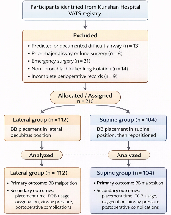
Figure 1: Flow diagram of patient selection and group allocation. Please click here to view a larger version of this figure.
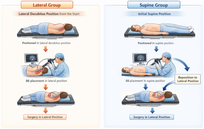
Figure 2: Schematic illustration of bronchial blocker placement in the lateral and supine groups. In the lateral group (left panel), patients were positioned in the surgically required lateral decubitus position before induction of anesthesia. After induction, the bronchial blocker was placed under fiberoptic bronchoscopy guidance while the patient remained in the lateral position, and surgery proceeded without subsequent repositioning. In the supine group (right panel), bronchial blocker placement was initially performed in the supine position after induction of anesthesia. Patients were then repositioned to the lateral decubitus position for surgery, after which fiberoptic bronchoscopy was repeated to reconfirm or adjust the blocker position before surgical incision. The schematic illustrates the procedural differences in patient positioning and bronchial blocker placement between the two study groups. The illustration was created by the authors using vector-based presentation software. Abbreviations: BB = bronchial blocker; FOB = fiberoptic bronchoscopy. Please click here to view a larger version of this figure.
| Variable | Lateral Group (n = 112) | Supine Group (n = 104) | Statistic | P value |
| Age (years) | 58.4 ± 5.3 | 59.1 ± 5.9 | t = -0.463 | 0.644 |
| Male sex (%) | 64 (57.1) | 61 (58.7) | χ2 = 0.050 | 0.822 |
| Height (cm) | 168.2 ± 6.4 | 167.9 ± 6.1 | t = 0.359 | 0.72 |
| Weight (kg) | 67.3 ± 8.9 | 66.8 ± 9.2 | t = 0.417 | 0.677 |
| BMI (kg/m2) | 23.8 ± 1.6 | 23.7 ± 1.8 | t = 0.274 | 0.784 |
| ASA physical status I/II/III, n | 18/64/30 | 16/60/28 | χ2 = 0.019 | 0.99 |
| Smoking history (%) | 35 (31.3) | 34 (32.7) | χ2 = 0.052 | 0.82 |
| Hypertension (%) | 41 (36.6) | 40 (38.5) | χ2 = 0.079 | 0.778 |
| Diabetes mellitus (%) | 19 (17.0) | 17 (16.3) | χ2 = 0.015 | 0.903 |
| COPD (%) | 13 (11.6) | 12 (11.5) | χ2 = 0.001 | 0.987 |
| FEV1 (L) | 2.34 ± 0.45 | 2.31 ± 0.48 | t = 0.472 | 0.637 |
| FEV1/FVC (%) | 78.5 ± 6.2 | 77.9 ± 6.5 | t = 0.731 | 0.466 |
| Preoperative SpO2 (%) | 98.3 ± 1.1 | 98.0 ± 1.2 | t = 0.274 | 0.801 |
| PaO2 (mmHg) | 91.4 ± 4.8 | 92.1 ± 5.3 | t = -0.473 | 0.637 |
| PaCO2 (mmHg) | 38.6 ± 3.2 | 38.9 ± 3.5 | t = -0.521 | 0.603 |
| Right-sided surgery (%) | 63 (56.3) | 60 (57.7) | χ2 = 0.046 | 0.831 |
| Lobectomy (%) | 71 (63.4) | 65 (62.5) | χ2 = 0.018 | 0.892 |
| Segmentectomy (%) | 29 (25.9) | 27 (26.0) | χ2 = 0.000 | 0.991 |
| Wedge resection (%) | 12 (10.7) | 12 (11.5) | χ2 = 0.037 | 0.847 |
| Expected surgery time (min) | 128.5 ± 24.6 | 130.1 ± 25.2 | t = -0.471 | 0.638 |
| Actual surgery time (min) | 136.2 ± 28.9 | 138.7 ± 30.1 | t = -0.642 | 0.521 |
| Total intravenous anesthesia, n (%) | 69 (61.6) | 63 (60.6) | χ2 = 0.024 | 0.877 |
| Endotracheal tube size (mm) | 7.4 ± 0.3 | 7.4 ± 0.4 | t = 0.000 | 1.000 |
| Mallampati class III, n (%) | 15 (13.4) | 14 (13.5) | χ2 = 0.001 | 0.988 |
| Limited neck mobility, n (%) | 8 (7.1) | 7 (6.7) | χ2 = 0.014 | 0.905 |
| Target mainstem bronchus (left/right), n | 49/63 | 44/60 | χ2 = 0.046 | 0.831 |
Table 1: Baseline demographic, clinical, pulmonary, anesthetic, and surgical characteristics of patients in the lateral and supine groups. Data are presented as mean ± standard deviation for normally distributed continuous variables, median (interquartile range) for nonnormally distributed continuous variables, or number (percentage) for categorical variables. Between-group comparisons were performed using the independent-samples t-test or Mann–Whitney U test for continuous variables and the chi-square test or Fisher’s exact test for categorical variables, as appropriate. All tests were two-sided, and p < 0.05 was considered statistically significant. Abbreviations: ASA = American Society of Anesthesiologists; BMI = body mass index; COPD = chronic obstructive pulmonary disease; FEV₁ = forced expiratory volume in 1 s; FVC = forced vital capacity; SpO₂ = peripheral oxygen saturation; PaO₂ = arterial partial pressure of oxygen; PaCO₂ = arterial partial pressure of carbon dioxide; TIVA = total intravenous anesthesia.
| Outcome | Lateral group (n = 112) | Supine group (n = 104) | Effect size (95% CI) | Statistic | P value |
| Any BB malposition (%) | 14 (12.5) | 32 (30.8) | RR 0.41 (0.23–0.72) | χ2 = 10.738 | 0.001 |
| Proximal migration (%) | 9 (8.0) | 20 (19.2) | RR 0.42 (0.20–0.87) | χ2 = 5.815 | 0.016 |
| Distal migration (%) | 5 (4.5) | 12 (11.5) | RR 0.39 (0.14–1.05) | χ2 = 3.722 | 0.054 |
| BB placement time (min) | 3.8 ± 0.9 | 5.6 ± 1.2 | MD -1.8 (-2.1 to -1.4) | t = -12.084 | <0.001 |
| FOB examinations (n) | 1.3 ± 0.3 | 2.1 ± 0.5 | MD -0.8 (-1.0 to -0.6) | t = -11.764 | <0.001 |
| Total FOB duration (min) | 2.6 ± 0.7 | 4.3 ± 1.0 | MD -1.7 (-2.0 to -1.4) | t = -14.022 | <0.001 |
| Anesthesia-to-incision time (min) | 18.4 ± 3.5 | 24.7 ± 4.2 | MD -6.3 (-7.4 to -5.2) | t = -12.337 | <0.001 |
Table 2: Primary and procedural outcomes comparing lateral versus supine positioning for bronchial blocker placement. Data are presented as mean ± standard deviation or number (percentage), as appropriate. Effect sizes are reported as relative risk for categorical outcomes and mean difference for continuous outcomes, with corresponding 95% confidence intervals. Between-group comparisons were performed using the chi-square test for categorical variables and the independent-samples t-test for continuous variables. All tests were two-sided, and p < 0.05 was considered statistically significant. Abbreviations: BB = bronchial blocker; FOB = fiberoptic bronchoscopy; RR = relative risk; MD = mean difference; CI = confidence interval.
| Parameter | Lateral group (n = 112) | Supine group (n = 104) | Effect size (95% CI) | Statistic | P value |
| Lowest SpO2 (%) | 93.6 ± 2.8 | 90.9 ± 3.4 | MD 2.7 (1.9 to 3.5) | t = 6.596 | <0.001 |
| PaO2 during OLV (mmHg) | 186.5 ± 22.4 | 158.3 ± 25.1 | MD 28.2 (17.5 to 38.9) | t = 6.125 | <0.001 |
| Peak airway pressure (cm H2O) | 23.4 ± 3.1 | 26.8 ± 3.6 | MD -3.4 (-4.3 to -2.5) | t = -7.298 | <0.001 |
| Intraoperative hypoxemia (%) | 9 (8.0) | 21 (20.2) | RR 0.40 (0.19–0.83) | χ2 = 6.663 | 0.01 |
| Repeated FOB required (%) | 11 (9.8) | 29 (27.9) | RR 0.35 (0.18–0.67) | χ2 = 11.661 | 0.001 |
Table 3: Intraoperative physiological parameters and airway management outcomes in the lateral and supine groups. Continuous variables are presented as mean ± standard deviation, and categorical variables are presented as number (percentage). Effect sizes are reported as mean difference or relative risk with 95% confidence intervals, as appropriate. Between-group comparisons were performed using the independent-samples t-test for continuous variables and the chi-square test for categorical variables. All tests were two-sided, and p < 0.05 was considered statistically significant. Intraoperative hypoxemia was defined as peripheral oxygen saturation <90% during one-lung ventilation lasting longer than 1 min or requiring clinical intervention. Abbreviations: SpO₂ = peripheral oxygen saturation; PaO₂ = arterial partial pressure of oxygen; OLV = one-lung ventilation; RR = relative risk; MD = mean difference; CI = confidence interval; FOB = fiberoptic bronchoscopy.
| Complication | Lateral group (n = 112) | Supine group (n = 104) | Effect size (95% CI) | Statistic | P value |
| Postoperative sore throat (%) | 12 (10.7) | 25 (24.0) | RR 0.45 (0.24–0.84) | χ2 = 6.744 | 0.009 |
| Hoarseness (%) | 7 (6.3) | 18 (17.3) | RR 0.36 (0.16–0.81) | χ2 = 6.443 | 0.011 |
| Postoperative hypoxemia (%) | 5 (4.5) | 14 (13.5) | RR 0.33 (0.12–0.88) | χ2 = 5.441 | 0.02 |
| Airway mucosal injury (%) | 3 (2.7) | 10 (9.6) | RR 0.28 (0.08–0.95) | χ2 = 4.588 | 0.032 |
Table 4: Postoperative airway-related complications in the lateral and supine groups. Data are presented as numbers (percentages). Effect sizes are expressed as relative risk with 95% confidence intervals. Between-group comparisons were performed using the chi-square test or Fisher’s exact test, as appropriate. All tests were two-sided, and p < 0.05 was considered statistically significant. Postoperative hypoxemia was defined as peripheral oxygen saturation <90% within the first 24 h after surgery. Abbreviations: RR = relative risk; CI = confidence interval; SpO₂ = peripheral oxygen saturation.
Supplementary File 1: De-identified participant-level dataset, data dictionary, and statistical analysis files. This file contains the de-identified participant-level data used in the analyses, definitions and coding rules for all variables, and information on statistical analyses. Please click here to download this file.
Discussion
The findings demonstrated that bronchial blocker (BB) placement performed directly in the lateral decubitus position was associated with greater device stability, improved procedural efficiency, more favorable intraoperative physiological parameters, and fewer airway-related complications during video-assisted thoracic surgery (VATS). Compared with conventional supine placement followed by patient repositioning, lateral placement was associated with a lower incidence of BB malposition, fewer fiberoptic bronchoscopy (FOB) interventions, shorter anesthesia-to-incision intervals, and improved oxygenation parameters. These observations supported further evaluation of a position-sensitive approach in which BB placement was aligned with the final surgical position31.
Published evidence supported the clinical rationale for avoiding patient repositioning after initial BB placement. In a multicenter randomized trial involving 306 patients, Li et al reported BB malposition in 39 of 154 patients (25.3%) following conventional supine placement compared with 1 of 152 patients (0.7%) following lateral placement (p < 0.001)31. Chen et al. similarly reported malposition rates of 29% and 11% in the supine and right lateral positions, respectively, in a randomized trial involving 90 patients32. Among patients with unsuccessful first attempts, the median placement time was 201 s in the supine group and 97 s in the lateral group. However, that trial evaluated left-sided blockade using an initial blind insertion attempt followed by bronchoscopic verification and adjustment, whereas the present study used continuous fiberoptic guidance. These procedural differences limited direct comparison of event rates. Nevertheless, the findings collectively supported positioning as a relevant determinant of blocker stability and placement efficiency.
Complications associated with conventional BB placement extended beyond displacement, although not all were attributable to positioning. Cano et al reported that four of 134 patients required conversion to a double-lumen tube because anatomical alterations prevented adequate right upper lobe collapse1. This finding indicated that inadequate lung isolation could reflect both bronchial anatomy and device position. Airway-related morbidity also warranted balanced interpretation. Chen et al32 found no between-group difference in postoperative sore throat and reported no hoarseness or clinically apparent airway trauma in either group. Thus, the lower rates of airway-related complications observed in the present cohort did not establish that supine positioning itself caused airway injury. Fewer corrective manipulations provided a plausible explanation, but the mechanism remained unconfirmed. The present findings complement existing randomized evidence by examining device stability, procedural efficiency, and perioperative outcomes in routine clinical practice. The applicability of these findings to pediatric lung resection and patients with tracheobronchial anomalies remained uncertain because these settings involved distinct anatomical and anesthetic considerations33,34.
The clinical implications were considerable. Improved BB stability contributed to more reliable one-lung ventilation, fewer interruptions for bronchoscopic verification, and a lower likelihood of hypoxemia or elevated airway pressures35. Consistent improvements in procedural efficiency, including shorter placement time, fewer bronchoscopic examinations, reduced cumulative bronchoscopy duration, and shorter anesthesia-to-incision intervals, were closely aligned with enhanced recovery after surgery principles36. These benefits extended beyond technical success, potentially improving operating room workflow by reducing procedural interruptions and streamlining perioperative processes. The parallel reductions in postoperative sore throat, hoarseness, hypoxemia, and airway mucosal injury further suggested that minimizing repeated airway manipulation could reduce procedure-related trauma and enhance postoperative recovery37. Although this approach did not inherently require additional equipment, its implementation depended on operator proficiency, coordinated positioning, and readiness to manage airway difficulties.
Several practical considerations could limit the broader adoption of lateral-position placement. The technique evaluated in the present study involved anesthesia induction and tracheal intubation in the lateral position, as well as BB placement, and therefore required proficiency in lateral airway management. This approach differed from techniques in which the trachea was intubated in the supine position before lateral positioning and blocker insertion32. The unfamiliar orientation, restricted access to the head after surgical preparation, and the need to coordinate the bronchoscope, blocker, and endotracheal tube could be challenging for less experienced teams. All procedures in the present study were performed by experienced thoracic anesthesiologists, and patients with predicted or documented difficult airways were excluded. Consequently, these findings did not establish equivalent feasibility during the learning phase or in patients with challenging airways. Although adoption patterns were not assessed, established supine workflows, training requirements, and uncertainty about performance across operators and airway conditions could help explain why promising results have not translated into universal adoption as a standard approach.
Lateral placement also did not eliminate the possibility of subsequent BB displacement. When displacement occurred, bronchoscopic adjustment could be attempted in the lateral position, but restricted access, surgical drapes, and the need to stabilize the endotracheal tube could complicate manipulation. If satisfactory positioning or ventilation could not be restored, returning the patient to the supine position could become necessary. Subsequent lateral repositioning would require additional personnel, renewed securing of the patient and airway devices, and repeat bronchoscopic verification, potentially offsetting the initial time savings and creating further opportunities for displacement. These challenges could be more pronounced in less experienced teams. The net procedural benefit, therefore, depended on both successful initial placement and the ability to manage subsequent malposition efficiently. Accordingly, lateral placement was best regarded as an option for appropriately selected patients and adequately trained teams rather than as a universal replacement for conventional practice.
The findings also highlighted the importance of considering patient positioning as a modifiable procedural variable rather than a passive background characteristic. Positioning influenced airway anatomy, bronchial alignment, and device–airway interactions, all of which could affect the performance of lung-isolation techniques18,31. Although the present study focused on BBs, the broader concept of aligning device placement with the final operative position may be relevant to other airway and thoracic procedures, including selective lobar blockade, tracheobronchial interventions, and endobronchial therapeutic procedures34,38. In minimally invasive and robotic-assisted thoracic surgery, where optimal lung collapse and stable airway management are essential for surgical exposure, improvements in device stability could have a particularly meaningful impact on clinical workflow and procedural reliability.
Several avenues for future investigation remained. Prospective randomized studies could further evaluate the relationship between positioning strategy and BB performance while standardizing operator training, placement criteria, and physiological endpoints39. Advanced imaging techniques and three-dimensional airway modeling could help characterize the anatomical changes that occur during repositioning and clarify the mechanisms responsible for device migration40,41. Multicenter studies involving different patient populations, surgical procedures, and BB designs could help determine the generalizability of the findings and identify subgroups that might derive the greatest benefit from lateral placement. Such investigations could further refine airway management protocols and strengthen the evidence base supporting position-specific deployment strategies.
Several limitations should be acknowledged. The analysis was conducted at a single institution, which could limit generalizability to centers with different patient populations, perioperative workflows, or levels of operator experience. The comparisons were unadjusted, and confounding by patient characteristics, surgical factors, and clinician selection of the positioning strategy could not be excluded. The observed associations should therefore not be interpreted as independent or causal effects of lateral positioning. Objective displacement distances were not consistently available for all malposition events; therefore, displacement severity based on a numerical distance threshold was not analyzed. All procedures were performed by experienced thoracic anesthesiologists using a single BB system within a standardized clinical workflow, thereby enhancing internal consistency but potentially limiting extrapolation to other devices or practice environments.
The present analysis did not establish the learning curve, the frequency of rescue conversions to supine positioning, or the additional time associated with such conversions, thereby limiting the assessment of the technique's practical efficiency in less-experienced teams. Induction- and intubation-specific rescue interventions and complications were not separately reported, preventing a definitive assessment of the safety of lateral induction and tracheal intubation. Furthermore, the comparison involved different positioning strategies throughout induction, intubation, and blocker placement; therefore, the observed outcomes could not be attributed solely to the position used for blocker insertion. Finally, longer-term outcomes, including postoperative pulmonary complications, hospital length of stay, and patient-reported recovery metrics, were not systematically evaluated and warrant further investigation.
In summary, BB placement performed directly in the lateral decubitus position was associated with greater device stability, improved procedural efficiency, more favorable intraoperative physiological parameters, and fewer airway-related complications than conventional supine placement followed by repositioning. These findings identified patient positioning as an important and potentially modifiable determinant of BB performance. Lateral-position placement could offer a useful alternative in selected patients when performed by experienced teams, but broader implementation would require further evaluation of training needs, rescue procedures, and overall procedural efficiency.
Disclosures
The authors declare that they have no conflicts of interest.
Materials
| Name | Company | Catalog Number | Comments |
|---|---|---|---|
| Anesthesia information management system | The First People's Hospital of Kunshan information center | AIMS-KS-2025 | Used to retrieve anesthesia induction time, surgical incision time, airway management records, intraoperative monitoring values, ventilator parameters, and bronchoscopy-related documentation. |
| Anesthesia machine with integrated ventilator | Drägerwerk AG & Co. KGaA | Fabius Plus XL | Used for general anesthesia delivery, two-lung ventilation, one-lung ventilation, airway pressure monitoring, and adjustment of inspired oxygen concentration during VATS. |
| Arterial blood gas analyzer | Radiometer Medical ApS | ABL800 FLEX | Used to measure arterial oxygen tension, arterial carbon dioxide tension, pH, and related gas-exchange variables during one-lung ventilation. |
| ASA physical status classification form | American Society of Anesthesiologists | ASA-PS-2020 | Used to classify preoperative physical status as I–III during eligibility assessment and baseline characteristic extraction. |
| Bronchial blocker displacement record sheet | Department of Anesthesiology, The First People's Hospital of Kunshan | BB-DRS-2025 | Used to document proximal migration, distal migration, cuff protrusion, loss of occlusion, timing of displacement, and whether repositioning exceeded 15 mm. |
| Bronchial blocker placement checklist | Department of Anesthesiology, The First People's Hospital of Kunshan | BB-CHECK-2025 | Used to standardize pre-insertion inspection, fiberoptic guidance, target bronchus confirmation, cuff positioning, occlusion verification, and post-placement documentation. |
| Bronchial blocker, 9 Fr | Zhejiang Tappa Medical Technology Co., Ltd. | T2504EBT005 | Disposable endobronchial blocker used for bronchial blocker-assisted one-lung ventilation; the same size and device type were used for all included procedures. |
| Capnography module | Philips Medical Systems | M3015A | Used to continuously monitor end-tidal carbon dioxide and confirm successful tracheal intubation and ventilation during two-lung and one-lung ventilation. |
| Case report form | Department of Anesthesiology, The First People's Hospital of Kunshan | CRF-BB-VATS-2025 | Used to collect demographic characteristics, airway assessment variables, surgical details, bronchial blocker procedure variables, outcomes, complications, and statistical coding information. |
| Cisatracurium besylate injection | Jiangsu Hengrui Pharmaceuticals Co., Ltd. | H20123320 | Neuromuscular blocking agent used for maintenance of muscle relaxation during general anesthesia according to institutional practice. |
| Continuous electrocardiography monitor | Philips Medical Systems | IntelliVue MX700 | Used for continuous electrocardiography, heart rate, noninvasive blood pressure, oxygen saturation, temperature, and integrated perioperative monitoring. |
| Data management spreadsheet software | Microsoft Corporation | Microsoft Excel 2021 | Used for data entry, data cleaning, table preparation, variable checking, calculation of derived fields, and preparation of summary tables; RRID: SCR_016137. |
| Dedicated study database | Department of Anesthesiology, The First People's Hospital of Kunshan | BB-VATS-DB-2025 | Locked anonymized analytical database containing eligibility variables, baseline variables, procedural outcomes, physiological parameters, and complication variables. |
| Disposable airway suction catheter | Winner Medical Co., Ltd. | SC-14F | Used to remove airway secretions when bronchoscopic visualization of the carina or target mainstem bronchus was impaired. |
| Disposable anesthesia breathing circuit | Intersurgical Ltd. | BC-ADULT-22MM | Used to connect the single-lumen endotracheal tube to the anesthesia machine during two-lung and one-lung ventilation. |
| Disposable medical gloves | Winner Medical Co., Ltd. | GL-2022 | Used during airway preparation, bronchoscopy handling, bronchial blocker insertion, and routine infection-control procedures. |
| Disposable syringe, 10 mL | Becton, Dickinson and Company | 309604 | Used to inflate the bronchial blocker cuff gradually under fiberoptic visualization. |
| Electronic medical record system | The First People's Hospital of Kunshan information center | EMR-KS-2025 | Used to retrieve demographic information, preoperative assessment, comorbidities, operative records, postoperative recovery records, and complication documentation. |
| Endotracheal tube fixation tape | 3M Health Care | 1530-1 | Used to secure the single-lumen endotracheal tube and reduce migration during bronchial blocker insertion and patient repositioning. |
| Fiberoptic bronchoscope | Olympus Medical Systems Corp. | BF-P60 | Used to visualize the carina and main bronchi, guide bronchial blocker placement, confirm cuff position, and reassess suspected malposition. |
| Fiberoptic bronchoscopy documentation form | Department of Anesthesiology, The First People's Hospital of Kunshan | FOB-DOC-2025 | Used to record bronchoscopy timing, indications, number of examinations, cumulative bronchoscopy duration, blocker position, and corrective maneuvers. |
| Flow diagram preparation software | Microsoft Corporation | Microsoft PowerPoint 2021 | Used to prepare the patient selection flow diagram and procedural schematic for lateral and supine bronchial blocker placement. |
| Intravenous catheter | Becton, Dickinson and Company | 382523 | Used to establish peripheral venous access before induction of general anesthesia. |
| Laboratory information system | The First People's Hospital of Kunshan information center | LIS-KS-2025 | Used to retrieve validated laboratory values and arterial blood gas results included in the analysis. |
| Laryngoscope | Heine Optotechnik GmbH & Co. KG | Classic+ Miller/Macintosh | Used for direct laryngoscopy when placing the single-lumen endotracheal tube. |
| Mallampati airway classification record | Department of Anesthesiology, The First People's Hospital of Kunshan | AIR-MALL-2025 | Used during preoperative airway assessment to identify Mallampati class IV anatomy for exclusion screening. |
| Noninvasive blood pressure cuff | Philips Medical Systems | M1574A | Used for repeated noninvasive blood pressure measurements during anesthesia induction, positioning, and intraoperative management. |
| One-lung ventilation monitoring record sheet | Department of Anesthesiology, The First People's Hospital of Kunshan | OLV-MON-2025 | Used to document oxygen saturation, airway pressures, end-tidal carbon dioxide, arterial oxygen tension, hypoxemia events, and ventilation adjustments during one-lung ventilation. |
| Operating table with lateral-positioning supports | Maquet GmbH | Alphamaxx 1133 | Used to maintain the patient in the surgically required lateral decubitus position and support safe repositioning in the supine group. |
| Operative report system | The First People's Hospital of Kunshan information center | ORS-KS-2025 | Used to retrieve operative side, surgical procedure type, pleural opening information, surgical incision time, and intraoperative surgical notes. |
| Opioid analgesic injection | Humanwell Healthcare Group Co., Ltd. | Sufentanil citrate H20054172 | Used as part of general anesthesia induction and maintenance according to patient characteristics and anesthesiologist preference. |
| Peripheral oxygen saturation sensor | Philips Medical Systems | M1191B | Used for continuous peripheral oxygen saturation monitoring and detection of intraoperative and postoperative hypoxemia. |
| Post-anesthesia care unit record | The First People's Hospital of Kunshan nursing department | PACU-KS-2025 | Used to document immediate postoperative oxygenation status, sore throat, hoarseness, airway discomfort, and recovery-room respiratory support. |
| Postoperative airway complication assessment form | Department of Anesthesiology, The First People's Hospital of Kunshan | AIR-COMP-2025 | Used to record sore throat, hoarseness, postoperative hypoxemia, airway mucosal injury, and additional airway-related events within 24 h after surgery. |
| PowerPoint | Microsoft Corporation | Version 2021; RRID: SCR_023631 | Used by the authors to create the schematic illustration in Figure 2 |
| Propofol injection | Fresenius Kabi | Propofol-Lipuro 1% | Intravenous hypnotic agent used for induction of general anesthesia according to institutional thoracic anesthesia practice. |
| Pulse oximeter module | Philips Medical Systems | M3001A | Used with the oxygen saturation sensor to continuously monitor oxygen saturation during induction, positioning, one-lung ventilation, and recovery. |
| Quality-control verification log | Department of Anesthesiology, The First People's Hospital of Kunshan | QC-BB-2025 | Used to document duplicate checking, missing-value review, source-document verification, discrepancy resolution, and final dataset locking. |
| Single-lumen endotracheal tube | Covidien Medtronic | Mallinckrodt Hi-Lo Oral/Nasal Tracheal Tube | Used as the primary airway conduit through which the bronchial blocker and fiberoptic bronchoscope were introduced. |
| Statistical analysis software | IBM Corp. | SPSS Statistics Version 26.0 | Used for Shapiro–Wilk testing, t tests, Mann–Whitney U tests, chi-square tests, Fisher exact tests, multivariable regression, propensity score matching, and effect estimate reporting; RRID: SCR_002865. |
| Succinylcholine injection | Shanghai Xudong Haipu Pharmaceutical Co., Ltd. | H31020546 | Neuromuscular blocking agent used to facilitate tracheal intubation during induction of general anesthesia. |
| Suction system | Allied Healthcare Products, Inc. | G180 | Used to provide airway suction and maintain bronchoscopic visualization during airway management and blocker placement. |
| Temperature probe | Philips Medical Systems | 21075A | Used for intraoperative body temperature monitoring during VATS and one-lung ventilation. |
| Thoracic surgery database | The First People's Hospital of Kunshan information center | TSDB-KS-2025 | Used to identify adult patients undergoing elective unilateral VATS between June 2022 and August 2025 and support consecutive case screening. |
| VATS thoracoscope system | Karl Storz SE & Co. KG | IMAGE1 S H3-Z | Used by the surgical team for video-assisted thoracic surgery and visual assessment of operative lung collapse after pleural opening. |
| Ventilator breathing filter | Intersurgical Ltd. | Clear-Guard 200 | Used between the airway circuit and endotracheal tube according to routine infection-control and anesthesia practice. |
| Video laryngoscope | Shenzhen Mindray Bio-Medical Electronics Co., Ltd. | UEscope VL300M | Used when video-assisted laryngoscopy was selected for single-lumen endotracheal intubation. |
References
- Cano PA et al. One-lung ventilation with a bronchial blocker in thoracic patients. BMC Anesthesiol. 2023;23(1):398.
- Gelzinis T. The Anesthetic Management of Patients Undergoing Nonintubated Video-Assisted Thoracic Surgery. Curr Anesthesiol Rep. 2021;11(4):437-445.
- Gonsette K, Tuna T, Szegedi LL. Anesthesia for robotic thoracic surgery. Saudi J Anaesth. 2021;15(3):356-361.
- Li Y, Huang H, Hang L. Comparison of efficacy and safety of different suction pressure for speeding non-ventilated lung collapse in uniport video-assisted thoracoscopic surgery: a randomized-controlled trial. BMC Surg. 2024;24(1):247.
- Li YT et al. Efficacy of rigid-angle bronchial blockers for uniportal video-assisted thoracoscopic tumor resection. Ann Card Anaesth. 2023;26(3):303-308.
- Nakanishi T et al. Combined use of the ProSeal laryngeal mask airway and a bronchial blocker vs. a double-lumen endobronchial tube in thoracoscopic surgery: a randomized controlled trial. J Clin Anesth. 2023;88:111136.
- Wang J et al. Extraluminal placement of a bronchial blocker compared with carbon dioxide artificial pneumothorax in infants undergoing video-assisted thoracoscopic surgery. Ann Thorac Cardiovasc Surg. 2022;28(1):48-55.
- Wang Y et al. Predicting postoperative complications after pneumonectomy using machine learning: a 10-year study. Ann Med. 2025;57(1):2487636.
- Xiang YY, Chen Q, Tang XX, Cao L. Comparison of the effect of double-lumen endotracheal tubes and bronchial blockers on lung collapse in video-assisted thoracoscopic surgery: a systematic review and meta-analysis. BMC Anesthesiol. 2022;22(1):330.
- Zhang Z et al. Study protocol for a randomized controlled trial assessing the effect of lateral position intubation on bronchial blocker placement during unilateral video-assisted thoracic surgery. Trials. 2025;26(1):553.
- Amjad MA, Khan A, Khan AR. Use of an endobronchial blocker where a double-lumen tube failed to ventilate: a case report of a distorted tracheobronchial anatomy. Cureus. 2022;14(11):e32047.
- Bhende VV et al. The enigma of recurrent lung abscess: management and outcomes in a school-aged child with a review of literature. Cureus. 2024;16(7):e63579.
- Gnass M et al. Transbronchial lung cryobiopsy guided by radial mini-probe endobronchial ultrasound in interstitial lung diseases—a multicenter prospective study. Adv Respir Med. 2020;88(2):123-128.
- Koo BS et al. A case of one-lung ventilation using a single-lumen tube placed under fiberoptic bronchoscopic guidance in a 4-year-old child: a case report. Medicine (Baltimore). 2020;99(34):e21737.
- Onimaru T, Kamata M, Nakagawa H. One-lung ventilation and 3D image analysis in a case of tracheal bronchus with steeply angled branching of left main bronchus: a case report. JA Clin Rep. 2022;8(1):55.
- Pollard JE, Smith DW, Morgan DE, Skaggs JD. Combined use of a Total Control Introducer and a hyperangulated video laryngoscope to place a left-sided double lumen endotracheal tube in a patient with a history of difficult laryngoscopy. BMJ Case Rep. 2021;14(10):e243407.
- Schiena CA et al. Case report: thoracic duct ligation for left-sided chylothorax after pneumonectomy with contralateral VATS procedure using indocyanine green fluorescence. Front Surg. 2025;12:1558519.
- She S et al. Transbronchial cryobiopsy in interstitial lung disease: safety of a standardized procedure. J Bronchology Interv Pulmonol. 2020;27(1):36-41.
- Somma J et al. Non-ventilated lung deflation during one-lung ventilation with a double-lumen endotracheal tube: a randomized-controlled trial of occluding the non-ventilated endobronchial lumen before pleural opening. Can J Anaesth. 2021;68(6):801-811.
- Wang YC et al. A survey of one-lung ventilation device in lung resection surgeries in Taiwan—a population-based nationwide cohort study. J Formos Med Assoc. 2020;119(1 Pt 3):449-454.
- Zhang Y et al. Laryngeal mask airway combined with visual bronchial blocker versus double-lumen tube for lung isolation in video-assisted thoracoscopic surgery: a protocol for a multicentre randomised controlled trial. BMJ Open. 2025;15(12):e110539.
- Jia H, Tan WF, Ma H, Cui Y. A novel method using a single lumen tube and extraluminal bronchial blocker for one-lung ventilation in severe tracheal stenosis: a case report. Ann Palliat Med. 2021;10(1):749-753.
- Koegelenberg CFN et al. South African Thoracic Society consensus statement on transbronchial lung cryobiopsy for interstitial lung disease. Afr J Thorac Crit Care Med. 2025;31(3):e3748.
- Liu Z, Ren S, Liu N, Luo Y. Clinical application of intubation-free anesthesia in radical resection of lung cancer. Front Med (Lausanne). 2023;10:1175437.
- Marraro GA. Selective bronchial intubation for one-lung ventilation and independent-lung ventilation in pediatric age: state of the art. Zhongguo Dang Dai Er Ke Za Zhi. 2020;22(6):543-554.
- Moreault O et al. Double-lumen endotracheal tubes and bronchial blockers exhibit similar lung collapse physiology during lung isolation. Can J Anaesth. 2021;68(6):791-800.
- Ng B, Thangavelautham S, Aman M, Harikrishnan S, Kothandan H. One-lung ventilation via end tracheostomy for thoracic surgery: a case report. Cureus. 2025;17(1):e77539.
- Ren S, Che J, Yan H, Liu Z. Accidental transection of bronchial blocker during thoracoscopic radical resection of lung cancer in two cases. Can J Anaesth. 2024;71(5):679-680.
- Templeton TW et al. Hypoxemia in young children undergoing one-lung ventilation: a retrospective cohort study. Anesthesiology. 2021;135(5):842-853.
- Horvath B et al. The evolution, current value, and future of the American Society of Anesthesiologists Physical Status Classification System. Anesthesiology. 2021;135(5):904-919.
- Li H et al. Lung isolation with a bronchial blocker placed in the lateral position for patients undergoing thoracic surgery: a multicenter, randomized clinical trial. J Clin Anesth. 2025;104:111869.
- Chen S et al. Right lateral decubitus as a strategy to improve left bronchial blocker placement in thoracic operations: a randomized clinical trial. BMC Anesthesiol. 2026;26(1):201.
- Gonçalves P et al. Anesthetic management for pediatric lung resection: a single-center retrospective analysis. Cureus. 2024;16(12):e76138.
- Hu W, Sun Z, Zhang K, Zhang X. Discussion on the application of double bronchial blocker in tracheal bronchus. Asian J Surg. 2024;47(7):3106-3107.
- Kapoor R, Owusu-Agyemang P, Thakar DR, Heir JS. External tracheal manipulation for bronchial blocker placement in children undergoing thoracic surgery requiring one lung ventilation: a case report. Ann Card Anaesth. 2021;24(1):105-107.
- Niedmers H et al. Current approaches to anesthetic management in thoracic surgery—an evaluation from the German Thoracic Registry. Anaesthesiologie. 2022;71(8):608-617.
- Weber J, Defosse JM. From two to one—techniques for one-lung ventilation. Anasthesiol Intensivmed Notfallmed Schmerzther. 2025;60(7-8):401-414.
- Xiao H et al. Effect of lung isolation with different airway devices on postoperative pneumonia in patients undergoing video-assisted thoracoscopic surgery: a propensity score-matched study. BMC Pulm Med. 2024;24(1):165.
- Zhang TH et al. A randomised comparison of the efficacy of a Coopdech bronchial blocker and a double-lumen endotracheal tube for minimally invasive esophagectomy. Transl Cancer Res. 2020;9(8):4686-4692.
- Zhao L et al. A comparison of bronchial blocker under artificial pneumothorax and double-lumen endobronchial tube for lung isolation in thoracoscopic enucleation of oesophageal leiomyoma. J Cardiothorac Surg. 2021;16(1):322.
- Cheng Q et al. The disconnection technique with the use of a bronchial blocker for improving nonventilated lung collapse in video-assisted thoracoscopic surgery. J Thorac Dis. 2020;12(3):876-882.