This protocol describes a goggle- and eyelid-suturing-free rodent model of myopia induced by subconjunctival TGF-β2 injections, which drives the NLRP3/IL-1β-MMP2 axis to cause axial elongation, while a split biometric recording regimen minimizes anesthesia artifacts for high-fidelity data.
Method Article
Establishment of a TGF-β-Induced Rodent Myopia Model Using Subconjunctival Injections
In This Article
Summary
Abstract
Myopia is a major global public health concern characterized by excessive axial elongation and scleral extracellular matrix (ECM) remodeling. While experimental models are essential for investigating these mechanisms, traditional optical defocus methods in small rodents present technical limitations. Here, a standardizable pharmacological myopia model was established and evaluated via repeated subconjunctival injections of transforming growth factor-beta (TGF-β). To achieve targeted delivery, a dual-handed surgical stabilization technique was developed to evert the upper eyelid, allowing a 33-gauge needle to target the palpebral conjunctiva at a shallow 20°–30° angle. Through in vivo screening in rats, TGF-β2 (500 pg/eye) was identified as a prominent driver of posterior eye growth. Subsequent dose-response titration in mice revealed an inverted U-shaped profile. In the primary validation cohort, the lowest dose (250 pg/eye) induced pronounced axial elongation (0.197 ± 0.012 mm) and myopic refractive shifts (−1.58 ± 0.63 D). At the molecular level, repetitive TGF-β2 administration activated the retinal NOD-like receptor family pyrin domain-containing 3 (NLRP3) / interleukin-1β (IL-1β) inflammasome cascade and upregulated matrix metalloproteinase 2 (MMP2) expression in both species. Coupled with a two-day split biometric recording regimen that minimizes anesthesia-associated artifacts, this protocol provides a reliable, scalable platform for evaluating anti-myopia interventions.
Introduction
Myopia has become a major global public health concern, with its prevalence projected to affect nearly 50% of the world's population by 20501,2. The primary pathological driver of progressive myopia is the excessive axial elongation of the eye, which causes active extracellular matrix remodeling and tissue degeneration, leading to structural thinning of the posterior ocular coats3. These dynamic alterations encompass both structural remodeling (characterized by scleral ECM degradation, collagen loss, and posterior layer thinning) and functional remodeling (marked by reduced biomechanical rigidity, altered choroidal perfusion, and aberrant retino-RPE inflammatory signaling), which collectively increase the lifelong risk of vision-threatening complications and irreversible visual impairment, including myopic maculopathy, retinal detachment, glaucoma, and premature cataracts4,5,6,7. Despite the substantial clinical and socioeconomic burdens of this condition, the precise upstream molecular mechanisms governing aberrant ocular growth remain incompletely understood. Therefore, elucidating the signaling pathways that drive axial elongation and developing reproducible, standardized animal models are critical steps toward identifying novel therapeutic targets and managing the global myopia epidemic.
While form-deprivation myopia (FDM) and lens-induced myopia (LIM) serve as gold standards for investigating optical defocus and emmetropization8,9,10,11, implementing these optical defocus models in small rodents—particularly mice—presents substantial technical challenges. The active grooming behavior of mice frequently leads to goggle detachment, necessitating stressful countermeasures like protective collars12. Furthermore, mechanical friction, eyelid suturing, and altered microenvironments beneath the occluder routinely cause secondary corneal complications, such as opacity or corneal ulceration13,14,15. While FDM and LIM remain foundational paradigms for investigating visual defocus, secondary ocular surface complications—such as corneal opacity, mechanical friction, or eyelid suture irritation—may interfere with optical biometric measurements15. These procedural artifacts compromise tear-film stability and corneal transparency, leading to higher experimental exclusion rates and increased measurement noise13,16. In contrast, pharmacological induction bypasses physical occluders, preserving corneal clarity and facilitating reliable, reproducible optical and ultrasonic biometry. In myopia, excessive ocular elongation is driven by altered extracellular matrix (ECM) remodeling of the posterior sclera17, where transforming growth factor-beta (TGF-β) isoforms act as key regulators of collagen and proteoglycan synthesis18,19. Emerging evidence suggests that TGF-β signaling may intersect with low-grade ocular inflammation and matrix metalloproteinase 2 (MMP2)-mediated matrix degradation20, potentially contributing to scleral remodeling and axial elongation21,22,23,24. This is supported by elevated levels of transforming growth factor-beta 2 (TGF-β2), interleukin-6 (IL-6), and MMP2 reported in the aqueous humor of myopic eyes25,26,27, as well as the activation of the TGF-β / nuclear factor kappa B (NF-κB) / MMP2 axis in experimental models20,21. While these findings reflect dynamic intraocular molecular signaling during myopic progression, the relative anatomical contributions to aqueous humor markers—whether from local anterior secretion, subconjunctival depot flux, or posterior vitreous diffusion—remain to be fully elucidated. However, the scope and inherent limitations of this pharmacological model must be recognized. Because subconjunctival TGF-β delivery bypassed optical defocus and visual processing pathways, this model is most appropriate for dissecting downstream biochemical cascades—specifically TGF-β2 / NOD-like receptor family pyrin domain-containing 3 (NLRP3) / interleukin-1β (IL-1β) / MMP2-mediated scleral ECM remodeling—and screening candidate therapeutic agents targeting inflammatory or matrix degradation pathways. Conversely, it is not appropriate for investigating upstream visual signal transduction, retina-to-sclera optical signaling, or environmental light-driven emmetropization mechanisms, where traditional FDM or LIM models remain indispensable.
To address these methodological gaps, a myopia model was established and validated using repeated subconjunctival injections of TGF-β in rats and mice, offering a refined approach that minimizes mechanical trauma and ocular surface irritation. By circumventing the need for physical occluders, this protocol provides precise control over pharmacological dosing and experimental timelines while substantially reducing potential behavioral confounders associated with head-mounted hardware. In this study, the potency of three recombinant human TGF-β isoforms was evaluated (which have validated functional cross-reactivity in rodent tissues supported by sequence conservation28) in rats and mapped the non-linear dose-response kinetics of TGF-β2 in mice. Importantly, this standardized regimen efficiently induced phenotypic myopia—characterized by concurrent axial elongation and myopic refractive shifts—and activated the retinal NOD-like receptor family pyrin domain-containing 3 (NLRP3) / interleukin-1β (IL-1β) inflammasome and MMP2 axis. Ultimately, this protocol provides a viable, molecularly defined framework for targeting downstream scleral remodeling while detailing its specific operational boundaries.
Protocol
All animal experiments were conducted in accordance with the guidelines and regulations of the Institutional Animal Care and Use Committee (IACUC) of China Medical University (CMU). All procedures complied with the Association for Research in Vision and Ophthalmology (ARVO) Statement for the Use of Animals in Ophthalmic and Vision Research. Full approval from the CMU-IACUC was obtained prior to the initiation of all experiments. Animals were euthanized at the experimental endpoint in strict accordance with the guidelines of the Institutional Animal Care and Use Committee (IACUC) of China Medical University (CMU) and local animal welfare regulations.
1. Animals and ethical approval
- Obtain male Brown Norway (BN) rats (3–4 weeks of age, 60–80 g) and male C57BL/6 (B6) mice (3–4 weeks of age, 15–20 g) from an appropriate laboratory animal supplier.
- House all animals in a controlled environment (12 h light/dark cycle, 22 °C ± 2 °C, 50% ± 10% relative humidity) with ad libitum access to standard chow and water.
- Allow a minimum of 3 days of acclimatization prior to initiating any experimental procedure.
- Assign rats or mice randomly to treatment groups: vehicle control group and TGF-β2 injection group. Use a minimum of N = 5 animals per cohort to ensure adequate statistical power.
NOTE: A between-subjects design was used. Both eyes of an individual animal received identical treatments (either bilateral vehicle control [1–2 µL BSS] or bilateral active rhTGF-β2).- Measure bilateral ocular parameters (axial length and refractive error) for each animal.
NOTE: Ensure written CMU-IACUC approval is secured and that all personnel performing procedures are appropriately trained and certified.
- Measure bilateral ocular parameters (axial length and refractive error) for each animal.
2. Preparation of TGF-β2 solution
NOTE: Perform all reconstitution and dilution steps inside a certified biosafety cabinet using strict aseptic techniques.
- Reconstitute lyophilized recombinant human TGF-β2 in sterile 50% glycerol containing 0.1% bovine serum albumin (BSA) to prepare a 100 µg/mL stock solution.
- Prepare working solutions by performing serial dilutions of the stock solution in sterile Balanced Salt Solution (BSS).
- For rat cohorts (500 pg dose), dilute the stock solution to a final concentration of 250 ng/mL (250 pg/µL) for a 2 µL injection volume per eye.
- For mouse dose-response cohorts, prepare three independent working solutions at concentrations of 250 ng/mL, 375 ng/mL, and 500 ng/mL (250, 375, and 500 pg/µL) for a 1 µL injection volume per eye.
- Prepare single-use 15–20 µL aliquots in sterile microcentrifuge tubes, snap-freeze in liquid nitrogen, and store at -20 °C. Thaw aliquots on ice immediately prior to use.
CAUTION: Liquid nitrogen can cause severe cold burns and presents an asphyxiation hazard. Wear appropriate cryogenic PPE and handle it in a well-ventilated area in accordance with institutional safety procedures. - Prepare the vehicle control solution in parallel using sterile BSS supplemented with an equivalent matrix of 50% glycerol and 0.1% BSA.
3. Subconjunctival injection procedure
- Place animals in an induction chamber with 3–4% isoflurane for anesthetic induction. Subsequently, transfer the animal to a nose cone delivering 1.5–2% isoflurane for anesthetic maintenance.
CAUTION: Isoflurane is a volatile anesthetic. Use an appropriate scavenging system and handle it in accordance with institutional occupational health and safety procedures. - Verify an adequate depth of anesthesia by confirming the absence of a toe-pinch withdrawal reflex.
- Position the animal prone under a fiber-optic spotlight. Gently grasp, elevate, and slightly evert the superior eyelid using fine-tipped microforceps to fully expose the palpebral conjunctiva and conjunctival fornix (Figure 1B).
- Pre-wet a 10 µL glass micro-syringe and 33-gauge needle 2–3 times with the working solution to eliminate dead space. Load the target volume (2 µL for rats or 1 µL for mice) and purge all microbubbles.
CAUTION: Handle needles and other sharps carefully and dispose of them in an approved sharps container in accordance with institutional regulations. - Orient the 33-G needle with the bevel facing upward (bevel-up) and target the designated insertion site on the exposed palpebral conjunctival surface (Figure 1B,C).
- Insert the needle tip through the conjunctival epithelium at a shallow 20°–30° angle (Figure 1C).
- Gently advance the needle through the narrow submucosal space past the conjunctival fornix into the palpebral subconjunctival domain (Figure 1C).
CRITICAL STEP (structures to avoid): Strictly avoid penetrating the underlying meibomian glands or accidentally perforating deeper ocular structures. Visually confirm proper superficial placement under the spotlight by ensuring the needle bevel remains visible beneath the translucent conjunctival surface throughout advancement. - Slowly inject the solution over 5–10 s. Hold the needle in place for an additional 5 s before slowly withdrawing to prevent fluid backflow.
NOTE: Proper subconjunctival delivery is confirmed by immediate localized conjunctival elevation and blanching (bleb formation).- If a bleb fails to form or significant leakage occurs, perform a single re-injection at the inferior palpebral conjunctival space and record the deviation in the experimental log.
NOTE: Limit re-injections strictly to one attempt per eye per session; exclude animals with persistent delivery failure from longitudinal analysis.
- If a bleb fails to form or significant leakage occurs, perform a single re-injection at the inferior palpebral conjunctival space and record the deviation in the experimental log.
- Sanitize the micro-syringe cavity between individual animal injections within the same group by flushing 3 times with sterile 70% ethanol, followed by 5 times with sterile saline.
- Inspect the 33-G needle tip under magnification every 3–5 injections, and replace the needle immediately if tip dulling or bending occurs.
- Inject all vehicle-control animals first, before handling or injecting treatment cohorts, to eliminate the risk of pharmacological cross-contamination. Repeat the syringe sanitization cycle prior to transitioning to treatment groups.
- Rinse the syringe barrel and plunger with a dedicated micro-syringe cleaning solution, then with ultrapure water and absolute ethanol after each experimental session, and store dry.
- Repeat subconjunctival injections once weekly for 3 consecutive weeks (Days 0, 7, and 14). Perform longitudinal biometric assessments at baseline (Days −1 and 0) and endpoint (Days 20 and 21) as detailed in Section 4.
- Place the animal in a warm recovery cage lined with paper towels following the injection. Monitor the animal continuously until full sternal recumbency and normal ambulatory activity are regained.
- Inspect eyes under a stereomicroscope post-recovery to exclude intraocular hemorrhage, corneal opacity, or acute mechanical trauma. Monitor animals daily for signs of distress or localized infection.
4. Model validation methods
- Ocular refractive error assessment
- Place rodents on a thermostatically controlled heating pad to maintain physiological body temperature and prevent anesthetic cataracts. Maintain animals under light isoflurane anesthesia (1–1.5%) via a nose cone to ensure consistent anesthetic depth.
- Instill 1 drop of Mydrin-P (0.5% tropicamide and 0.5% phenylephrine hydrochloride) into each eye 10–15 min prior to measurement to induce complete cycloplegia and pupillary dilation.
- Position the animal in sternal recumbency under dim, indirect ambient lighting. Apply sterile BSS continuously right up to the moment of optical acquisition to preserve corneal transparency.
- Measure ocular refraction using an automated infrared photorefractometer within a strict 5–10 min window post-anesthetic induction. For BN rats, gently retract the eyelids manually to fully expose the dilated pupil during acquisition.
- Acquire three independent optical readings per eye and average the values to determine the baseline or endpoint refractive error.
CRITICAL STEP (Timing & Sequence): Conduct photorefraction on Day 1 of the evaluation sequence (Day –1 for baseline; Day 20 for endpoint) under dim lighting prior to any contact procedures. Performing photorefraction after contact ultrasonography or acoustic gel application severely disrupts corneal optics, scatters infrared light, and invalidates automated refractive readings.
- Axial length measurement via high-frequency B-scan ultrasonography
- On the following day (Day 0 for baseline or Day 21 for endpoint), evaluate ocular axial length under light isoflurane anesthesia (1–1.5%) using a handheld 16 MHz linear ultrasound probe calibrated to rodent tissue acoustic velocity (1532–1550 m/s).
- For B6 mice (direct corneal interface): Apply a baseline protective layer of Ginpol carbomer gel (0.2%) directly onto the cornea, followed by a layer of standard acoustic gel.
NOTE: Ensure the gel layer completely covers the corneal surface without introducing trapped air bubbles, which cause acoustic attenuation. - For BN Rats (transpalpebral interface): Position the animal with closed eyelids, apply ultrasound gel onto the outer eyelid surface, and acquire measurements transpalpebrally.
NOTE: Transpalpebral measurement eliminates direct corneal contact, significantly reducing the risk of corneal abrasion in larger rodent eyes.
- For B6 mice (direct corneal interface): Apply a baseline protective layer of Ginpol carbomer gel (0.2%) directly onto the cornea, followed by a layer of standard acoustic gel.
- Align the ultrasound probe and measure axial length along the central optical axis from the anterior ocular interface to the posterior scleral echo wall.
- Acquire ten independent replicate B-scan traces per eye and calculate the mean axial length.
- Gently flush residual acoustic gel from the eye using sterile BSS immediately following acquisition to prevent post-procedural corneal irritation.
CRITICAL STEP (zero-pressure touch): Place the ultrasound probe onto the gel interface with an extremely light, zero-pressure touch. Any downward physical pressure on the corneal apex artificially compresses the anterior chamber depth and introduces false axial length shortening. - Record the averaged axial length and refractive error measurements for subsequent analysis.
- On the following day (Day 0 for baseline or Day 21 for endpoint), evaluate ocular axial length under light isoflurane anesthesia (1–1.5%) using a handheld 16 MHz linear ultrasound probe calibrated to rodent tissue acoustic velocity (1532–1550 m/s).
Results
Establishment and anatomical validation of the subconjunctival injection model
To address technical variability associated with optical goggling or eyelid suturing, the subconjunctival space was validated between the palpebral and bulbar conjunctivae as a targeted localized drug reservoir (Figure 1A). Anatomical access via the superior fornix provided consistent exposure of the target site while maintaining ocular surface integrity (Figure 1B). Successful model establishment was confirmed by specific operational and biometric criteria, including the immediate transillumination verification of a localized, fluid-filled submucosal bleb (chemosis) over the target palpebral subconjunctival space without scleral perforation, tissue hemorrhage, or meibomian gland injury (Figure 1C), as well as longitudinal biometric verification of concurrent axial elongation (Δ AL > 0.15 mm in mice; >0.40 mm in rats) accompanied by a myopic refractive shift (Δ Refraction < −1.0 D) at Day 21 relative to vehicle-treated controls. Conversely, unsuccessful or suboptimal outcomes were identified by specific technical and physiological failure modes. Technical failure—such as premature fluid reflux upon needle withdrawal or inadvertent deep eyelid/orbital tissue penetration—resulted in minimal or absent bleb formation, leading to inadequate pharmacological retention and failure to induce myopic shifts; animals displaying persistent leakage were documented and excluded according to Protocol Step 3.8.1. Additionally, ocular surface complications from deep needle misplacement or excessive mechanical traction caused localized subconjunctival hemorrhage or corneal trauma, introducing confounding inflammatory responses. Suboptimal phenotypic outcomes were also observed during dose-response evaluations, where administration of suprathreshold concentrations (e.g., 500 pg/eye in mice) yielded an attenuated U-shaped response with reduced axial elongation, underscoring the physiological requirement for strict dose optimization. Verified localized drug retention under these standardized criteria ensured high-fidelity tissue targeting for downstream biometric and molecular analyses.
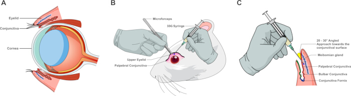
Figure 1: Anatomical landmarks and schematic illustration of the subconjunctival injection procedure in rodents. (A) Sagittal cross-section of the rodent eye depicting primary anatomical landmarks, including the eyelid, cornea, conjunctival sac, and targeted subconjunctival space. (B) Surgical demonstration of tissue stabilization and palpebral conjunctiva exposure using microforceps; the blue dot indicates the target needle insertion site for microinjection using a 33-gauge syringe. (C) High-magnification schematic illustrating the needle orientation and insertion trajectory relative to the palpebral conjunctival surface, conjunctival fornix, and underlying ocular structures. Please click here to view a larger version of this figure.
Screening of TGF-β isoforms in rats and dose-response titration of TGF-β2 in mice for axial elongation
Experiments were conducted in sequential, independent experimental cohorts comprising the dose-screening cohorts (Figure 2) and primary validation cohorts (Figure 3). Bilateral parameters were measured across cohorts, with individual animals (N) designated as the primary biological replicate. Inter-eye dependencies (OD versus OS within the same animal) were accounted for using two-way repeated-measures ANOVA or mixed-effects models, where appropriate. Continuous data are presented as mean ± SD. Differences between control and treatment groups were evaluated using two-tailed Student's t-tests, one-way ANOVA, or two-way repeated-measures ANOVA, where appropriate. A p-value < 0.05 was considered statistically significant.
To determine the most potent transforming growth factor-β (TGF-β) isoform and establish an optimal dosage regimen for inducing axial elongation, the effects of repeated subconjunctival injections in rat and mouse cohorts from baseline (Day 0) to Day 21 (Figure 2) were evaluated. Biometric recordings were performed using the two-day split regimen to prevent anesthesia-induced artifacts.
Three recombinant human TGF-β isoforms (TGF-β1, TGF-β2, and TGF-β3) were first screened in the rat cohort at a standardized concentration of 500 pg/eye (Figure 2A). One-way ANOVA, followed by post hoc multiple comparisons, revealed that all three isoforms significantly stimulated posterior ocular growth compared with the vehicle-treated control group over the 21-day period. While both TGF-β1 (0.444 ± 0.042 mm) and TGF-β3 (0.454 ± 0.049 mm) promoted significant axial elongation (p < 0.05 and p < 0.01 versus the control of 0.328 ± 0.071 mm, respectively), TGF-β2 induced the greatest axial length increase (0.486 ± 0.074 mm) and demonstrated the highest statistical significance (p < 0.001), establishing it as the most effective inducer of the myopic phenotype.
Based on the rat screening results, a dose-response titration of TGF-β2 in the mouse cohort was performed, testing three concentrations: 250, 375, and 500 pg/eye (Figure 2B). Subconjunctival TGF-β2 delivery resulted in an inverted U-shaped dose-response profile. The lowest dose (250 pg/eye) yielded the most significant acceleration in axial elongation compared to vehicle controls (0.177 ± 0.038 mm versus 0.044 ± 0.011 mm; p < 0.0001). At higher doses (375 and 500 pg/eye), the magnitude of axial length change progressively diminished, although it remained significantly higher than controls (0.123 ± 0.029 mm; p < 0.001 and 0.095 ± 0.024 mm; p < 0.01, respectively).
Together, these data establish a weekly subconjunctival regimen of 500 pg/eye for rats and 250 pg/eye for mice as the optimal parameters for subsequent structural and molecular investigations.
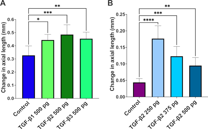
Figure 2: Dose-selection screening of transforming growth factor-β (TGF-β) isoforms and concentrations on axial length in rat and mouse eyes. (A) Change in axial length in rats (initial screening cohort), calculated as the difference between baseline (Day 0, before the first injection) and the final endpoint (Day 21), after three sequential subconjunctival injections of TGF-β1, TGF-β2, or TGF-β3 (500 pg/eye administered on Days 0, 7, and 14), compared with vehicle-treated controls. (B) Change in axial length in mice (dose-screening cohort), calculated as the difference between baseline (Day 0) and Day 21, after three sequential subconjunctival injections of TGF-β2 at three distinct concentrations (250, 375, or 500 pg/eye administered on Days 0, 7, and 14), compared with vehicle-treated controls. Data are presented as mean ± SD (N = 5 animals/group, representing 10 eyes per group). Statistical significance was evaluated by one-way ANOVA followed by post hoc multiple comparisons; *p < 0.05, **p < 0.01, ***p < 0.001, ****p < 0.0001 versus the vehicle control group. Please click here to view a larger version of this figure.
Repeated subconjunctival TGF-β2 injections induce ocular axial elongation and myopic shifts in rats and mice
To evaluate the biometric impact of repetitive subconjunctival TGF-β2 delivery (3 weekly injections), longitudinal changes in ocular axial length and refractive error from baseline to Week 3 in both rat and mouse models (Figure 3) were monitored. The two-day split protocol (refractive status captured on Day 20, followed by B-scan ultrasonography on Day 21) minimized potential anesthetic artifacts and ocular stress. Both species exhibited phenotypic shifts consistent with experimental myopia development following this multi-dose regimen.
In the rat cohort, animals receiving repeated subconjunctival injections of 500 pg/eye TGF-β2 showed a significantly greater increase in axial length over the 21-day period compared to vehicle controls (0.466 ± 0.045 mm versus 0.388 ± 0.032 mm; p < 0.01; Figure 3A). This elongation was accompanied by a significant myopic shift in refractive error (p < 0.05; Figure 3B). While vehicle-control eyes exhibited an expected slight hyperopic drift (+0.31 ± 0.93 D), TGF-β2–treated eyes transitioned into a myopic refractive state (−1.38 ± 0.91 D).
Consistent with the rat data, mice treated with 250 pg/eye TGF-β2 also demonstrated distinct structural and refractive changes. Axial elongation was significantly enhanced in TGF-β2–treated mice compared to vehicle controls (0.197 ± 0.012 mm versus 0.110 ± 0.014 mm; p < 0.0001; Figure 3C). Furthermore, this structural elongation resulted in a significant myopic shift (p < 0.05; Figure 3D), effectively reversing the natural hyperopic refraction seen in control mice (+2.25 ± 1.77 D) to a myopic profile (−1.58 ± 0.63 D).
To verify inter-ocular response symmetry and confirm that pooling bilateral ocular measurements does not introduce statistical bias, axial elongation dynamics were evaluated separately for left (OS, n = 5) and right (OD, n = 5) eyes within each group (Supplementary Figure 1). In both rats (Supplementary Figure 1A) and mice (Supplementary Figure 1B), no statistically significant difference was observed between OS and OD eyes within either the vehicle-control or TGF-β2–treated cohorts, confirming symmetrical response dynamics across eyes. Furthermore, both OS and OD eyes in TGF-β2–treated animals demonstrated consistent and robust axial elongation relative to their corresponding vehicle controls (p < 0.05 for rats; p < 0.0001 for mice), supporting the validity of accounting for bilateral measurements in the primary biometric analysis.
Collectively, these findings demonstrate that repeated subconjunctival administration of TGF-β2 effectively drives posterior ocular growth and a corresponding myopic refractive shift in both rodent models (Figure 3 and Supplementary Figure 1).
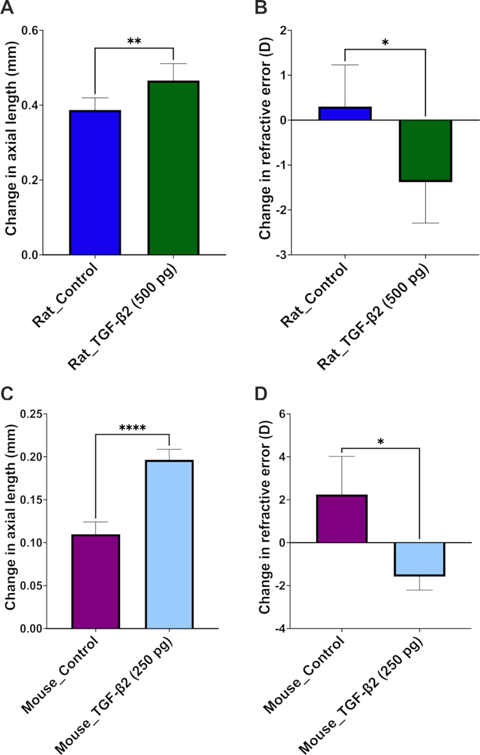
Figure 3: Primary validation of TGF-β2 effects on axial elongation and refractive error in independent rat and mouse cohorts. (A) Change in axial length in rats, calculated as the difference between baseline and the Week 3 final endpoint in eyes receiving subconjunctival TGF-β2 (500 pg/eye administered sequentially on Days 0, 7, and 14) or vehicle control. (B) Change in refractive error in the same rat eyes, delineating a significant myopic shift in the TGF-β2-treated group compared with the vehicle-control group. (C) Change in axial length in mice (independent primary validation cohort), calculated as the difference between baseline and the Week 3 final endpoint in eyes receiving subconjunctival TGF-β2 (250 pg/eye administered on Days 0, 7, and 14) or vehicle control. (D) Change in refractive error in the same mouse eyes, revealing a corresponding negative dioptric shift under optimal TGF-β2 induction. Data are presented as mean ± SD (N = 5 animals/group, representing 10 eyes per group). Statistical significance was rigorously evaluated using unpaired two-tailed t-tests; *p < 0.05, **p < 0.01, ****p < 0.0001 versus the corresponding vehicle control group. Please click here to view a larger version of this figure.
Subconjunctival TGF-β2 administration upregulates scleral MMP2 expression during experimental myopia progression
To assess downstream tissue remodeling along the retino-scleral axis, matrix metalloproteinase 2 (MMP2) expression in scleral cross-sections following subconjunctival TGF-β2 administration (Figure 4) was evaluated. Quantitative immunofluorescence analysis of posterior ocular sections in the rat model demonstrated a significant upregulation of MMP2 protein levels within the scleral stroma of TGF-β2–treated eyes (500 pg/eye) at Day 21 compared with vehicle controls (p < 0.0001; Figure 4A, B).
A prominent MMP2 fluorescent signal was localized throughout the scleral matrix underlying the retinal pigment epithelium (RPE) and choroidal layers, confirming localized MMP2-associated extracellular matrix (ECM) remodeling in the outer ocular coat during experimental myopia development. These structural changes in the sclera prompted further investigation into upstream molecular alterations within the adjacent retina.
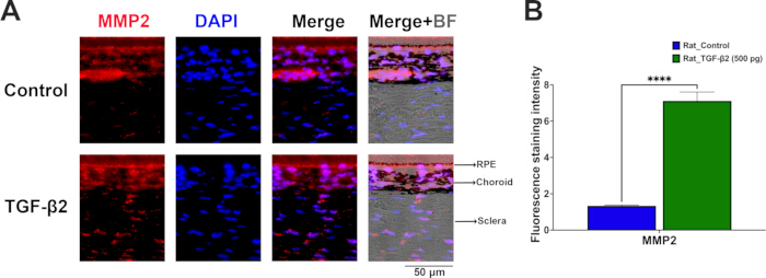
Figure 4: Subconjunctival TGF-β2 administration upregulates MMP2 expression in the rat sclera. (A) Representative immunofluorescence images of posterior ocular cross-sections showing scleral MMP2 expression (red) in vehicle-control and TGF-β2–treated (500 pg/eye) rats at Day 21. Cell nuclei were counterstained with DAPI (blue). Brightfield overlay (Merge+BF) delineates the anatomical boundaries of the RPE, choroid, and sclera. Scale bar = 50 µm. (B) Quantitative analysis of fluorescence staining intensity confirming a significant increase in scleral MMP2 expression in TGF-β2–treated rat eyes. Data are presented as mean ± SD (N = 3 independent biological replicates per group, 1 eye per animal). Statistical significance was evaluated using an unpaired two-tailed t-test; ****p < 0.0001 versus the vehicle control group. Abbreviations: RPE, retinal pigment epithelium; BF, brightfield. Please click here to view a larger version of this figure.
TGF-β2 induction upregulates MMP2 and inflammasome-related proteins in both rat and mouse retinas
To investigate the downstream molecular alterations induced by repetitive subconjunctival injections, immunofluorescence staining on posterior ocular sections from both Brown Norway rats and C57BL/6 mice at Day 21 (Figure 5A–D) was performed. All evaluations were performed on sagittal retinal sections strictly aligned to the optic nerve head to ensure consistent spatial orientation. Quantitative analysis revealed that weekly TGF-β2 administration significantly altered specific protein expression profiles across the retinal laminae in both species compared to vehicle controls (Figure 5E,F).
In the rat model, administration of 500 pg/eye TGF-β2 led to a significant increase in retinal TGF-β2 levels (p < 0.0001; Figure 5A,E). This upregulation was accompanied by elevated expression of matrix metalloproteinase 2 (MMP2) (p < 0.0001; Figure 5B,E), indicating active extracellular matrix remodeling. Furthermore, TGF-β2 treatment activated the retinal inflammatory cascade, evidenced by the upregulation of the inflammasome sensor NLRP3 (p < 0.0001; Figure 5C,E) and the pro-inflammatory cytokine IL-1β (p < 0.0001; Figure 5D,E).
Similarly, mice treated with 250 pg/eye TGF-β2 exhibited consistent upward trends in these biomarkers (Figure 5F). Retinal TGF-β2 (p < 0.01; Figure 5A,F) and MMP2 (p < 0.001; Figure 5B,F) expressions were significantly elevated. The inflammasome pathway was also activated in the mouse retina, with significant increases in NLRP3 expression (p < 0.01; Figure 5C,F) and a pronounced upregulation of IL-1β (p < 0.0001; Figure 5D,F) compared to near-undetectable baseline levels in the control group.
Together, these findings demonstrate that subconjunctival TGF-β2 administration promotes retinal MMP2-mediated matrix remodeling drivers and activates the NLRP3/IL-1β inflammasome pathway in the posterior retina of both rodent species.
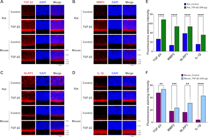
Figure 5: Repeated subconjunctival TGF-β2 administration upregulates MMP2 and inflammasome-related protein expression in the posterior retina of rats and mice. (A–D) Representative immunofluorescence images of retinal sagittal sections from control and TGF-β2–treated groups in rats (upper panels) and mice (lower panels), stained for (A) TGF-β2, (B) MMP2, (C) NLRP3, and (D) IL-1β (red), with nuclei counterstained by DAPI (blue). Scale bar = 50 µm. The representative fields selectively showcase the alterations within the outer retinal layers, highlighting prominent protein expression spanning from the outer plexiform layer (OPL) down to the basal retinal pigment epithelium (RPE) complex. (E) Quantification of fluorescence staining intensity for TGF-β2, MMP2, NLRP3, and IL-1β in rat retinas from the control group and the TGF-β2–treated group (500 pg). (F) Quantification of fluorescence staining intensity for TGF-β2, MMP2, NLRP3, and IL-1β in mouse retinas from the control group and the TGF-β2–treated group (250 pg). Data are presented as mean ± SD (N = 3 independent biological replicates per group, 1 eye per animal). Statistical significance was rigorously evaluated using unpaired two-tailed t-tests; **p < 0.01, ***p < 0.001, ****p < 0.0001 versus the respective vehicle-control group. Abbreviations: OPL, outer plexiform layer; ONL, outer nuclear layer; PIS, photoreceptor inner segments; POS, photoreceptor outer segments; RPE, retinal pigment epithelium. Please click here to view a larger version of this figure.
Supplementary Figure 1: Inter-ocular symmetry and evaluation of bilateral data pooling following subconjunctival TGF-β2 administration. (A) Comparative analysis of axial length changes between left eyes (OS, n = 5) and right eyes (OD, n = 5) in rats receiving vehicle control or bilateral subconjunctival TGF-β2 (500 pg/eye). (B) Corresponding inter-ocular comparison between OS (n = 5) and OD (n = 5) eyes in mice under vehicle control or bilateral TGF-β2 treatment (250 pg/eye). Statistical testing confirmed no significant difference between OS and OD eyes within the same treatment cohort, validating that bilateral measurement pooling does not introduce interocular bias. Simultaneously, both OS and OD eyes in TGF-β2–treated animals exhibited significant axial elongation relative to their respective vehicle controls. Data are presented as mean ± SD (N = 5 animals per group, representing 5 OS and 5 OD eyes per group). Statistical significance was evaluated using two-way repeated-measures ANOVA followed by Sidak’s post hoc test; *p < 0.05, ****p < 0.0001 versus the corresponding vehicle control. Abbreviations: OS, oculus sinister (left eye); OD, oculus dexter (right eye). Please click here to download this file.
Supplementary Figure 2: Safety evaluation of structural retinal integrity and electrophysiological function following subconjunctival TGF-β2 administration. (A) Representative hematoxylin and eosin (H&E)-stained sagittal retinal cross-sections from control and TGF-β2–treated mice (250 pg/eye) and rats (500 pg/eye) at Day 21, demonstrating preserved retinal layer architecture across species (N = 3 independent biological replicates per group). Scale bars = 20 µm. (B) Representative scotopic electroretinography (ERG) waveforms recorded from mice at Day 21, comparing vehicle control eyes (grey line) and TGF-β2–treated eyes (red line). Comparable a-wave and b-wave response dynamics confirm preserved retinal light responsiveness and electrophysiological safety (N = 3 mice per group, representing 6 eyes per group). Abbreviations: GCL, ganglion cell layer; INL, inner nuclear layer; ONL, outer nuclear layer. Please click here to download this file.
Discussion
A notable finding in the study is the non-linear, inverted U-shaped dose-response profile elicited by TGF-β2 in the mouse cohort, where the lowest administered dose (250 pg/eye) induced the most prominent axial elongation (Figure 2B). This biphasic phenomenon is characteristic of many potent cytokine signaling pathways, including the TGF-β superfamily29,30. Excessively high concentrations of exogenous TGF-β2 may lead to receptor saturation or trigger intracellular negative feedback loops—such as the upregulation of inhibitory SMAD family member 7 (Smad7) or receptor-mediated endocytosis—thereby attenuating downstream biological responses31,32. The immunofluorescence data further support this pharmacological window; while the 250 pg dose in mice (and 500 pg in rats) resulted in a moderate elevation in retinal TGF-β2 retention, it simultaneously triggered a robust downstream inflammatory cascade (Figure 5F). This suggests that the optimized low-dose threshold resides within a highly sensitive physiological window, maximizing targeted tissue efficacy while avoiding ligand-induced desensitization or localized suppression33. Therefore, these findings highlight the necessity of rigorous dose titration when establishing reproducible rodent models of chemical-induced myopia.
Compared with traditional form-deprivation myopia (FDM) or hyperopic defocus lens-induced myopia (LIM) models—which require customized hardware and are prone to lens displacement, grooming-induced detachment, and corneal complications—the subconjunctival protocol offers direct pharmacological control and minimal equipment costs13,34. Consistent with prior work showing that periocular delivery modulates scleral and retinal TGF-β signaling35, these data demonstrate posterior-segment biological responses following anterior subconjunctival TGF-β2 administration (Figure 4 and Figure 5). This efficient posterior targeting is attributed to the unique ocular scale and fluid dynamics of small rodents. Given the compact axial length of rodent eyes (~3 mm in mice and ~6 mm in rats), subconjunctival administration bathes a relatively large proportion of the episcleral plane. Driven by high transscleral permeability and the anatomical continuity of the sub-Tenon space, the injected formulation may diffuse across the sclera and choroid toward the posterior pole. Crucially, this route is associated with biological effects in posterior ocular tissues while avoiding the severe risks of globe perforation and optic nerve injury inherent to deep posterior sub-Tenon or intravitreal injections in small rodents. This approach avoids the mechanical and behavioral confounds of head-mounted lenses13,36, as well as the well-documented risks of retinal detachment and endophthalmitis associated with intravitreal injection37,38. Furthermore, partitioning the biometric measurements into two brief sessions improves upon conventional single-session paradigms. Prolonged anesthetic exposure frequently causes ocular disruptions—such as anesthetic cataracts in mice and spontaneous palpebral closure in rats—which compromise the accuracy of refractive and axial measurements39,40. By separating the refractive error (Days –1 and 20) and axial length (Days 0 and 21) assessments, the protocol is designed to minimize potential cumulative ocular stress and maintain consistent imaging conditions, which may help mitigate intra-experimental variability. To further eliminate potential confounding variables arising from systemic cross-contamination—such as systemic vascular absorption or lymphatic transport affecting non-target tissue—as well as contralateral sympathetic responses, a strictly between-subject design was implemented. By administering bilateral vehicle control or bilateral active rhTGF-β2 within individual animals, this paradigm prevents interocular contamination while ensuring robust pharmacological evaluation. Consistently, separate biometric analyses of the paired left and right eyes confirmed high inter-ocular symmetry, validating that bilateral data pooling did not introduce statistical or physiological bias across treatment cohorts (Supplementary Figure 1).
This rodent model effectively recapitulates the TGF-β2–driven inflammatory and matrix-remodeling axis previously established in studies by the authors in tree shrews and Brown Norway rats21,41. Previous investigations by the authors showed that exogenous TGF-β or TGF-β2 amplified intraocular inflammation—specifically via NF-κB and IL-1β activation—while upregulating MMP2 expression, depleting scleral collagen I, and driving axial elongation42,43. Conversely, dampening complement and inflammasome activity via complement decay-accelerating factor (CD55) suppressed both localized inflammation and myopia progression41,44. Similar chronic inflammatory signatures, marked by elevated MMP2 activation and subsequent collagen I loss, are also evident in classical form-deprivation models and in patients with pathological high myopia42,43,45,46. Importantly, rather than functioning as an isolated or universal primary inducer operating uniformly across all tissues, TGF-β (specifically the TGF-β2 isoform) acts as a context-dependent signaling bridge. It links early-stage retinal/RPE inflammatory signaling—specifically via NLRP3 inflammasome and complement activation—to downstream scleral extracellular matrix (ECM) execution21,41. Recognizing this spatiotemporal transition resolves apparent literature inconsistencies regarding TGF-β expression, as its dynamics shift between early local inflammatory amplification in the retina/RPE and late MMP-mediated matrix degradation in the sclera47,48. Specifically, while the structural execution of scleral ECM degradation ultimately dictates physical eye elongation (Figure 4), the molecular findings highlight the outer retina and RPE as the primary upstream signaling origin (Figure 5). Here, while the present study exclusively utilized quantitative immunofluorescence (IF) to map the in situ spatial distribution and laminar upregulation of these markers, the overall gene and protein dynamics (via RT-qPCR and Western blot, respectively) of this retinal phosphorylated nuclear factor kappa B (p-NF-κB)/IL-1β/tumor necrosis factor-alpha (TNF-α)/NLRP3 cascade under identical TGF-β2 induction paradigms have been independently established in the previous studies by the authors46,49. Subconjunctival TGF-β2 activates the NLRP3/IL-1β–MMP2 cascade within the retina/RPE, which propagates across the retino-choroidal-scleral axis to drive downstream scleral MMP2 expression and matrix remodeling. These parallels strongly support the central role of low-grade inflammation and MMP-mediated remodeling of the scleral extracellular matrix (ECM) in accelerated eye growth.
Despite these procedural advantages, several limitations of the present protocol warrant consideration. First, as a purely pharmacological model driven by exogenous cytokine delivery, this approach bypasses the optical defocus and retina-to-sclera light signal transduction pathways characteristic of traditional form-deprivation myopia (FDM) or lens-induced myopia (LIM) models16,50. Consequently, while highly suitable for targeting downstream biochemical cascades—specifically the inflammatory-MMP2 remodeling axis—it is complementary to, rather than a replacement for, models investigating upstream visual processing or optical manipulations51. Second, although subconjunctival injection avoids systemic toxicity and intravitreal risks, the requirement for repeated micro-injections (once weekly for 3 weeks) introduces transient localized mechanical perturbation and brief anesthesia exposure, which could subtly influence ocular surface homeostasis if not rigorously executed38,52. Achieving reproducible induction of experimental myopia via this route relies heavily on precise technical execution during subconjunctival delivery. The primary technical determinant of success is the correct shallow bevel insertion beneath the palpebral conjunctiva to establish a localized, well-circumscribed submucosal bleb (chemosis). Verification via immediate transillumination ensures that fluid remains within the tissue plane, preventing inadvertent scleral perforation, deep orbital extravasation, or microvascular laceration. To prevent post-injection fluid reflux or active leakage—a common failure mode that compromises effective pharmacological dosing—the micro-needle should be held in situ for 5 s post-delivery before slow, steady withdrawal under visual control. In cases where immediate reflux, insufficient bleb height, or inadvertent tissue hemorrhage occurs upon withdrawal, animals should be documented and excluded according to pre-specified criteria to avoid confounding variability. Adherence to these strict technical benchmarks guarantees high-fidelity, targeted posterior drug retention while minimizing ocular surface trauma. Furthermore, while the absolute refractive status (e.g., -1.58 D in mice and -1.38 D in rats) in rodent models may appear modest relative to their high baseline dioptric power, the net myopic shifts relative to vehicle controls are substantial (+3.83 D in mice and +1.69 D in rats; both p < 0.05) and strictly concordant with structural axial elongation and downstream scleral MMP2 upregulation across both species. Importantly, safety and physiological tolerability evaluations confirmed the non-cytotoxic nature of this low-dose subconjunctival administration paradigm. Histological cross-sections (H&E) revealed well-preserved retinal layer architecture across both mice and rats, while scotopic electroretinography (ERG) at Day 21 demonstrated intact a-wave and b-wave responses without implicit time delays or amplitude depression (Supplementary Figure 2), confirming that localized TGF-β2 delivery induces targeted axial elongation without causing structural retinopathy or electrophysiological impairment. Third, the endpoint evaluations at Week 3 capture acute-to-subacute inflammatory responses and matrix remodeling; long-term studies are needed to determine whether these phenotypic changes persist post-treatment or exhibit recovery dynamics following washout41,53. Finally, while direct pharmacokinetic and ocular tissue penetration analyses of exogenous TGF-β2 were beyond the scope of this model validation study, the robust and consistent biometric shifts (axial elongation and refractive error change), alongside the specific retinal/RPE molecular activations established in the previous studies by authors46,49, support effective biological action in posterior ocular structures. Future studies incorporating further protein expression profiling (e.g., Western blotting), RT-qPCR, or fluorescently tagged TGF-β2 tracking will further clarify its precise tissue distribution and clearance kinetics. Recognizing these boundaries will guide researchers in selecting this model for appropriate therapeutic screening and mechanistic validation.
By demonstrating that subconjunctival TGF-β2 activates the NLRP3/IL-1β–MMP2 axis within the outer retina and retinal pigment epithelium (RPE) of both Brown Norway rats and C57BL/6 mice, the protocol provides a pharmacologically defined framework that validates these inflammatory and matrix-remodeling pathways across rodent species. Consequently, the TGF-β2–induced model serves as a reliable platform for standardizing the induction of axial elongation, dissecting retino-scleral signaling cascades, and screening preclinical interventions targeting TGF-β2, complement components, the inflammasome, or MMP2.
Disclosures
The authors have no disclosures or conflicts of interest to declare relevant to this study. AI-assisted language tools were utilized solely for grammar editing and English syntax refinement during manuscript preparation. No generative AI tools were used in data acquisition, image processing, quantitative analysis, or figure construction.
Acknowledgements
Experiments and data analysis were performed in part through the use of the Medical Research Core Facilities, Office of Research & Development, at China Medical University, Taichung, Taiwan, R.O.C. The authors sincerely thank the National Center for Biomodels (NCB, NIAR, Taiwan) and BioLASCO Taiwan Co., Ltd. for their exceptional technical support in contract breeding and animal supply services. Additionally, the schematic architecture and microsurgical procedures illustrated in Figure 1 were designed and custom-tailored using high-resolution vector assets from Mind the Graph (mindthegraph.com, Premium License) and were professionally compiled and refined utilizing Affinity Designer (Serif Europe Ltd.). Research funding was provided in part by the National Science and Technology Council, Taiwan, R.O.C. (NSTC-112-2314-B-039-029; 115-2320-B-039-056), China Medical University Hospital, Taichung, Taiwan (DMR-114-091; DMR-115-117), and China Medical University, Taichung, Taiwan (CMU114-MF-78; CMU-114-ASIA-10). The funders had no role in the study design, data collection and analysis, the decision to publish, or preparation of the manuscript. The sponsor or funding organization had no role in the design or conduct of this research.
Materials
| Name | Company | Catalog Number | Comments |
|---|---|---|---|
| Automated Infrared Photorefractor | StriaTech GmbH (Tübingen, Germany) | ST-PR.01 | With mouse/rat species-specific presets |
| Balanced Salt Solution (BSS) | Alcon Laboratories, Inc. (Fort Worth, USA) | N/A | Sterile intraocular irrigating solution |
| Bovine Serum Albumin (BSA) | Sigma-Aldrich (St. Louis, USA) | A3059 | Lyophilized powder, 0.1% fraction V |
| Brown Norway (BN) rats | BioLASCO Taiwan Co., Ltd. and/or the National Center for Biomodels | https://www.biolasco.com.tw/product/brown-norway-rats/ | |
| C57BL/6 (B6) mice | BioLASCO Taiwan Co., Ltd. and/or the National Center for Biomodels | https://www.ncb.niar.org.tw/p3_animal2_detail.asp?cid=1&nid=3&ppage=0&type=1 | |
| DAPI Solution (1 mg/mL) | Thermo Scientific (Thermo Fisher Scientific, Waltham, USA) | 62248 | Used at a working dilution of 1:500 for immunofluorescence. |
| Ethanol Absolute (≥99.5%) | J.T.Baker (Avantor Performance Materials, Radnor, USA) | N/A | Reagent grade, 75% for rinse, 20%–100% for dehydration |
| Fiber-Lite Illuminator (150W, Dual Gooseneck) | Stoelting Co. (Wood Dale, USA) | N/A | High-intensity light source |
| Fine-tipped Microforceps | Stephens Instruments (Lexington, USA) | S5-1375 | Straight, serrated, with a guide pin |
| Ginpol Ophthalmic Gel | Winston Medical Supply Co., Ltd. (Tainan, Taiwan) | N/A | Carbomer 0.2%, corneal protection |
| Glycerol (Molecular Biology, ≥99.0%) | Sigma-Aldrich (St. Louis, USA) | G5516 | Reagent grade, 50% working matrix |
| Goat anti-Rabbit IgG (H+L) Secondary Antibody, Alexa Fluor Plus 555 | Invitrogen (Thermo Fisher Scientific, Waltham, USA) | A32732 | Used at a working dilution of 1:800 for immunofluorescence. |
| Hand-held Multi-species Electroretinograph System (HMSERG) | OcuScience (Henderson, USA) | Model 2000 (HMsERG-2000), Full-field flash ERG system for rodents with ERGView software. | |
| Hartman′s Fixative (Davidson's Fixative) | Sigma-Aldrich (St. Louis, USA) | H0290 | Rapid fixation cocktail for ocular geometry |
| High-frequency Ultrasound Probe | ASUSTeK Computer Inc. (Taipei, Taiwan) | LU710LH | Handheld, 16 MHz frequency |
| Micro-injection Needle (33-G) | Hamilton Company (Reno, USA) | 7803-05 | Point style 4, shallow bevel angle |
| Micro-injection Syringe (10 µL) | Hamilton Company (Reno, USA) | 80014 (#1701) | Gastight micro-liter syringe |
| Mydrin-P Ophthalmic Solution | Santen Pharmaceutical Co., Ltd. (Osaka, Japan) | N/A | 0.5% Tropicamide / 0.5% Phenylephrine |
| Normal Goat Serum | Jackson ImmunoResearch Laboratories, Inc. (West Grove, USA) | 005-000-121 | Used as a 10% blocking solution for immunofluorescence. |
| Photorefractor Acquisition Software | StriaTech GmbH (Tübingen, Germany) | N/A | Automated Mean Spherical Equivalent processing |
| Rabbit anti-IL1 beta Monoclonal Antibody (HL1421) | GeneTex, Inc. (Irvine, USA) | GTX636887 | Used at a working dilution of 1:500 for immunofluorescence. |
| Rabbit anti-MMP2 Polyclonal Antibody | GeneTex, Inc. (Irvine, USA) | GTX104577 | Used at a working dilution of 1:500 for immunofluorescence. |
| Rabbit anti-NLRP3 Polyclonal Antibody | Abcam (Cambridge, UK) | ab214185 | Used at a working dilution of 1:400 for immunofluorescence. |
| Rabbit anti-TGF-beta 2 Polyclonal Antibody | AbboMax, Inc. (San Jose, USA) | 500-3784 | Used at a working dilution of 1:250 for immunofluorescence. |
| Recombinant Human TGF-β1 | PeproTech (Thermo Fisher Scientific, Cranbury, USA) | 100-21 | Lyophilized powder, high purity |
| Recombinant Human TGF-β2 | PeproTech (Thermo Fisher Scientific, Cranbury, USA) | 100-35 | Lyophilized powder, high purity |
| Recombinant Human TGF-β3 | PeproTech (Thermo Fisher Scientific, Cranbury, USA) | 100-36 | Lyophilized powder, high purity |
| Sterile Aquasonic 100 Ultrasound Gel | Parker Laboratories, Inc. (Fairfield, USA) | N/A | Standard acoustic coupling matrix |
| Ultrasound Imaging Software | ASUSTeK Computer Inc. (Taipei, Taiwan) | N/A | For live B-scan visualization and calipers |
References
- Holden BA, et al. Myopia: a growing global problem with sight-threatening complications. Community eye health. 2015;28(90):35.
- Holden BA, et al. Global prevalence of myopia and high myopia and temporal trends from 2000 through 2050. Ophthalmology. 2016;123(5):1036-42.
- McMonnies CW. An examination of the relation between intraocular pressure, fundal stretching, and myopic pathology. Clinical and Experimental Optometry. 2016;99(2):113-9.
- Fledelius HC. Myopia and significant visual impairment: global aspects [Presentation]. Presented at: 7th International Conference on Myopia; Taipei, Taiwan; 1998.
- Park SC, Liebmann JM, Ritch R. Glaucoma in myopia. Pathologic Myopia: Springer; 2013. p. 283-92.
- Gale J, Ikuno Y. Ii. B. Myopic Vitreopathy. Vitreous: In Health and Disease: Springer; 2014. p. 113-29.
- Saw SM, Gazzard G, Shih-Yen EC, Chua WH. Myopia and associated pathological complications. Ophthalmic and Physiological Optics. 2005;25(5):381-91.
- Tkatchenko TV, Shen Y, Tkatchenko AV. Mouse Experimental Myopia Has Features of Primate Myopia. Investigative Ophthalmology & Visual Science. 2010;51(3):1297-303.
- Morgan IG, Ashby RS, Nickla DL. Form deprivation and lens-induced myopia: are they different? Ophthalmic Physiol Opt. 2013;33(3):355-61.
- Barathi VA, Boopathi VG, Yap EP, Beuerman RW. Two models of experimental myopia in the mouse. Vision Res. 2008;48(7):904-16.
- Zhao HL, Wang RQ, Wu MQ, Jiang J. Dynamic changes of ocular biometric parameters: a modified form-deprivation myopia model of young guinea pigs. Int J Ophthalmol. 2011;4(5):484-8.
- Wu XH, et al. Unaltered retinal dopamine levels in a C57BL/6 mouse model of form-deprivation myopia. Invest Ophthalmol Vis Sci. 2015;56(2):967-77.
- Faulkner AE, Kim MK, Iuvone PM, Pardue MT. Head-mounted goggles for murine form deprivation myopia. J Neurosci Methods. 2007;161(1):96-100.
- Wang Y, Zhang Y, Tian N. Cause and Management of Suture-related Ocular Complications after Buried-suture Double-eyelid Blepharoplasty. J Plast Reconstr Aesthet Surg. 2021;74(12):3431-6.
- Pang K, Lennikov A, Yang M. Hypoxia adaptation in the cornea: Current animal models and underlying mechanisms. Animal Model Exp Med. 2021;4(4):300-10.
- Schaeffel F, Feldkaemper M. Animal models in myopia research. Clinical and Experimental Optometry. 2015;98(6):507-17.
- Harper AR, Summers JA. The dynamic sclera: extracellular matrix remodeling in normal ocular growth and myopia development. Exp Eye Res. 2015;133:100-11.
- Jobling AI, Nguyen M, Gentle A, McBrien NA. Isoform-specific changes in scleral transforming growth factor-beta expression and the regulation of collagen synthesis during myopia progression. J Biol Chem. 2004;279(18):18121-6.
- Vranka JA, Kelley MJ, Acott TS, Keller KE. Extracellular matrix in the trabecular meshwork: intraocular pressure regulation and dysregulation in glaucoma. Exp Eye Res. 2015;133:112-25.
- Tien P-T, et al. Diacerein inhibits myopia progression through lowering inflammation in retinal pigment epithelial cell. Mediators of Inflammation. 2021;2021(1):6660640.
- Ku H, et al. The role of transforming growth factor beta in myopia development. Molecular immunology. 2024;167:34-42.
- Yin X, Ge J. The role of scleral changes in the progression of myopia: a review and future directions. Clinical Ophthalmology. 2025:1699-707.
- Schippert R, Brand C, Schaeffel F, Feldkaemper MP. Changes in scleral MMP-2, TIMP-2 and TGFβ-2 mRNA expression after imposed myopic and hyperopic defocus in chickens. Experimental eye research. 2006;82(4):710-9.
- Xin J, et al. Crosstalk between myopia and inflammation: a mini review. International journal of medical sciences. 2024;21(9):1589.
- Zhang K, et al. Elevated Transforming Growth Factor-β2 in the Aqueous Humor: A Possible Explanation for High Rate of Capsular Contraction Syndrome in High Myopia. Journal of Ophthalmology. 2016;2016(1):5438676.
- Zhang M, et al. Complement and local ocular inflammatory response: Chronic immune factors in myopia. Science Progress. 2025;108(3):00368504251383046.
- Meng L, et al. Aqueous Humour Cytokines as Predictors of Axial Elongation in High Myopia: A 2-Year Longitudinal Cohort Study. Clinical & Experimental Ophthalmology. 2026;54(6):756-768.
- Miller DA, et al. Murine transforming growth factor-β2 cDNA sequence and expression in adult tissues and embryos. Molecular Endocrinology. 1989;3(7):1108-14.
- Langer JC, Henckaerts E, Orenstein J, Snoeck HW. Quantitative trait analysis reveals transforming growth factor-beta2 as a positive regulator of early hematopoietic progenitor and stem cell function. J Exp Med. 2004;199(1):5-14.
- Shinar DM, Rodan GA. Biphasic effects of transforming growth factor-beta on the production of osteoclast-like cells in mouse bone marrow cultures: the role of prostaglandins in the generation of these cells. Endocrinology. 1990;126(6):3153-8.
- Heldin CH, Moustakas A. Signaling Receptors for TGF-β Family Members. Cold Spring Harb Perspect Biol. 2016;8(8).
- Zhao B, Chen YG. Regulation of TGF-β Signal Transduction. Scientifica (Cairo). 2014;2014:874065.
- Akhurst RJ. Targeting TGF-β Signaling for Therapeutic Gain. Cold Spring Harb Perspect Biol. 2017;9(10).
- Pardue MT, Stone RA, Iuvone PM. Investigating mechanisms of myopia in mice. Exp Eye Res. 2013;114:96-105.
- Cordeiro MF, Gay JA, Khaw PT. Human Anti-Transforming Growth Factor-β2 Antibody: A New Glaucoma Anti-Scarring Agent. Investigative Ophthalmology & Visual Science. 1999;40(10):2225-34.
- Guo ZV, et al. Procedures for Behavioral Experiments in Head-Fixed Mice. PLOS ONE. 2014;9(2):e88678.
- Jager RD, Aiello LP, Patel SC, Cunningham ET, Jr. Risks of intravitreous injection: a comprehensive review. Retina. 2004;24(5):676-98.
- Lommatzsch AP, et al. Endophthalmitis als schwere Komplikation der intravitrealen Medikamentengabe. Klin Monbl Augenheilkd. 2013;230(11):1130-4.
- Li XT, Qin Y, Zhao JY, Zhang JS. Acute lens opacity induced by different kinds of anesthetic drugs in mice. Int J Ophthalmol. 2019;12(6):904-8.
- Tkatchenko TV, Tkatchenko AV. Ketamine-xylazine anesthesia causes hyperopic refractive shift in mice. J Neurosci Methods. 2010;193(1):67-71.
- Lin S-C, et al. The Function of Transforming Growth Factor 2 in Facilitating Inflammas ome Activation to Enhance the Development of Myopia via Complement Sys tem. Cells. 2025;14(16):1295.
- Wei CC, et al. Allergic Conjunctivitis-induced Retinal Inflammation Promotes Myopia Progression. EBioMedicine. 2018;28:274-86.
- Wei CC, et al. Corrigendum to "Allergic Conjunctivitis-induced Retinal Inflammation Promotes Myopia Progression" [EBioMedicine 28 (2018) 274-286]. EBioMedicine. 2019;41:717-8.
- Lin HJ, et al. Role of Chronic Inflammation in Myopia Progression: Clinical Evidence and Experimental Validation. EBioMedicine. 2016;10:269-81.
- Lin HJ, et al. Sclera-related gene polymorphisms in high myopia. Mol Vis. 2009;15:1655-63.
- Lin CF, et al. Harnessing Lactiplantibacillus plantarum EP21 and its membrane vesicles to inhibit myopia development. Gut Microbes. 2025;17(1):2534677.
- Chou Y-L, et al. Complement decay-accelerating factor inhibits inflammation-induced myopia development. Molecular Immunology. 2024;171:47-55.
- Ku H, et al. Myopia development in tree shrew is associated with chronic inflammatory reactions. Current Issues in Molecular Biology. 2022;44(9):4303-13.
- Chou Y-L, et al. Vitamin D3 Reshapes Gut Microbiota and Metabolite Profiles in a Rat Model of Inflammation-Induced Myopia. Biomolecules. 2026;16(7):939.
- Wallman J, Winawer J. Homeostasis of eye growth and the question of myopia. Neuron. 2004;43(4):447-68.
- Morgan IG, et al. IMI Risk Factors for Myopia. Investigative Ophthalmology & Visual Science. 2021;62(5).
- Wildsoet C, et al. IMI-interventions for controlling myopia onset and progression report. Investigative Ophthalmology and Visual Science. 2019;60(3):M106-M31.
- Rada JAS, Shelton S, Norton TT. The sclera and myopia. Experimental eye research. 2006;82(2):185-200.
Reprints and Permissions
This article has been published
Video Coming Soon