This study demonstrates that miR-1298 is closely associated with invasion and migration in CRC and may function as a tumor suppressor by inhibiting CRC progression, highlighting its potential as a novel therapeutic target for CRC treatment.
Research Article
Role of the miR-1298/Bone Morphogenetic Protein 7 Axis in Colorectal Cancer
* These authors contributed equally
In This Article
Summary
Abstract
The incidence of colorectal cancer (CRC) has been on the rise in recent years. We explored the clinical significance and molecular mechanisms of miR-1298 in CRC. A total of 85 patients with CRC were enrolled. All gene levels were evaluated by RT-qPCR. In CRC cells, the levels of miR-1298 and bone morphogenetic protein 7 (BMP7) were regulated using cell transfection. The target of miR-1298 was predicted using bioinformatics analysis and was identified by the dual-luciferase reporter system. CRC cell function was assessed utilizing the CCK-8 assay, the Transwell assay, and flow cytometry. The correlation between miR-1298 and BMP7 in CRC tissues was analyzed utilizing Pearson's correlation. miR-1298 was substantially downregulated in CRC and was associated with malignant clinicopathological features. In CRC cells, miR-1298 suppressed proliferation, migration, and invasion, and elevated apoptosis. BMP7 was a downstream target of miR-1298 and was negatively correlated with miR-1298. Overexpression of BMP7 partially reversed the suppressive effect of miR-1298 on proliferation, migration, and invasion in CRC cells, as well as its promotion of apoptosis. miR-1298 was related to invasion and migration and may suppress the progression of CRC. BMP7 may be a potential functional mediator of miR-1298 in CRC.
Introduction
CRC imposes a significant worldwide health burden, ranking as a primary contributor to cancer-related deaths worldwide1. CRC arises from the epithelial cells of the colon-rectum, usually developing gradually through a multi-stage process known as adenoma-to-cancer progression, a process that takes several years2. The consequences of CRC are far-reaching and multifaceted. In addition to the risk of death, CRC can also have a significant impact on the daily lives of patients. Treatment typically requires major surgery, which may result in complications such as a permanent colostomy, bowel dysfunction, or sexual and urinary dysfunction3. In addition, the financial burden of chemotherapy, targeted therapy, and long-term monitoring places a significant strain on both patients and the healthcare system. Therefore, investigating the molecular mechanisms underlying CRC remains a key priority in the field of public health.
In CRC, the dysregulation of specific microRNAs (miRNAs) is a prominent feature of tumorigenesis and influences key processes4. The identification of these differentially expressed miRNAs not only enhances our understanding of the pathogenesis of CRC but also offers potential avenues for new diagnostic biomarkers and therapeutic targets. Among the many cancer-associated miRNAs, miR-1298 has emerged as a key target of research. Current evidence suggests that miR-1298 frequently acts as a tumor suppressor in various cancers. For example, in non-small cell lung cancer (NSCLC), it inhibits cell growth and invasion by targeting key oncogenic pathways5. Similarly, in brain tumors, its downregulation is associated with tumor development, while its restoration inhibits tumor progression6. Given its established tumor-suppressive role in other cancers, the specific role of miR-1298 in colorectal carcinogenesis—particularly its downstream targets and functional consequences in CRC—remains largely unexplored. This knowledge gap is significant, given the distinct molecular landscape of CRC compared with other malignancies.
BMP7 is a secreted, functionally versatile cytokine that belongs to the transforming growth factor-β (TGF-β) superfamily7. In CRC, BMP7 primarily plays a tumor-promoting role. BMP7 is commonly upregulated in CRC tissues and cells8. BMP7 elevates the invasion, migration, and stem cell-like properties of cancer cells, likely by activating pro-invasive signaling cascades and modulating the tumor microenvironment to promote migration9. In various types of cancer, specific miRNAs have been identified as direct post-transcriptional repressors of BMP7. For example, miR-137 targets BMP7 in NSCLC to inhibit cell growth10. In light of these findings, we hypothesize that BMP7 may be involved in CRC. However, whether miR-1298 directly targets BMP7 in CRC has not been experimentally validated. Furthermore, no study has comprehensively characterized the miR-1298/BMP7 axis in CRC by integrating mechanistic validation, functional phenotyping, and clinical correlation.
Therefore, we aimed to: (1) investigate the regulatory pattern of miR-1298 in CRC and its association with clinicopathological features; (2) functionally characterize the effects of miR-1298 on CRC proliferation, apoptosis, invasion, and migration; (3) experimentally validate BMP7 as a target of miR-1298 in CRC; and (4) evaluate the diagnostic and prognostic value of miR-1298 in CRC patients. By integrating these complementary approaches, we sought to provide a comprehensive understanding of the miR-1298/BMP7 axis in CRC and to discover emerging biomarkers and therapy targets for this disease.
Protocol
This study was approved by the Ethics Committee of Wuhan Brain Hospital (approval number: 20220826). All participants provided written informed consent before enrollment. This study was conducted in accordance with the Declaration of Helsinki. Consent for publication was obtained from all participants whose data were included. Details of the reagents, kits, antibodies, cell lines, plasmids, and equipment used in this study, including their manufacturers and catalog information, are provided in the Table of Materials.
Patients and specimens
From May 2022 to August 2024, we collected paired cancerous and adjacent noncancerous tissue samples from 85 patients with CRC. None of the participants had a history of cancer, chemotherapy, or radiation therapy. In addition, 71 healthy subjects undergoing physical examination were included in this study.
Prognosis
The critical values for the high/low expression groups of miR-1298 were determined based on the median expression level in CRC tissue samples. The follow-up period was 12 months, with follow-up conducted by telephone or re-examination. The endpoint event was death. Among the 85 patients, 14 deaths were recorded during the follow-up period, and there were no censored patients.
Cell culture and cell transfection
The normal human colon cell line FHC, as well as the human CRC cell lines SW480 and HCT-116, were obtained. Cells were cultured in Dulbecco's Modified Eagle Medium (DMEM) supplemented with 10% fetal bovine serum (FBS). miR-1298 mimic or inhibitor was transfected into SW480 and HCT-116 cells using Lipofectamine 3000. Then, miR-1298 mimic and the pcDNA3.1-BMP7 (OE-BMP7) or the pcDNA3.1 vector (OE-NC) were co-transfected into SW480 cells and HCT-116 cells. The specific steps were as follows: The cells were cultured at 37 °C with 5% CO₂ until they reached 70%–80% confluence. Cells were seeded at 5 x 10⁴ cells per well in a 24-well plate and incubated overnight. A transfection mixture containing 250 µL of Opti-MEM, 1.25 µL of Lipofectamine 3000, 1.5 µL of P3000, 50 nM miRNA, and 1 µg of plasmid was prepared and incubated for 15 min before being added to the cells. The medium was replaced with complete DMEM after 6 h, and the samples were collected at 48 h.
Bioinformatics analysis
The target of miR-1298 was predicted using the TargetScan 8.0 database (https://www.targetscan.org/vert_80/). The predicted binding site between miR-1298 and BMP7 was then identified.
Dual-luciferase reporter assay
The wild-type and mutant 3′-UTR sequences of BMP7 containing the predicted miR-1298 binding site were cloned into the pmirGLO dual-luciferase reporter vector to generate the BMP7 Wt and BMP7 Mut reporter plasmids, respectively. For the luciferase assay, SW480 and HCT-116 cells were co-transfected with either the BMP7 Wt or BMP7 Mut plasmid along with the miR-1298 mimic, inhibitor, or their corresponding negative controls. The specific steps were as follows: Cells were seeded in 24-well plates at 4 x 10⁴ cells per well. Each well was co-transfected with 0.8 µg of reporter plasmid, 50 nM miR-1298 mimic, inhibitor, or corresponding negative control, and Lipofectamine 3000. After 8 h, the culture medium was aspirated, and the cells were lysed by adding 100 µL of passive lysis buffer (PLB) for 15 min. A 20 µL aliquot of the lysate was transferred to a white assay plate, 50 µL of LAR II was added, and firefly luciferase activity was measured. An additional 50 µL Stop&Glo reagent was added to measure Renilla. Finally, the normalization of the data was completed.
RT-qPCR
RNA was extracted from cultured cells, serum, and frozen CRC tissues using TRIzol reagent. Briefly, cells were lysed directly in 1 mL of TRIzol per 10 cm dish; tissue fragments were homogenized in TRIzol on ice. After chloroform phase separation and isopropanol precipitation, RNA pellets were washed with 75% ethanol, air-dried, and dissolved in RNase-free water. RNA purity and concentration were determined using a microvolume spectrophotometer (A260/A280 = 1.8–2.1, A260/A230 > 2.0). For mRNA analysis, residual genomic DNA was removed with gDNA Eraser, and 500 ng of RNA was reverse-transcribed using the PrimeScript RT Reagent Kit with oligo(dT) and random hexamers (37 °C for 15 min and 85 °C for 5 s). For miRNA analysis, 100 ng of RNA was reverse-transcribed with a stem-loop primer specific for miR-1298-5p or U6 using the Mir-X miRNA First-Strand Synthesis Kit (16 °C for 30 min, 42 °C for 30 min, and 85 °C for 5 s). No-RT and no-template controls were included in every run. cDNA was diluted appropriately and stored at −80 °C until qPCR. The levels of miR-1298 and BMP7 were quantified using SYBR Green chemistry. U6 and GAPDH were used as endogenous controls for miRNA and mRNA normalization, respectively. The relative expression was calculated using the comparative 2−ΔΔCt method. The primer sequences (5′–3′) were as follows: miR-1298-forward: TCGGCAGGTTCATTCGGCTGTC, reverse: CTCAACTGGTGTCGTGGA; U6-forward: CTCGCTTCGGCAGCACA, reverse: AACGCTTCACGAATTTGCGT; BMP7-forward: CAAGAACCAGGAAGCCCTGC, reverse: TTCTTACAGGCCTGCCTCTGG; and GAPDH-forward: GACATGCCGCCTGGAGAAAC, reverse: AGCCCAGGATGCCCTTTAGT. The reaction conditions for qPCR were as follows: 95 °C for 2 min, 35 cycles of 95 °C for 30 s, 60 °C for 30 s, and 72 °C for 60 s. cDNA templates were serially diluted (5-fold) to generate standard curves. Amplification efficiency E was calculated using the formula, E = (10-1/slope-1) x 100%. The amplification efficiency of the primers was calculated as follows: EmiR-1298 = 102.1%, EBMP7 = 96.3%, EGAPDH = 100.3%, EU6 = 99.8%. All primers met the acceptable efficiency range of 90–110%. All RT-qPCR experiments included three independent biological replicates, each with 3 technical replicates. The stability of U6 and GAPDH was evaluated using the geNorm algorithm. The M values of both genes were below the recommended threshold of 1.5, confirming their suitability.
Cell counting kit-8 (CCK-8) assay
SW480 and HCT-116 cells were seeded in 96-well plates at 2 x 10³ cells/well in 100 µL of complete DMEM and incubated for 24 h. At 48 h post-transfection, plates were incubated for 1 h at 37 °C. Absorbance at 450 nm was measured using a microplate reader. Six replicate wells were used per group, and a blank containing medium plus CCK-8 without cells was subtracted.
Flow cytometry
Apoptosis was evaluated by flow cytometry utilizing an Annexin V-FITC/PI Apoptosis Detection Kit. At 48 h post-transfection, SW480 and HCT-116 cells were harvested by trypsinization, washed twice with cold PBS, and resuspended in 1x binding buffer at 1 x 10⁶ cells/mL. Aliquots of 1 x 10⁵ cells were transferred to FACS tubes, stained with 5 µL of Annexin V-FITC and 5 µL of PI for 15 min at room temperature in the dark, and diluted with 400 µL of binding buffer before acquisition. Unstained, single-stained, and staurosporine-treated positive controls were included for compensation and gate setting. Data were analyzed utilizing FlowJo software (FlowJo 10.2).
Cell migration and invasion
Cell migration and invasion were evaluated using 24-well Transwell chambers with 8 µm pore-size PET membranes. For invasion assays, the upper membrane was pre-coated with 50 µL of Matrigel at 37 °C for 2 h and rehydrated with serum-free DMEM for 30 min before seeding; migration assays omitted this coating step. Transfected SW480 and HCT-116 cells were starved in serum-free DMEM for 12 h, after which 2 x 10⁴ cells in 100 µL of serum-free DMEM containing 0.1% BSA were seeded into the upper chamber. The lower chamber was filled with 600 µL DMEM containing 10% FBS as a chemoattractant. After 24 h (migration) or 48 h (invasion) at 37 °C, non-migrated cells on the upper surface were removed with a cotton swab. Cells on the lower surface were fixed with 4% paraformaldehyde for 15 min, stained with 0.1% crystal violet for 20 min, rinsed, and air-dried. Five random fields per membrane were imaged at 200x magnification using an inverted microscope and counted manually by two independent observers.
Western blot
Total cellular proteins were extracted by homogenizing cells in RIPA lysis buffer supplemented with a protease inhibitor cocktail. Protein concentrations were determined using a BCA Protein Assay Kit. Equivalent amounts of protein lysates (50 µg) were separated by 10% SDS-PAGE and subsequently transferred onto PVDF membranes. After blocking non-specific binding sites with 2% BSA, the membranes were probed with primary antibodies against BMP7 (1:1,000) and GAPDH (1:2,000) for 90 min at 37 °C. Following three washes with PBST, the membranes were incubated with a horseradish peroxidase (HRP)-conjugated secondary antibody. Protein bands were visualized using an ECL chemiluminescence reagent.
Statistical analysis
Data were analyzed using SPSS 27.0. All values were reported as mean ± standard deviation (SD), except where noted. The Shapiro-Wilk test was applied to check for normal distribution. Intergroup differences between the two groups were determined using an unpaired two-tailed Student's t-test. The comparison of miR-1298 expression between CRC tumor tissues and adjacent non-cancerous tissues was conducted using a paired Student's t-test. Before conducting the parameter test, the Levene test was utilized to assess the homogeneity of variances. For multiple-group comparisons, parametric data were analyzed via one-way ANOVA, with Tukey's HSD test used for post hoc pairwise comparisons. The correlation between miR-1298 and BMP7 in CRC tissues was evaluated utilizing Pearson's correlation analysis. p < 0.05 was considered statistically significant.
Results
miR-1298 was substantially reduced in CRC
In CRC tissues, miR-1298 levels were lower than in adjacent noncancerous tissues (Figure 1A). Analysis of lymph node metastasis revealed that miR-1298 levels were lower in patients with lymph node metastasis than in those without lymph node metastasis (Figure 1B). In patients with CRC whose tumor size was > 5 cm, miR-1298 was significantly lower than in patients whose tumor size was ≤ 5 cm (p = 0.017; Figure 1C). miR-1298 decreased with advancing tumor stage, with significant differences between stage 1 and stage 3 (p = 0.047) and between stage 1 and stage 4 (p = 0.009; Figure 1D). In the serum of patients with CRC, miR-1298 was substantially reduced (Figure 1E), and ROC curve analysis showed an AUC of 0.889, with a sensitivity of 78.82% and specificity of 83.10% (Figure 1F).
Based on the median expression level of miR-1298, patients with CRC were divided into low- and high-expression groups. In patients with CRC, miR-1298 was closely linked to TNM stage (p < 0.001), tumor size (p = 0.003), lymph node metastases (p = 0.003), and tumor differentiation (p = 0.035). However, miR-1298 was not linked to age (p = 0.832) or sex (p = 0.165) (Table 1). The Kaplan–Meier analysis showed that patients with CRC with low miR-1298 expression had shorter overall survival than those with high miR-1298 expression (log-rank p = 0.039; Figure 1G). Cox regression analysis indicated that reduced miR-1298 expression may be a potential prognostic indicator in patients with CRC (p = 0.035, Table 2).
miR-1298 suppressed proliferation and promoted apoptosis in CRC cells
In SW480 cells, transfection with the miR-1298 mimic or inhibitor effectively altered miR-1298 expression, with a significant difference between the inhibitor NC and miR-1298 inhibitor groups (p = 0.005; Figure 2A). Overexpression of miR-1298 decreased cell viability and promoted apoptosis, whereas suppression of miR-1298 increased cell viability and decreased apoptosis; apoptosis differed significantly between the inhibitor NC and miR-1298 inhibitor groups (p = 0.008; Figure 2B,C). In HCT-116 cells, transfection similarly altered miR-1298 expression (p = 0.002; Figure 2D), and significant differences between the inhibitor NC and miR-1298 inhibitor groups were observed for cell viability (p = 0.007; Figure 2E) and apoptosis (p = 0.005; Figure 2F).
miR-1298 suppressed the migration and invasion of CRC cells
Overexpression of miR-1298 decreased the numbers of migrated and invasive SW480 and HCT-116 cells, whereas inhibition of miR-1298 increased cell migration and invasion (Figure 3A–D). In SW480 cells, the number of invasive cells was substantially lower in the miR-1298 mimic group than in the mimic NC group (p = 0.004; Figure 3B).
BMP7 was a target of miR-1298 in CRC
TargetScan denoted that BMP7 might be a target of miR-1298 (Figure 4A). Dual-luciferase reporter assays in SW480 and HCT-116 cells showed that miR-1298 overexpression or inhibition altered the luciferase activity of the BMP7 Wt reporter but not the BMP7 Mut reporter (Figure 4B,C). In HCT-116 cells, luciferase activity of the BMP7 Wt reporter was significantly lower in the miR-1298 mimic group than in the mimic NC group (p = 0.002; Figure 4C). In CRC tissues, BMP7 was substantially elevated (Figure 4D). In addition, BMP7 and miR-1298 exhibited a negative correlation (Figure 4E). In SW480 cells and HCT-116 cells, overexpression of miR-1298 inhibited BMP7 at both RNA and protein levels, while inhibition of miR-1298 upregulated BMP7 (Figure 4F,G).
miR-1298 may exert a tumor-suppressor effect in CRC through the regulation of BMP7
In SW480 cells, successful modulation of BMP7 expression was confirmed (Figure 5A). BMP7 overexpression partially reversed the decrease in cell viability and increase in apoptosis induced by miR-1298 overexpression, whereas BMP7 knockdown further decreased cell viability and increased apoptosis (Figure 5B,C). In HCT-116 cells, successful modulation of BMP7 expression was also confirmed (Figure 5D), with corresponding effects on cell viability and apoptosis (Figure 5E,F). In SW480 cells, BMP7 expression and cell viability were significantly lower in the miR-1298 mimic + si-BMP7 group than in the miR-1298 mimic + si-NC group (p = 0.002 and p = 0.005, respectively; Figure 5A,B). Apoptosis was significantly different between the miR-1298 mimic + OE-BMP7 and miR-1298 mimic + OE-NC groups (p = 0.001; Figure 5C). In HCT-116 cells, cell viability was significantly lower in the miR-1298 mimic + si-BMP7 group than in the miR-1298 mimic + si-NC group (p = 0.002; Figure 5E). Highly expressed BMP7 partially reversed the suppressive effects of miR-1298 on CRC cell migration and invasion, whereas BMP7 knockdown enhanced these inhibitory effects (Figure 6A–D). In SW480 cells, BMP7 knockdown significantly decreased the numbers of migrated and invasive cells compared with the si-NC group (p = 0.003 and p = 0.020, respectively; Figure 6A,B). In HCT-116 cells, BMP7 overexpression significantly altered the number of migrated cells compared with the OE-NC group (p = 0.030; Figure 6C), whereas BMP7 knockdown significantly decreased the number of invasive cells compared with the si-NC group (p = 0.007; Figure 6D).
DATA AVAILABILITY:
The data supporting the findings of this study are provided as a Supplementary File.
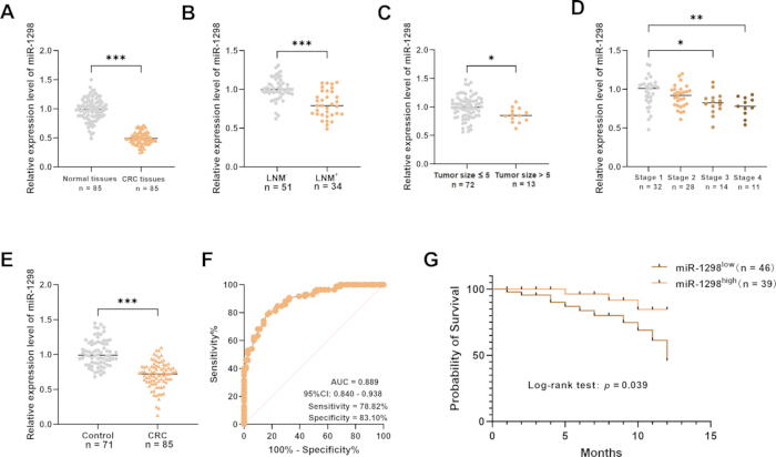
Figure 1: Expression of miR-1298 in CRC tissues. (A) Relative expression of miR-1298 in CRC tissues and adjacent noncancerous tissues. (B) Relative expression of miR-1298 in CRC tissues from patients with (LNM+) and without (LNM−) lymph node metastasis. (C) Relative expression of miR-1298 in CRC tissues according to tumor size. (D) Relative expression of miR-1298 in CRC tissues according to tumor stage. (E) Relative expression of serum miR-1298 in patients with CRC and healthy controls. (F) Receiver operating characteristic (ROC) curve for serum miR-1298 in distinguishing patients with CRC from healthy controls. (G) Kaplan–Meier overall survival curves for patients with CRC according to low and high miR-1298 expression. *p < 0.05; **p < 0.01; ***p < 0.001. Please click here to view a larger version of this figure.
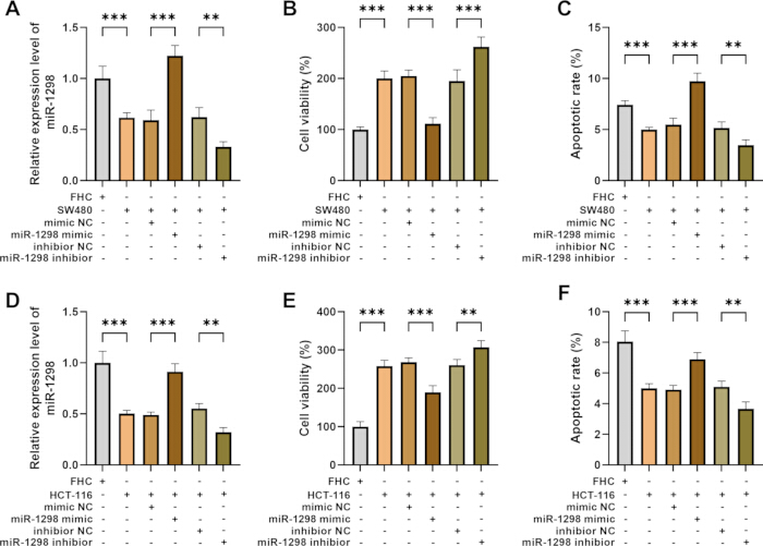
Figure 2: Effects of miR-1298 on the proliferation and apoptosis of CRC cells. (A) Relative expression of miR-1298 in FHC and SW480 cells following transfection with miR-1298 mimic, mimic NC, miR-1298 inhibitor, or inhibitor NC. (B) Cell viability of SW480 cells following miR-1298 overexpression or inhibition. (C) Apoptosis of SW480 cells following miR-1298 overexpression or inhibition. (D) Relative expression of miR-1298 in FHC and HCT-116 cells following transfection with miR-1298 mimic, mimic NC, miR-1298 inhibitor, or inhibitor NC. (E) Cell viability of HCT-116 cells following miR-1298 overexpression or inhibition. (F) Apoptosis of HCT-116 cells following miR-1298 overexpression or inhibition. Data are presented as mean ± SD. Error bars represent SD. **p < 0.01; ***p < 0.001. Please click here to view a larger version of this figure.
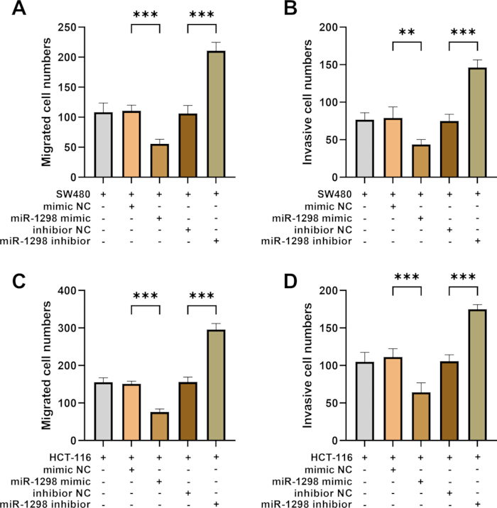
Figure 3: Effects of miR-1298 on migration and invasion of CRC cells. (A) Migrated cell numbers in SW480 cells following transfection with miR-1298 mimic, mimic NC, miR-1298 inhibitor, or inhibitor NC. (B) Invasive cell numbers in SW480 cells following transfection with miR-1298 mimic, mimic NC, miR-1298 inhibitor, or inhibitor NC. (C) Migrated cell numbers in HCT-116 cells following transfection with miR-1298 mimic, mimic NC, miR-1298 inhibitor, or inhibitor NC. (D) Invasive cell numbers in HCT-116 cells following transfection with miR-1298 mimic, mimic NC, miR-1298 inhibitor, or inhibitor NC. Data are presented as mean ± SD. Error bars represent SD. **p < 0.01; ***p < 0.001. Please click here to view a larger version of this figure.
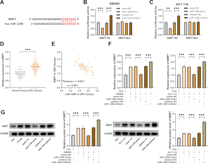
Figure 4: Targeting relationship between miR-1298 and BMP7. (A) Predicted binding site between miR-1298 and the BMP7 3′ UTR. (B) Relative luciferase activity of BMP7 Wt and BMP7 Mut reporters following miR-1298 overexpression or inhibition in SW480 cells. (C) Relative luciferase activity of BMP7 Wt and BMP7 Mut reporters following miR-1298 overexpression or inhibition in HCT-116 cells. (D) Relative expression of BMP7 in CRC tissues and adjacent noncancerous tissues. (E) Correlation between miR-1298 and BMP7 expression in CRC tissues. (F) Relative BMP7 expression in SW480 and HCT-116 cells following transfection with miR-1298 mimic, mimic NC, miR-1298 inhibitor, or inhibitor NC. (G) Relative BMP7 protein levels in SW480 and HCT-116 cells following transfection with miR-1298 mimic, mimic NC, miR-1298 inhibitor, or inhibitor NC. **p < 0.01; ***p < 0.001. Please click here to view a larger version of this figure.
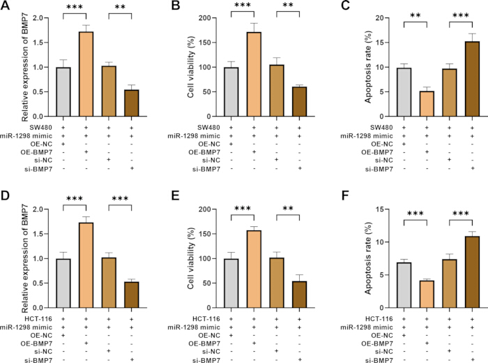
Figure 5: Co-regulation of miR-1298 and BMP7 on proliferation and apoptosis in CRC cells. SW480 and HCT-116 cells were co-transfected with miR-1298 mimic and OE-NC, OE-BMP7, si-NC, or si-BMP7. (A) Relative BMP7 expression in SW480 cells. (B) Cell viability in SW480 cells. (C) Apoptosis in SW480 cells. (D) Relative BMP7 expression in HCT-116 cells. (E) Cell viability in HCT-116 cells. (F) Apoptosis in HCT-116 cells. Data are presented as mean ± SD. Error bars represent SD. **p < 0.01; ***p < 0.001. Abbreviations: OE-NC = overexpression negative control; OE-BMP7 = BMP7 overexpression; si-NC = small interfering RNA negative control; si-BMP7 = BMP7-targeting small interfering RNA. Please click here to view a larger version of this figure.
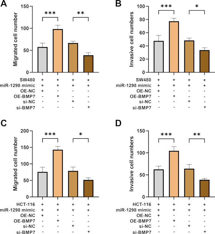
Figure 6: Co-regulation of miR-1298 and BMP7 on migration and invasion of CRC cells. SW480 and HCT-116 cells were co-transfected with miR-1298 mimic and OE-NC, OE-BMP7, si-NC, or si-BMP7. (A) Migrated cell numbers in SW480 cells. (B) Invasive cell numbers in SW480 cells. (C) Migrated cell numbers in HCT-116 cells. (D) Invasive cell numbers in HCT-116 cells. Data are presented as mean ± SD. Error bars represent SD. *p < 0.05; **p < 0.01; ***p < 0.001. Abbreviations: OE-NC = overexpression negative control; OE-BMP7 = BMP7 overexpression; si-NC = small interfering RNA negative control; si-BMP7 = BMP7-targeting small interfering RNA. Please click here to view a larger version of this figure.
| Variable | Total | miR-1298 | p value | |
| (n = 85) | Low (n = 46) | High (n = 39) | ||
| Age (years) | ||||
| ≤50 | 49 | 27 | 22 | 0.832 |
| >50 | 36 | 19 | 17 | |
| Sex (male/female) | ||||
| Male | 44 | 27 | 17 | 0.165 |
| Female | 41 | 19 | 22 | |
| TNM stage | ||||
| 1–2 | 60 | 41 | 19 | <0.001*** |
| 3–4 | 25 | 5 | 20 | |
| Tumor size (cm) | ||||
| ≤5 | 72 | 34 | 38 | 0.003** |
| >5 | 13 | 12 | 1 | |
| Lymph node metastasis | ||||
| Yes | 34 | 25 | 9 | 0.003** |
| No | 51 | 21 | 30 | |
| Tumor differentiation | ||||
| Low-grade | 53 | 24 | 29 | 0.035* |
| High-grade | 32 | 22 | 10 | |
Table 1: Association between clinicopathological features and miR-1298 expression levels in patients with CRC. Patients were categorized into low (n = 46) and high (n = 39) miR-1298 expression groups, and associations between miR-1298 expression and clinicopathological characteristics were evaluated. *p < 0.05; **p < 0.01; ***p < 0.001. Abbreviations: CRC = colorectal cancer; TNM = tumor node metastasis.
| Variable | Hazard ratio | 95% CI | p value | |
| Lower | Upper | |||
| Age | 0.776 | 0.186 | 3.242 | 0.728 |
| Sex | 0.614 | 0.152 | 2.474 | 0.492 |
| TNM stage | 4.62 | 1.181 | 18.078 | 0.028* |
| Tumor size | 8.078 | 1.833 | 35.587 | 0.006** |
| Lymph node metastasis | 6.584 | 1.134 | 38.245 | 0.036* |
| Tumor differentiation | 6.392 | 1.748 | 23.377 | 0.005** |
| miR-1298 | 0.145 | 0.024 | 0.87 | 0.035* |
Table 2: Cox regression analysis of prognostic factors in patients with CRC. Cox regression analysis was performed to evaluate the association of clinicopathological characteristics and miR-1298 expression with prognosis in patients with CRC. *p < 0.05; **p< 0.01. Abbreviations: CI = confidence interval; CRC = colorectal cancer; TNM = tumor node metastasis.
Supplementary file. Please click here to download this File.
Discussion
CRC imposes a significant burden on individuals and society because of its high incidence and high mortality rates. Despite screening programs such as colonoscopy, a large number of CRC patients are still diagnosed at an advanced stage, highlighting the urgent need for more effective, non-invasive biomarkers for early detection11. Furthermore, the prognosis of metastatic CRC remains poor, with drug resistance and disease recurrence being relatively common12. Therefore, a thorough understanding of the molecular mechanisms underlying the onset, progression, and metastasis of CRC is essential for developing new targeted therapies and overcoming treatment resistance. CRC is highly heterogeneous, meaning that tumors that appear similar may exhibit significant differences in behavior13. Ongoing research is therefore important for improving early detection, advancing personalized treatment, and ultimately improving survival in patients with CRC.
Abnormalities in miRNA are implicated in the development, staging, and prognosis of CRC14. Due to their stability in body fluids, miRNAs also hold great potential as non-invasive diagnostic and prognostic biomarkers15. In this study, miR-1298 was downregulated in CRC tissues, and its expression was associated with TNM stage and lymph node metastasis, suggesting a potential role in CRC progression. Notably, our study is the first to comprehensively characterize miR-1298 expression in a clinical CRC cohort and to demonstrate its association with key clinicopathological parameters, including TNM stage, lymph node metastasis, and tumor differentiation.
Dysregulation of core cellular processes—including proliferation, apoptosis, migration, and invasion—is a fundamental driver of cancer development and mortality16. Uncontrolled proliferation enables tumor cells to undergo initial clonal expansion. At the same time, evasion of apoptosis allows these cells to survive and accumulate in the face of internal damage or external stressors. This combination leads to the growth of the primary tumor17,18. Therefore, targeted therapies that address the molecular drivers of these characteristics are key to developing effective treatments for CRC. miRNAs contribute to tumor development by regulating the malignant phenotype of cancer cells19. miR-1298 is one such miRNA. For example, downregulation of miR-1298-5p promotes the progression of gastric cancer20. Upregulated miR-1298 suppresses proliferation of breast cancer cells, induces apoptosis, and causes cell cycle arrest21. In addition, miR-1298-5p also acts as a tumor suppressor in bladder cancer22. The results of this study indicated that overexpression of miR-1298 suppressed the proliferation, migration, and invasion and elevated apoptosis, suggesting that it may suppress the malignant phenotype of CRC. Importantly, our study extended beyond previous reports by systematically evaluating major malignant phenotypes in CRC cells, providing a more complete functional characterization of miR-1298 in this specific cancer type.
In terms of its tumor-promoting effects, BMP7 can promote cancer cell proliferation by regulating signaling pathways such as PI3K/AKT23. By modulating the level of anti‑apoptotic factors like Bcl‑2, BMP7 confers resistance to apoptosis in cancer cells24. In addition, BMP7 primarily enhances the migration and invasion of cancer cells by inducing epithelial-mesenchymal transition (EMT)25. The involvement of BMP7 in promoting cancer is consistent with our finding that BMP7 was elevated in CRC tissue. Bioinformatics analysis suggested that BMP7 may be a target of miR-1298 and identified a predicted binding site between miR-1298 and BMP7. Furthermore, rescue experiments indicated that BMP7 likely acted as a functional mediator of miR-1298 in CRC. Considering the signaling pathways associated with BMP7 in cancer, we further hypothesized that the miR-1298/BMP7 axis regulates the downstream signaling pathways (SMAD, PI3K/AKT, EMT) that control the behavior of CRC cells. Crucially, we experimentally validated this prediction through luciferase reporter assays, confirming direct binding of miR-1298 to the 3′ UTR of BMP7. Furthermore, rescue experiments demonstrated that BMP7 restoration partially reversed the tumor-suppressive effects of miR-1298 overexpression, providing functional evidence that BMP7 was a downstream mediator of miR-1298 in CRC. To our knowledge, this is the first study to establish the miR-1298/BMP7 regulatory axis in colorectal cancer.
In addition to mechanistic insights, our study provided translational evidence supporting the clinical utility of miR-1298. ROC curve analysis demonstrated that miR-1298 may have diagnostic value in CRC. Survival analysis further revealed that low miR-1298 expression was linked to poorer prognosis, and Cox regression analysis denoted that reduced miR-1298 may be a prognostic factor for CRC, which supports the latent value of miR-1298 as a diagnostic and prognostic biomarker for CRC.
This study has some limitations. Regarding clinical samples, because this study primarily utilized data from a single center, future multicenter validation studies are needed to more robustly confirm the tumor-suppressive role of miR-1298 in CRC. This study focused on in vitro cellular experiments, which did not fully capture the complexity of the in vivo tumor microenvironment. The lack of xenograft tumor models was one of the limitations of this study. Future research should include in vivo validation to further elucidate the role of the miR-1298/BMP7 axis in the tumor microenvironment. Additionally, future experiments could further validate the upstream and downstream pathways of the miR-1298/BMP7 axis through bioinformatics predictions and in vitro and in vivo experiments. The follow-up period was relatively short, which limited the maturity of the long-term survival estimation. Although Kaplan–Meier and Cox regression analyses were conducted, the results may have been affected by the relatively short follow-up period. An extended follow-up is currently underway to verify the prognostic value of miR-1298 in CRC. Furthermore, the downstream signaling pathways (SMAD, PI3K/AKT, EMT) regulated by the miR-1298/BMP7 axis in controlling the behavior of CRC cells have not been experimentally verified in this study and require further exploration. This study did not conduct reverse rescue experiments (miR-1298 inhibitor + BMP7 siRNA). Such experiments would provide complementary evidence as to whether BMP7 knockdown could reverse the tumor-promoting effects of miR-1298 inhibition. The absence of this type of rescue experiment weakens our mechanistic conclusions. Future studies that incorporate this reverse rescue experimental design and in vivo validation are needed to fully establish the function of the miR-1298/BMP7 axis in CRC.
In summary, miR-1298 might be linked to CRC progression. Functionally, miR-1298 may inhibit proliferation, invasion, and migration of CRC cells. BMP7 may be a potential functional mediator of miR-1298 in CRC.
Disclosures
The authors have no relevant financial or non-financial interests to disclose.
Acknowledgements
The authors declare that no funds, grants, or other support were received during the preparation of this manuscript.
Materials
| Name | Company | Catalog Number | Comments |
|---|---|---|---|
| 0.1% crystal violet solution | Sigma-Aldrich | C0775 | |
| 10% SDS-PAGE Kit | Epizyme | PG112 | |
| 4% paraformaldehyde | Sigma-Aldrich | P6148 | |
| 6-well cell culture plate | Corning | 3516 | |
| 96-well cell culture plate | Corning | 3599 | |
| Annexin V-FITC/PI Apoptosis Detection Kit | Various (e.g., Elabscience, BD Biosciences) | E-CK-A211 (Elabscience) or 556547 (BD) | |
| Anti-BMP7 | Abcam | ab176707 | |
| Anti-GAPDH | Abcam | ab181602 | |
| BCA Kit | Sangon | C503021 | |
| BMP7 Mutant 3‘-UTR reporter plasmid | Custom constructed (cloned into pmirGLO) | Custom | |
| BMP7 primers | Sangon Biotech (Shanghai, China) | Custom synthesis | |
| BMP7 Wild-type 3‘-UTR reporter plasmid | Custom constructed (cloned into pmirGLO) | Custom | |
| Cell Counting Kit-8 (CCK-8) | Dojindo (Japan) | CK04 | |
| Cell culture flask (T-25) | Corning | 430639 | |
| DMEM (Dulbecco‘s Modified Eagle Medium) | Invitrogen (Thermo Fisher Scientific) | 11995065 | |
| Fetal Bovine Serum (FBS) | Invitrogen (Thermo Fisher Scientific) | 16140071 | |
| FHC cell line (normal human colon epithelial) | ATCC | CRL-1831 | |
| Flow cytometer | BD Biosciences | FACSCanto™ II | |
| FlowJo V10 software | FlowJo, LLC | — | |
| GAPDH primers (endogenous control for mRNA) | Sangon Biotech (Shanghai, China) | Custom synthesis | |
| HCT-116 cell line (human colorectal carcinoma) | ATCC | CCL-247 | |
| HRP-conjugated secondary antibody | Beyotime | A0208 | |
| Inverted microscope | Nikon (or equivalent) | Eclipse Ts2 | |
| Lipofectamin 3000 Transfection Reagent | Invitrogen (Thermo Fisher Scientific) | L3000015 | |
| Luciferase Assay System | Promega | E1500 | |
| Matrigel Basement Membrane Matrix | Corning | 354230 | |
| Microplate reader | BioTek (Agilent) | Synergy H1 | |
| miR-1298 inhibitor | GenePharma (Shanghai, China) | Custom (synthesized upon order) | |
| miR-1298 mimic | GenePharma (Shanghai, China) | Custom (synthesized upon order) | |
| miR-1298 primers | Sangon Biotech (Shanghai, China) | Custom synthesis | |
| Mir-X miRNA First-Strand Synthesis Kit | Takara Bio | 638313 | |
| OE-NC | GenePharma (Shanghai, China) | Custom (synthesized upon order) | |
| Opti-MEM | Gibco | 31985070 | |
| Passive Lysis Buffer | Promega | E194A | |
| pcDNA3.1 vector (empty vector control) | Invitrogen (Thermo Fisher Scientific) | V79020 | |
| pcDNA3.1-BMP7 (OE-BMP7 overexpression plasmid) | GenePharma (Shanghai, China) | Custom (synthesized upon order) | |
| pmirGLO Dual-Luciferase miRNA Target Expression Vector | Promega | E1330 | |
| PMSF | Beyotime | ST506 | |
| PrimeScript RT Reagent Kit (Perfect Real Time) | Takara (Japan) | RR037A | |
| PrimeScript RT reagent Kit with gDNA Eraser | Takara Bio | RR047A | |
| Protease inhibitor cocktail | Thermo | 78429 | |
| PVDF | Absin | abs935 | |
| RIPA | Abcam | ab156034 | |
| RNase-free microcentrifuge tubes (1.5 mL) | Axygen (Corning) | MCT-150-C | |
| RNase-free pipette tips | Axygen (Corning) | T-300 (10 µL), T-200-Y (200 µL), T-1000-B (1000 µL) | |
| si-BMP7 | GenePharma (Shanghai, China) | Custom (synthesized upon order) | |
| si-NC | GenePharma (Shanghai, China) | Custom (synthesized upon order) | |
| Staurosporine (apoptosis positive control) | Sigma-Aldrich | S5921 | |
| SW480 cell line (human colorectal adenocarcinoma) | ATCC | CCL-228 | |
| SYBR Green qPCR Master Mix | Takara (Japan) | RR036A | |
| Transwell chamber (24-well, 8.0 µm pore size) | Corning (Costar) | 3422 | |
| TRIzol Reagent | Invitrogen (Thermo Fisher Scientific) | 15596026 | |
| U6 primers (endogenous control for miRNA) | Sangon Biotech (Shanghai, China) | Custom synthesis |
References
- Ali EAM, Altaie AM, Talaat IM, Hamoudi R. Putative role of Tie2-expressing monocytes/macrophages in colorectal cancer progression through enhancement of angiogenesis and metastasis. Cancers (Basel). 2025;17(17):2856.
- Golfinopoulou R, Hatziagapiou K, Mavrikou S, Kintzios S. Unveiling colorectal cancer biomarkers: harnessing biosensor technology for volatile organic compound detection. Sensors (Basel). 2024;24(14):4712.
- Islind AS, et al. Individualized blended care for patients with colorectal cancer: the patient's view on informational support. Support Care Cancer. 2021;29(6):3061-67.
- Sun Y, et al. IGF2BP3 enhances ferroptosis resistance in colon cancer by stabilizing SLC7A11 and is regulated by miR-98-5p. Front Oncol. 2025;15:1576895.
- Du Z, et al. MicroRNA-1298 is downregulated in non-small cell lung cancer and suppresses tumor progression in tumor cells. Diagn Pathol. 2019;14(1):132.
- Qi Y, et al. The dual role of glioma exosomal microRNAs: glioma eliminates tumor suppressor miR-1298-5p via exosomes to promote immunosuppressive effects of MDSCs. Cell Death Dis. 2022;13(5):426.
- Vasan R, et al. High BMP7 expression is associated with poor prognosis in ovarian cancer. J Cell Mol Med. 2023;27(21):3378-87.
- Motoyama K, et al. Clinical significance of BMP7 in human colorectal cancer. Ann Surg Oncol. 2008;15(5):1530-37.
- Shi Q, et al. Analysis of the role of the BMP7-Smad4-Id2 signaling pathway in SW480 colorectal carcinoma cells. Mol Med Rep. 2011;4(4):627-31.
- Yang YR, et al. MicroRNA-137 inhibits cell migration and invasion by targeting bone morphogenetic protein-7 (BMP7) in non-small cell lung cancer cells. Int J Clin Exp Pathol. 2015;8(9):10847-53.
- Yan T, et al. Oxidative stress gene expression in ulcerative colitis: implications for colon cancer biomarker discovery. Sci Rep. 2025;15(1):22641.
- Zhou Y, et al. Interleukin-17F suppressed colon cancer by enhancing caspase 4 mediated pyroptosis of endothelial cells. Sci Rep. 2024;14(1):18363.
- Ku X, et al. In-depth characterization of mass spectrometry-based proteomic profiles revealed novel signature proteins associated with liver metastatic colorectal cancers. Anal Cell Pathol (Amst). 2019;2019:7653230.
- Wang Y, et al. Overexpression of microRNA-216a inhibits autophagy by targeting regulated MAP1S in colorectal cancer. Onco Targets Ther. 2019;12:4621-29.
- Correa-Gallego C, et al. Circulating plasma levels of microRNA-21 and microRNA-221 are potential diagnostic markers for primary intrahepatic cholangiocarcinoma. PLoS One. 2016;11(9):e0163699.
- Qin Y, Zhou Y. Heterogeneous nuclear ribonucleoprotein K in cancer biology and its therapeutic applications (review). Mol Med Rep. 2026;33(5):144.
- Louis L, Chee BS, McAfee M, Nugent M. Electrospun drug-loaded and gene-loaded nanofibres: the holy grail of glioblastoma therapy? Pharmaceutics. 2023;15(6):1649.
- Termini D, Den Hartogh DJ, Jaglanian A, Tsiani E. Curcumin against prostate cancer: current evidence. Biomolecules. 2020;10(11):1536.
- Wu SR, Wu Q, Shi YQ. Recent advances of miRNAs in the development and clinical application of gastric cancer. Chin Med J (Engl). 2020;133(15):1856-67.
- Li X, et al. MiR-1298-5p level downregulation induced by Helicobacter pylori infection inhibits autophagy and promotes gastric cancer development by targeting MAP2K6. Cell Signal. 2022;93:110286.
- Chen W, et al. microRNA-1298 inhibits the malignant behaviors of breast cancer cells via targeting ADAM9. Biosci Rep. 2020;40(12):BSR20201215.
- Zhang X, et al. Circular RNA TAF4B promotes bladder cancer progression by sponging miR-1298-5p and regulating TGFA expression. Front Oncol. 2021;11:643362.
- Leinhauser I, et al. Oncogenic features of the bone morphogenic protein 7 (BMP7) in pheochromocytoma. Oncotarget. 2015;6(36):39111-26.
- Guan J, et al. Bone morphogenetic protein-7 (BMP-7) mediates ischemic preconditioning-induced ischemic tolerance via attenuating apoptosis in rat brain. Biochem Biophys Res Commun. 2013;441(3):560-66.
- Lim M, Chuong CM, Roy-Burman P. PI3K, Erk signaling in BMP7-induced epithelial-mesenchymal transition (EMT) of PC-3 prostate cancer cells in 2- and 3-dimensional cultures. Horm Cancer. 2011;2(5):298-309.