This protocol describes a reproducible method for isolating and culturing primary murine aortic smooth muscle cells from thoracic and abdominal regions. The method yields viable cells suitable for in vitro studies of vascular remodeling and aneurysm-related biology.
Method Article
Isolation and Culture of Primary Murine Aortic Smooth Muscle Cells for In vitro Studies Relevant to Vascular Remodeling and Aortic Aneurysm Biology
In This Article
Summary
Abstract
Isolation and cultivation of primary aortic smooth muscle cells (SMCs) provide a valuable approach for investigating cellular mechanisms underlying vascular diseases, including aortic aneurysms. The present article describes a detailed and optimized protocol for the isolation and cultivation of murine aortic SMCs from thoracic and abdominal aortic segments. The current procedure includes careful aortic dissection, removal of surrounding tissue, and a two-step enzymatic digestion using collagenase, elastase, and soybean trypsin inhibitor, followed by mechanical dissociation and cell expansion under defined culture conditions. Particular emphasis is given on steps that improve cell yield, purity, and reproducibility, including adventitial removal and controlled digestion timing. Using this approach, the primary SMCs can be successfully isolated and expanded, exhibiting characteristic morphology and growth patterns. This protocol provides a reproducible and adaptable approach for generating primary aortic SMCs and serves as a platform for in vitro studies relevant to vascular remodeling and aortic aneurysm biology.
Introduction
Aortic smooth muscle cells (SMCs) are essential components of the vascular wall, contributing to both structural integrity and functional regulation of the aorta. In healthy vessels, SMCs maintain a contractile phenotype characterized by expression of contractile proteins such as α-smooth muscle actin (α-SMA) and smooth muscle myosin heavy chain (MYH11)1,2,3. This phenotype supports vascular tone and preserves extracellular matrix organization. However, in pathological conditions such as abdominal aortic aneurysm (AAA), SMCs undergo phenotypic modulation toward a synthetic and pro-inflammatory state1,2,3. This transition is associated with increased proliferation, secretion of cytokines, and production of matrix-degrading enzymes, ultimately contributing to weakening of the aortic wall4,5.
Animal models, including angiotensin II (Ang II) induced aortic aneurysm formation, have significantly advanced understanding of AAA pathogenesis1,6,7,8,9,10,11. Nevertheless, these models do not allow precise manipulation of cellular pathways or direct observation of SMC-specific mechanisms. In vitro systems using primary SMCs provide a complementary approach, enabling detailed investigation of signaling pathways, gene regulation, and cellular responses under controlled conditions12.
Despite their utility, isolation of primary murine aortic SMCs is technically challenging. The small size of the murine aorta, the presence of surrounding adipose and connective tissue, and the potential for contamination by adventitial fibroblasts can all compromise cell purity. Additionally, enzymatic digestion must be carefully controlled to balance tissue dissociation with preservation of cell viability. Variability in these steps often leads to inconsistent results across experiments and laboratories.
Several protocols describing the isolation and culture of primary murine aortic smooth muscle cells have been published, most of which rely on complete mechanical removal of the adventitia prior to enzymatic digestion and primarily focus on whole aortas or thoracic aortic segments13,14. The present protocol builds upon these methods by incorporating several technical refinements, including a brief enzymatic pre-digestion step to facilitate separation of the adventitial and medial layers before complete tissue dissociation, standardized digestion conditions with intermittent gentle mechanical dissociation, and a workflow for the separate isolation and culture of thoracic (THX) and abdominal (ABD) aortic smooth muscle cells. Together, these refinements are intended to improve the consistency and reproducibility of tissue processing while facilitating region-specific studies of vascular remodeling and aneurysm biology. The resulting SMC cultures are suitable for a wide range of downstream applications, including gene silencing, pharmacological studies, and mechanistic investigations relevant to vascular remodeling and aneurysm biology. While primary SMCs isolated from non-diseased aortas do not inherently recapitulate aneurysmal phenotypes, they provide a controlled in vitro system for investigating cellular pathways implicated in aneurysm development following targeted experimental manipulation.
Protocol
Adult 8- to 10-week-old female C57BL/6J mice weighing 18–20 g were used for the procedure described below. Two mice were used per isolation. All animal procedures were performed in accordance with the guidelines of the Institutional Animal Care and Use Committee (IACUC) at the University of Kentucky and were approved under protocol no. 2022-4153. The details of all the reagents and the equipment used are listed in the Table of Materials. A schematic overview of the complete isolation and culture workflow is shown in Figure 1.
CAUTION: Ketamine and xylazine are controlled substances and should be handled, stored, and disposed of in accordance with institutional policies and applicable regulations. Collagenase type II and elastase are proteolytic enzymes that may cause skin, eye, and respiratory irritation. Trypan blue is a hazardous dye and should be handled with appropriate personal protective equipment while avoiding skin and eye contact. All hazardous reagents should be handled and disposed of in accordance with institutional safety guidelines and the manufacturers' instructions.
1. Preparation of reagents and culture media
- Prepare 10 mL of enzyme digestion solution fresh on the day of isolation in a sterile 15 mL conical tube:
- Add 10 mg collagenase type II (final concentration of 1 mg/mL) (see Table of Materials).
- Add 10 mg soybean trypsin inhibitor (final concentration of 1 mg/mL) (see Table of Materials).
- Add 71 µL of elastase suspension (final concentration of 0.744 U/mL) (see Table of Materials).
- Prepare the solution in HBSS containing calcium and magnesium without phenol red (see Table of Materials).
- Add 100 µL of penicillin/streptomycin (1% of final volume) (see Table of Materials).
- Mix the solution thoroughly and filter sterilize using a 0.2 µm nylon syringe filter (see Table of Materials).
- Prepare complete SMC culture medium (500 mL)
- Add 385 mL of Dulbecco’s Modified Eagle Medium/Nutrient Mixture F-12 (DMEM/F12) without phenol red (see Table of Materials) as the base medium.
- Supplement with 100 mL of charcoal/dextran-treated fetal bovine serum (20% of final volume) (see Table of Materials).
- Add 10 mL of penicillin/streptomycin (2% of final volume) (see Table of Materials).
- Add 5 mL amphotericin B (1% of final volume) (see Table of Materials)
- Filter sterilize the media and store at 4 °C.
- Pre-warm all reagents to 37 °C prior to use.
2. Preparation for tissue processing
- Pre-aliquot enzyme digestion solution equally into 4 wells (2.5 mL each) of a 6-well culture plate and pre-warm in incubator at 37 °C with 5% CO₂ prior to tissue processing. 2 wells should be designated 'THX' and 2 'ABD' for the aortic segments. Multiple segments from the same anatomical region may be processed in a single well.
- Prepare 2 separate culture dishes (see Table of Materials) containing complete SMC culture medium and label them THX and ABD.
- Pre-warm culture dishes containing media in a humidified incubator.
- Prepare 20 mL of sterile HBSS in advance for vascular perfusion and tissue handling.
- Set up 2 additional culture dishes containing 3 mL of HBSS each for sequential cleaning of isolated aortas.
- Prepare labeled 15 mL conical tubes (see Table of Materials) containing 5 mL of HBSS each for temporary storage and separation of THX and ABD aortic segments.
- Sterilize surgical instruments between animals by applying detergent, rinsing with distilled water, disinfecting with 70% ethanol, and sterilizing using a bead sterilizer (see Table of Materials).
3. Animal preparation and aortic dissection
- Anesthetize mice by intraperitoneal (IP) injection of ketamine (300 mg/kg) and xylazine (30 mg/kg) (see Table of Materials) as approved under University of Kentucky IACUC protocol 2022-4153 for terminal procedures.
- Confirm adequate depth of anesthesia via absence of reflex response to toe pinch.
- Sterilize the surgical field using 70% ethanol.
- While under deep surgical anesthesia, euthanize the mice by exsanguination via cardiac puncture followed by transcardial perfusion with HBSS, in accordance with institutional and AVMA guidelines.
- Open the thoracic cavity and perform cardiac puncture to collect blood.
- Perfuse the vasculature through the left ventricle using a 10 mL syringe fitted with a 25-gauge needle to slowly inject 10 mL sterile HBSS. A perfusion rate of approximately 0.5 mL/s is recommended to ensure efficient removal of residual blood.
- Carefully remove surrounding organs (liver, lungs, spleen, gastrointestinal tract, kidneys). Leave a piece of the diaphragm connected to the aorta.
- Isolate the aorta from the aortic root to the iliac bifurcation with the intact diaphragm. The diaphragm serves as the anatomical boundary between the THX and ABD regions (Figure 2B).
- Transfer the aorta immediately into sterile HBSS (Figure 2A).
4. Aortic cleaning and segmentation
- Place the sterile culture dish containing the aorta under a dissecting microscope (see Table of Materials).
- Carefully remove adipose tissue and connective tissue using fine forceps (see Table of Materials).
- Separate the aorta into THX and ABD segments.
NOTE: The THX segment extends from the region of the aorta just above the diaphragm up to the aortic root, while the ABD segment extends from the region of the aorta just below the diaphragm down to the iliac bifurcation (Figure 2B). - Transfer cleaned segments into separate 15 mL conical tubes with fresh HBSS for further processing.
5. Enzymatic digestion and cell isolation
- Incubate aortic segments in enzyme solution at 37 °C for 8–10 min.
- Remove aortic segments from the enzyme solution.
- Transfer segments into separate culture dishes containing complete SMC media designated for THX and ABD samples.
- Remove the adventitial layer from the aorta using coarse and fine forceps. Complete adventitial removal is confirmed when the outer surface of the aorta appears smooth and translucent, with no visible residual connective tissue or adipose tissue remaining (Figure 2C).
- Transfer aortic segments into fresh enzyme solution and mince the medial layer into small fragments directly within the solution (Figure 3A).
- Incubate tissue fragments at 37 °C in a humidified incubator with 5% CO₂ for 1 h.
- Perform gentle mechanical dissociation by pipetting up and down approximately 10–15 times to facilitate cell release (Figure 3B).
- Repeat the mechanical dissociation step at 30–60 min intervals during continued incubation for a total digestion time of 2–3 h or until a homogeneous cell suspension is achieved.
6. Cell collection, washing, plating, and initial culture
- Transfer the enzyme digestion solution containing dissociated cells into separate 15 mL conical tubes (see Table of Materials) designated for THX and ABD samples.
- Rinse each digestion well with 5 mL of culture media and combine with the corresponding tubes.
- Centrifuge the cell suspension at 400 × g for 5 min at room temperature.
- Repeat centrifugation if pellet formation is insufficient.
- Remove the supernatant without disturbing the cell pellet (Figure 3C).
- Resuspend the cells with 5 mL of fresh culture media and perform an additional wash step followed by centrifugation under the same conditions.
- Remove the supernatant and resuspend the final cell pellet in an appropriate volume (typically 6 mL) of complete SMC culture medium.
- Plate the entire cell suspension obtained from the thoracic or abdominal aortic segment into two wells of a 6-well culture plate (3 mL suspended medium per well) and incubate at 37 °C with 5.0% CO₂. Cells are plated immediately following isolation without prior cell counting; therefore, no target seeding density is used at this stage.
- Maintain cultures undisturbed for 5–7 days to allow cell attachment and initial expansion.
PAUSE POINT: After plating, cultures may be maintained in the incubator for 5–7 days before proceeding to the cell maintenance and expansion steps in Section 7.
7. Cell maintenance and expansion
- Replace culture media every 3–4 days.
- Monitor cell morphology and confluence regularly.
- Passage cells when they reach approximately 80–90% confluence.
- Aspirate culture media and wash cells with pre-warmed phosphate-buffered saline (PBS) (see Table of Materials).
- Add 0.05% trypsin-EDTA and incubate at 37 °C for 5–10 min.
- Proceed when 80%–90% of cells are detached.
- Tap the culture plate gently to facilitate detachment.
- Add an equal volume of complete culture media to neutralize trypsin.
- Pipette gently to detach remaining cells.
- Transfer the cell suspension to conical tubes and centrifuge at 400 × g for 5 min.
- Remove the supernatant and resuspend the cell pellet in fresh culture media.
- Plate cells at a 1:2 split ratio (or the desired experimental density) and incubate at 37 °C with 5% CO₂. Allow cells to attach undisturbed for 24–48 h.
NOTE: For downstream experiments, use cells between passages 3 and 7 (P3–P7) to minimize culture-induced phenotypic drift. Cells beyond passage 7 are more prone to phenotypic drift and may exhibit altered morphology and function.
8. Validation by α-SMA immunofluorescence staining
- Grow cells on material conducive to fluorescent microscopy.
- Plate cells in a 96-well optical-bottom plate at 15,000 cells per well. Allow cells to grow to desired confluency.
- Prepare cells for fixation
- Aspirate the culture media from adherent cells. Wash 3 times with 200 µL per well of Tris-buffered saline with 0.1% Tween 20 (TBS-T) for 5 min per wash, then aspirate with a biological hood vacuum.
- Fix cells
- Add 200 µL per well of 4% paraformaldehyde (PFA) in 1x PBS. Incubate for 10–20 min at room temperature, then aspirate.
- Permeabilization
- Add 200 µL per well of 0.1% Triton X-100 in 1x PBS. Incubate for 10–15 min at room temperature, then aspirate.
- Blocking nonspecific binding
- Wash cells 3 times with 200 µL per well of TBS-T for 5 min per wash. Add 200 µL per well of 5% bovine serum albumin (BSA) in TBS-T.
- Place plate on orbital shaker at approximately 200 rpm for 1 h at room temperature.
- Primary antibody staining
- Prepare the primary α-SMA antibody at a 1:1,000 dilution in 5% BSA in TBS-T.
- Remove the plate from the orbital shaker and aspirate the blocking solution from wells designated for α-SMA staining. For the secondary-antibody-only control, omit the primary antibody and maintain the control well in 5% BSA in TBS-T.
- Add 200 µL per well of the diluted primary α-SMA antibody to the experimental wells. Incubate the experimental and secondary-only control wells overnight at 4 °C on a gentle orbital shaker.
- Secondary antibody staining
- Prepare the fluorescent secondary antibody at a 1:2,500 dilution in TBS-T containing 0.1% Tween 20.
- Remove the plate from the orbital shaker and aspirate all wells.
- Wash all wells 3 times with 200 µL per well of TBS-T for 5 min per wash, then aspirate.
- Add 200 µL per well of the diluted fluorescent secondary antibody to all wells, including the secondary-antibody-only control.
- Protect the plate from light and incubate on an orbital shaker at approximately 200 rpm for 1 h at room temperature.
- Nuclear staining and fluorescence imaging
- Prepare a 2 µg/mL solution of Hoechst 33342.
- Wash cells 3 times with 200 µL per well of TBS-T for 5 min per wash. Add sufficient Hoechst 33342 staining solution to cover the cells and incubate for 10 min at room temperature protected from light.
- Acquire fluorescence images using an inverted fluorescence microscope at 20x magnification for α-SMA-stained THX and ABD cultures and at 10x magnification for the secondary-antibody-only controls, using 385 nm excitation for Hoechst 33342 and 475 nm excitation for the fluorophore-conjugated secondary antibody. Secondary-antibody-only controls were imaged at 10x to include a sufficient number of nuclei within the field of view.
- Quantification of α-SMA-positive cells.
- Evaluate α-SMA positivity by counting Hoechst-positive nuclei associated with cells exhibiting clear α-SMA-positive spindle-shaped cytoplasmic staining. Score cells with ambiguous or non-defined fluorescent signal conservatively as α-SMA negative.
- Calculate α-SMA positivity as the number of α-SMA-positive cells divided by the total number of Hoechst-positive nuclei counted across the evaluated microscopic fields, multiplied by 100.
Results
Within 24–48 h post-isolation, a subset of cells begins to adhere to the culture surface and spread, initially exhibiting a rounded morphology. Cell attachment may be variable at early time points, and cultures may appear sparse due to the limited number of viable cells recovered during initial isolation. By days 3–5, adherent cells progressively adopt a characteristic spindle-shaped, elongated morphology typical of vascular smooth muscle cells (SMCs), and begin to proliferate, forming small clusters. A schematic overview of the experimental workflow, including aortic isolation, segmentation, enzymatic digestion, mechanical dissociation, and cell plating, is illustrated in Figure 1.
Representative images of aortic isolation, anatomical segmentation into THX and ABD regions, and removal of the adventitial layer are shown in Figure 2. The THX and ABD aortic regions were distinguished based on the anatomical location relative to the diaphragm, as indicated in Figure 2B. Representative images of enzymatic digestion, mechanical dissociation, and formation of a visible cell pellet following centrifugation are shown in Figure 3.
Cell count and viability were assessed at passage 3 using Trypan blue exclusion (1:1) and a Countess Automated Cell Counter. Data were obtained from 3 independent isolations using 6 mice total (2 mice per isolation). THX cultures had a mean cell count of 5.10 × 10⁵ ± 7.81 × 10⁴ cells (range, 4.20 × 10⁵–5.60 × 10⁵) and 66.33 ± 18.18% viability (range, 46–81%). ABD cultures had a mean cell count of 4.63 × 10⁵ ± 2.31 × 10⁴ cells (range, 4.50 × 10⁵–4.90 × 10⁵) and 61.66% ± 12.01% viability (range, 50%–74%) (Table 1).
By days 5–7, cultures typically reach approximately 70%–90% confluence, forming a dense and relatively uniform monolayer. Cells commonly align in parallel orientations, reflecting their contractile lineage. Differences in proliferation rate and cell density may be observed between THX and ABD SMCs, consistent with known regional heterogeneity of the aorta. Representative phase-contrast images of primary SMCs morphology from THX and ABD aortic segments at low (10x) and higher (20x) magnification are shown in Figure 4.
SMCs exhibited the characteristic elongated, spindle-shaped morphology, organized parallel alignment at confluence, and uniform growth pattern, whereas fibroblast-like cells typically exhibit a more flattened, irregular morphology with less organized orientation and heterogeneous spreading. These morphological and growth characteristics provide a practical and reproducible approach for identifying SMC populations in primary culture in the absence of molecular validation. Successful isolation is indicated by early cell attachment within 24–48 h, progressive acquisition of spindle-shaped morphology, and formation of a confluent monolayer by days 5–7. Cells typically align in parallel arrays at higher confluence, consistent with smooth muscle phenotype. Suboptimal results may include poor cell attachment, low cell yield, or the presence of flattened, irregularly shaped cells, which may indicate incomplete digestion, excessive enzymatic exposure, or contamination with non-smooth muscle cell populations.
Consistent cell viability and minimal fibroblast-like contamination further support successful isolation. To further validate SMC identity, representative primary cultures were immunostained for α-SMA (Figure 5) using the procedure described in Section 8. Both THX and ABD cultures demonstrated the characteristic filamentous α-SMA staining pattern expected of vascular SMCs. Quantification across four microscopic fields identified 69 α-SMA-positive cells among 72 Hoechst-positive cells evaluated, corresponding to an overall α-SMA positivity of 95.8% (95% CI, 88.5%–98.9%). Specifically, 20/20 THX cells and 49/52 ABD cells were α-SMA positive. Figure 5 shows representative fields from those evaluated.
During early passages, cells maintain a stable morphology and proliferative capacity. However, with extended culture, cells may gradually exhibit features of phenotypic modulation, including increased spreading, reduced alignment, and altered growth patterns.
Under these conditions, primary SMC cultures are suitable for downstream applications, including gene knockdown, pharmacological treatments, and mechanistic studies of pathways involved in vascular remodeling and aortic aneurysm development.
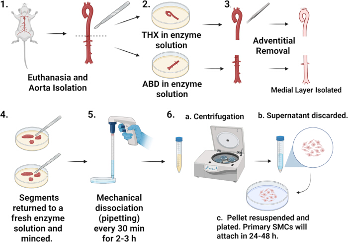
Figure 1: Schematic workflow overview. A schematic representation of the primary murine aortic smooth muscle cell isolation workflow. Panel 1 shows euthanasia and aorta isolation. Panel 2 shows separation of THX and ABD aortic segments into enzyme digestion solution. Panel 3 shows adventitial removal and isolation of the medial layer. Panel 4 shows return of tissue segments to fresh enzyme solution and mincing. Panel 5 shows mechanical dissociation by pipetting every 30 min for 2–3 h. Panel 6 shows cell recovery and plating, including 6a centrifugation, 6b supernatant removal, and 6c pellet resuspension and plating. Please click here to view a larger version of this figure.
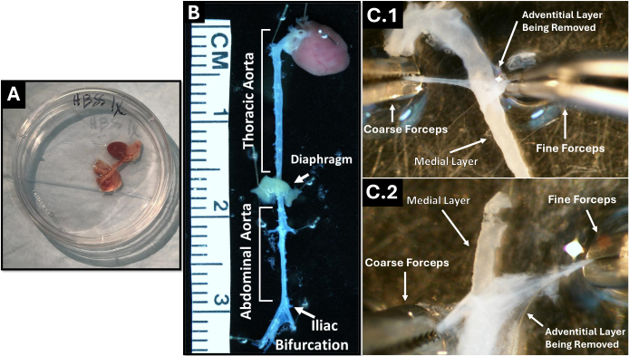
Figure 2: Isolation and preparation of mouse aorta segments. (A) Whole aorta isolated and placed in a culture dish. (B) Cleaned aorta with anatomical landmarks indicating THX and ABD regions, including the diaphragm and iliac bifurcation. (C) Representative images of adventitial removal from THX (top) and ABD (bottom) segments using fine and coarse forceps, revealing the underlying medial layer critical for smooth muscle cell isolation. Please click here to view a larger version of this figure.
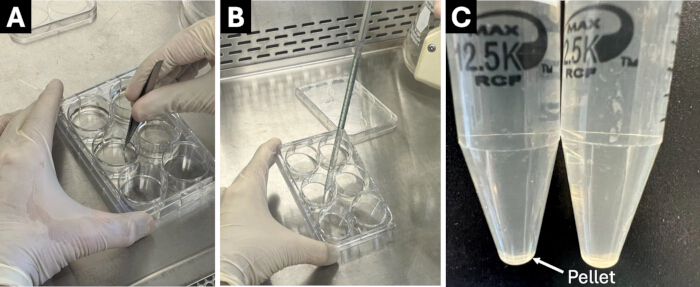
Figure 3: Enzymatic digestion and mechanical dissociation of aortic tissue. (A) Following removal of the adventitial layer, the medial layer is transferred into fresh enzyme solution and minced using small scissors. (B) Example of mechanical dissociation of aortic fragments via gentle pipetting. (C) After enzymatic digestion and centrifugation, a visible cell pellet is observed. Please click here to view a larger version of this figure.
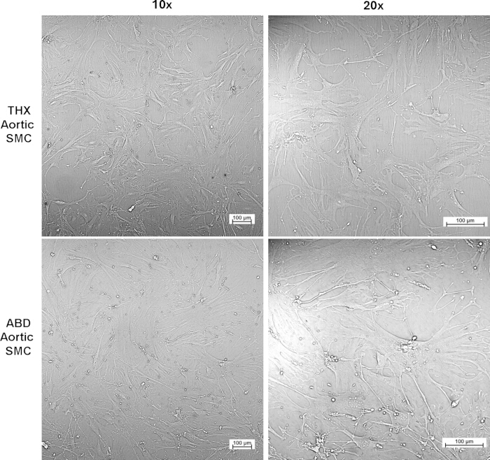
Figure 4: Representative morphology of primary murine aortic smooth muscle cells (SMCs). Primary SMCs isolated from thoracic and abdominal aortic segments were cultured under standard conditions and imaged at passage 4. Representative phase-contrast images are shown at low (10x) and higher (20x) magnification. Cells exhibit characteristic spindle-shaped, elongated morphology and alignment consistent with a contractile SMC phenotype. Differences in cell density and organization may be observed between THX- and ABD-derived SMCs, reflecting regional heterogeneity. Scale bars = 100 µm. Please click here to view a larger version of this figure.
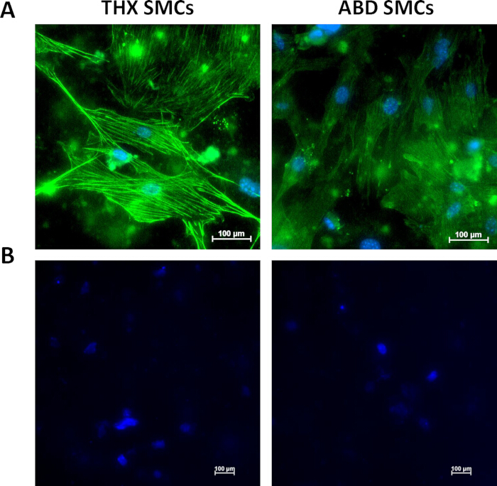
Figure 5: Immunofluorescence validation of primary murine aortic SMCs. (A) Representative immunofluorescence images of primary SMCs isolated from the THX (left) and ABD (right) aorta and stained for α-SMA (green). Nuclei were counterstained with Hoechst 33342 (blue). Images were acquired at 20x magnification. (B) Representative secondary-antibody-only control images of THX (left) and ABD (right) primary SMCs processed without the α-SMA primary antibody and counterstained with Hoechst 33342 (blue). Images were acquired at 10x magnification. Quantification across four microscopic fields identified 69/72 cells as α-SMA positive (95.8%). Scale bars = 100 µm. Please click here to view a larger version of this figure.
| Cell Count and Viability of Primary Murine Aortic Smooth Muscle Cell Cultures | |||
| Aortic segment | Independent isolations, n | Cell Count (Mean ± SD)* | Cell viability (Mean ± SD)† |
| THX | 3 | 5.10 x 10⁵ ± 7.81 x 10⁴ cells | 66.33 ± 18.18 % |
| ABD | 3 | 4.63 x 10⁵ ± 2.31 x 10⁴ cells | 61.66 ± 12.01 % |
| *Cell counts were obtained at passage 3 from 3 independent isolations. | |||
| †Cell count and viability were determined by 1:1 Trypan blue exclusion using a Countess Automated Cell Counter. | |||
Table 1: Cell count and viability of primary murine aortic smooth muscle cell cultures
| Step | Problem | Possible Cause | Recommended Solution |
| Aortic dissection | Low cell yield | Incomplete aorta recovery or tissue damage during dissection | Ensure full isolation from aortic root to iliac bifurcation; handle tissue gently to avoid damage |
| Aortic cleaning | Fibroblast contamination | Incomplete removal of adventitial tissue | Carefully strip adventitia after initial digestion; this is critical for SMC purity |
| Initial digestion | Difficulty removing adventitia | Insufficient enzymatic loosening | Ensure proper incubation (8-10 min) before attempting mechanical stripping |
| Enzymatic digestion | Low cell yield | Under-digestion of tissue | Extend digestion time slightly or increase frequency of mechanical dissociation |
| Enzymatic digestion | Poor cell viability | Over-digestion or excessive enzyme exposure | Reduce digestion time and avoid prolonged incubation |
| Mechanical dissociation | Poor cell release | Inadequate pipetting | Perform gentle but repeated pipetting every 30-60 min |
| Cell attachment | Poor adherence to plate | Cell damage or cold media | Use pre-warmed media and minimize handling stress |
| Early culture | No cell growth | Low viability or contamination | Confirm digestion conditions and sterility; repeat isolation if necessary |
| Cell morphology | Non-spindle shape (rounded/flat cells persist) | Contamination or phenotypic drift | Verify adventitia removal; avoid overgrowth and prolonged culture |
| Cell growth | Slow proliferation | Suboptimal serum concentration | Use 20% FBS during early expansion phase |
| Confluence | Over-confluent cells difficult to passage | Delayed splitting | Passage cells at 80-90% confluence |
| Passaging | Poor detachment | Cells are overly confluent or insufficient trypsinization | Increase trypsin exposure slightly and mechanically assist detachment |
| Long-term culture | Loss of SMC phenotype | High passage number or prolonged culture | Use cells between passages 3-7 |
| Reproducibility | Variability between isolations | Differences in technique or biological variability | Standardize enzyme prep, timing, and handling across experiments |
Table 2: Troubleshooting guide for the isolation and culture of primary murine aortic smooth muscle cells. Summary of common technical issues encountered during the isolation, digestion, and culture of primary murine aortic smooth muscle cells, including potential causes and recommended solutions to improve reproducibility.
Discussion
This protocol provides a robust and reproducible method for isolating and culturing primary murine aortic smooth muscle cells (SMCs) for in vitro studies. In addition to its technical utility, this approach is highly relevant for investigating mechanisms underlying vascular remodeling and aortic aneurysm development. Given the established role of the renin-angiotensin system (RAS) and Ang II in vascular inflammation and aneurysm formation15,16, isolation of primary aortic SMCs provides a valuable approach to interrogate these pathways at the cellular level.
A key determinant of success is the thorough removal of adventitial tissue. The adventitia contains fibroblasts and other non-SMC populations that can outgrow SMCs in culture if not adequately removed. The brief initial enzymatic digestion step facilitates mechanical separation of the adventitia from the medial layer, allowing more precise dissection. Failure to perform this step effectively is one of the most common causes of contamination and variability in SMC isolation protocols.
Enzymatic digestion conditions must also be carefully optimized. The combination of collagenase type II, elastase, and soybean trypsin inhibitor enables efficient breakdown of extracellular matrix components while preserving cell viability. However, digestion must be closely monitored. Insufficient digestion results in incomplete tissue dissociation and reduced cell yield, whereas excessive digestion can damage cells, impair attachment, and decrease viability. Mechanical dissociation through gentle, repeated pipetting is therefore an essential complementary step, promoting cell release while minimizing enzymatic overexposure. Together, these steps highlight the importance of balancing enzymatic and mechanical processing to optimize both cell yield and viability. Maintaining consistent digestion times, tissue handling, and mechanical dissociation throughout the procedure is equally important for improving reproducibility between isolations.
Culture conditions play a central role in maintaining SMC phenotype and function. High-serum media (20% fetal bovine serum) supports initial attachment and expansion of primary cells following isolation. Charcoal/dextran-treated serum was used to reduce variability from hormones and lipid components present in standard serum preparations. However, prolonged exposure to high serum or extended culture duration can promote phenotypic drift toward a synthetic or fibroblast-like state. To minimize these effects, it is recommended to use cells within early-to-mid passages (P3–P7) and avoid over-confluent conditions, which can alter morphology and reduce proliferative capacity.
The morphology of isolated cells provides an important initial indicator of culture quality. Healthy SMCs exhibit a characteristic spindle-shaped, elongated morphology and align in parallel arrays at higher confluence. Deviations from this morphology, including flattened or irregularly shaped cells, may indicate contamination, suboptimal culture conditions, or phenotypic drift. Morphological features suggestive of phenotypic switching from a contractile to a synthetic state include increased cell spreading, reduced spindle-shaped morphology, loss of parallel alignment, and more heterogeneous growth patterns.
While morphological assessment is informative, it is insufficient to definitively determine SMC phenotype and should be complemented by molecular validation using markers such as α-SMA and MYH11, depending on the application. In the present study, α-SMA immunofluorescence confirmed the identity of the isolated cells and demonstrated a high proportion of α-SMA-positive cells in both THX and ABD cultures (Figure 5). A key advantage of this protocol is the ability to isolate SMCs from distinct aortic regions, including THX and ABD segments. These regions exhibit intrinsic biological differences, including variation in extracellular matrix composition, embryological origin, and susceptibility to disease. Isolation of region-specific SMCs enables investigation of spatial heterogeneity in vascular biology and may provide insight into region-specific vulnerability to aneurysm formation16,17,18. Existing murine aortic VSMC isolation methods use either overnight collagenase digestion13 or 4–6 h of enzymatic digestion, with 5–6 h reported to provide better recovery14, compared with the 2–3 h digestion following an 8–10 min pre-digestion used in the present protocol, with a total processing time of approximately 4 h from euthanasia to cell plating. Adhikari et al. reported 25–35 × 103 viable cells/mL after 5 days of culture and 97%–99% positivity for sm22α and Acta2, whereas the present protocol produced passage-3 cell counts of 5.10 × 10⁵ ± 7.81 × 10⁴ cells for THX and 4.63 × 10⁵ ± 2.31 × 10⁴ cells for ABD, with an overall α-SMA positivity of 95.8%. Hubert et al. used overnight digestion but did not report a quantitative cell yield or purity percentage13,14. In contrast, the explant-based method of McMurray et al. required a mean lag period of approximately 7.6 days before SMC outgrowth from normal aortic explants, with >90% of explants showing outgrowth by day 10 and >95% by day 12, whereas the present enzymatic method permits direct plating of isolated cells on the day of isolation18. The present protocol combines a brief pre-digestion step that facilitates adventitial removal with standardized digestion and mechanical dissociation conditions to promote consistent tissue processing and cell recovery. Together with region-specific isolation of THX and ABD SMCs, the shorter processing time and quantitative characterization of cell recovery and α-SMA positivity provide practical advantages for reproducible in vitro studies.
This method is well suited for mechanistic studies involving genetic and pharmacological manipulation. Primary SMC cultures can be used for gene silencing approaches, including siRNA-mediated knockdown, as well as overexpression studies and drug treatments. These approaches allow direct interrogation of signaling pathways involved in SMC phenotypic switching, inflammation, and extracellular matrix remodeling, processes central to aortic aneurysm development and progression1,5.
It is important to consider the limitations of this protocol in the context of disease modeling. Smooth muscle cells in this protocol are isolated from non-diseased murine aortas and therefore do not inherently exhibit aneurysm-associated phenotypes. As such, additional experimental approaches, including pharmacological stimulation, genetic manipulation, or integration with in vivo models, are required to recapitulate disease-relevant processes such as inflammatory activation, extracellular matrix remodeling, and phenotypic switching. In addition to the considerations described above, this protocol has several limitations. Variability in cell yield and growth kinetics may occur between isolations due to biological differences between animals or minor variations in technique. Additionally, enzymatic digestion-based isolation may influence cell surface receptor expression or signaling pathways, which should be considered when interpreting downstream functional studies. Primary SMCs also gradually lose their contractile phenotype with increasing passage number, which may impact experimental outcomes. Careful standardization of procedures and use of early-passage cells can help mitigate these limitations. Common technical challenges and recommended solutions for this protocol are summarized in Table 2.
In summary, this protocol provides a reliable and adaptable framework for the isolation and culture of primary murine aortic SMCs. When combined with in vivo models, this approach enables comprehensive investigation of cellular and molecular mechanisms underlying vascular remodeling and aortic aneurysm pathogenesis.
Disclosures
The authors declare that they have no competing interests. Grammarly was used for language editing and grammar correction; all scientific content and interpretation were produced by the authors.
Acknowledgements
Financial support was provided, in part, by the University of Kentucky College of Medicine. The graphical abstract (Figure 1) was created using BioRender.com under an Academic Publication License, with publication agreement number GI2A3W9TY8.
Materials
| Name | Company | Catalog Number | Comments |
|---|---|---|---|
| 0.2 μm Nylon Syringe Filters | VWR | 76439-080 | Sterile filtration |
| 15 mL Conical Tubes | VWR | — | For aorta transfer after cleaning and for cell collection |
| 6-well / 12-well culture plates (Untreated) | Corning | — | Cell culture |
| 50 mL Conical Tubes | VWR | — | Cell collection |
| 96-well Optical-Bottom Microplate | Thermo Fisher Scientific | 165305 | For SMC Validation |
| α-Smooth Muscle Actin Antibody (Monoclonal) | Cell Signaling Technology | 19245S | For SMC validation; 1:1000 working dilution |
| Alexa Fluor 488 IgG secondary Antibody (Polyclonal) | Invitrogen | A-11008 | For SMC validation; 1:2500 working dilution |
| Amphotericin B | Thermo Fisher Scientific | 15290018 | Antifungal |
| Bovine Serum Albumin | Sigma-Aldrich | A7906 | For SMC Validation |
| CO2 Incubator (37 °C) | Various | — | Cell culture conditions |
| Collagenase Type II | Worthington Biochemical | LS004174 | For enzymatic digestion; 260 U/mg used |
| Countess Automated Cell Counter | Invitrogen | C10281 | For counting cells |
| Dissecting Microscope | Various | — | Tissue cleaning |
| DMEM/F12 Medium | Thermo Fisher Scientific | 21041-025 | Base culture media |
| Elastase | Worthington Biochemical | LS002279 | For matrix breakdown; 4.17 U/mg protein used |
| Fetal Bovine Serum (Charcoal/Dextran Treated) | Cytiva (Hyclone) | SH30068.03 | Used at 20% for culture |
| HBSS with Ca2+ and Mg2+ | Thermo Fisher Scientific | 14025-092 | Used for digestion buffer |
| Hoechst 33342 Solution | BD Pharmingen | 561908 | For SMC validation |
| Hot Bead Sterilizer | Fine Science Tools | 18000-45 | For aorta extraction |
| Ketamine | Dechra | — | For Mouse Anesthesia |
| Penicillin/Streptomycin | Thermo Fisher Scientific | 15140122 | Antibiotic |
| Phosphate Buffered Saline (PBS) | Thermo Fisher Scientific | 20012-027 | Washing buffer |
| Sodium Chloride | Fisher Scientific | BDH9286 | For SMC Validation; 150 mM working concentration (8.8 g for 1 L) |
| Soybean Trypsin Inhibitor | Worthington Biochemical | LS003570 | Prevents over-digestion |
| Tris Base | Fisher Scientific | BP152 | For SMC Validation; 20 mM working concentration (2.4 g for 1 L) |
| Triton X-100 | Fisher Scientific | BP151 | For SMC Validation; 0.1% working concentration |
| Trypan Blue Stain | Thermo Fisher Scientific | T10282 | For cell counting |
| Trypsin-EDTA (0.05%) | Thermo Fisher Scientific | 15400054 | For passaging |
| Tween 20 | Fisher Scientific | BP337 | For SMC Validation; 0.1% working concentation |
| Xylazine | Covetrus | 61035 | For Mouse Anesthesia |
| Optional Materials (if used) | |||
| Culture Flasks (T25/T75) | Corning | — | Cell expansion |
References
- Ensor M, et al. Nicotine promotes Ang II-induced abdominal aortic aortopathies in female and male mice: role of sex hormones. Clin Sci (Lond). 2025;139(8):411-429.
- Ailawadi G, et al. Smooth muscle phenotypic modulation is an early event in aortic aneurysms. J Thorac Cardiovasc Surg. 2009;138(6):1392-1399.
- Frismantiene A, Philippova M, Erne P, Resink TJ. Smooth muscle cell-driven vascular diseases and molecular mechanisms of VSMC plasticity. Cell Signal. 2018;52:48-64.
- Alsiraj Y, et al. Female mice with an XY sex chromosome complement develop severe angiotensin II-induced abdominal aortic aneurysms. Circulation. 2017;135(4):379-391.
- Thatcher SE, et al. Exogenous 17-β estradiol administration blunts progression of established angiotensin II-induced abdominal aortic aneurysms in female ovariectomized mice. Biol Sex Differ. 2015;6:12.
- Daugherty A, Manning MW, Cassis LA. Angiotensin II promotes atherosclerotic lesions and aneurysms in apolipoprotein E-deficient mice. J Clin Invest. 2000;105(11):1605-1612.
- AlSiraj Y, et al. Monosomy X in female mice influences the regional formation and augments the severity of angiotensin II-induced aortopathies. Arterioscler Thromb Vasc Biol. 2021;41(1):269-283.
- Alsiraj Y, et al. Sex chromosome complement defines diffuse versus focal angiotensin II-induced aortic pathology. Arterioscler Thromb Vasc Biol. 2018;38(1):143-153.
- Alsiraj Y, et al. Therapeutic assessment of combination therapy with a neprilysin inhibitor and angiotensin type 1 receptor antagonist on angiotensin II-induced atherosclerosis, abdominal aortic aneurysms, and hypertension. J Pharmacol Exp Ther. 2021;377(3):326-335.
- AlSiraj Y, et al. Serotonin 3 receptor antagonism reduces angiotensin II-induced abdominal aortic aneurysms: contribution of periaortic fat-derived serotonin. J Pharmacol Exp Ther. 2025;392(2):100533.
- AlSiraj Y et al. Role of adipocyte angiotensinogen or angiotensin type 1A receptors in the development of diet-induced atherosclerosis or angiotensin II-induced abdominal aortic aneurysms. JVS Vasc Sci. 2025;7:100397.
- Alexander MR, Owens GK. Epigenetic control of smooth muscle cell differentiation and phenotypic switching in vascular development and disease. Annu Rev Physiol. 2012;74:13-40.
- Hubert MO, Rodriguez-Vita J, Wiedmann L, Fischer A. Isolation of murine primary aortic smooth muscle cells. Bio Protoc. 2021;11(3):e3907.
- Adhikari N, et al. Guidelines for the isolation and characterization of murine vascular smooth muscle cells. A report from the International Society of Cardiovascular Translational Research. J Cardiovasc Transl Res. 2015;8(3):158-163.
- AlSiraj Y, Woolley C, Thatcher SE, Cassis LA. Sex differences and the role of the renin-angiotensin system in atherosclerosis and abdominal aortic aneurysms. In: Sex Differences in Cardiovascular Physiology and Pathophysiology. Academic Press; 2019:167-184.
- Boese AC, et al. Sex differences in vascular physiology and pathophysiology: estrogen and androgen signaling in health and disease. Am J Physiol Heart Circ Physiol. 2017;313(3):H524-H545.
- Majesky MW. Developmental basis of vascular smooth muscle diversity. Arterioscler Thromb Vasc Biol. 2007;27(6):1248-1258.
- McMurray HF, Parrott DP, Bowyer DE. A standardised method of culturing aortic explants, suitable for the study of factors affecting the phenotypic modulation, migration and proliferation of aortic smooth muscle cells. Atherosclerosis. 1991;86(2-3):227-237.
Reprints and Permissions
This article has been published
Video Coming Soon