This review presents fibrosis as the validated foundation of Metabolic dysfunction-associated steatotic liver disease (MASLD)-related hepatocellular carcinoma risk and evaluates evidence-graded metabolic, microbial, and immune modifiers for stratification, early detection, and prevention.
Review Article
Hepatocellular Carcinoma in Metabolic Dysfunction-Associated Steatotic Liver Disease Beyond Fibrosis: Metabolic, Microbial, and Immune Determinants
* These authors contributed equally
In This Article
Summary
Abstract
Metabolic dysfunction-associated steatotic liver disease (MASLD) is an increasingly important cause of hepatocellular carcinoma (HCC). Fibrosis and cirrhosis remain the best-validated predictors of HCC, yet a substantial minority of MASLD-related tumors are diagnosed without cirrhosis. This narrative review evaluates what ‘beyond fibrosis’ should mean for risk assessment rather than treating fibrosis as dispensable. We first separate the proportion of HCC cases that are noncirrhotic from the much lower absolute incidence of HCC in the noncirrhotic MASLD population. We then synthesize human, animal, and in vitro evidence for lipotoxic and oxidative injury, insulin and insulin-like growth factor signaling, gut-liver communication, and immune remodeling. We distinguish plausible mechanistic drivers from clinical risk modifiers, future-risk biomarkers, early detection tests, and disease-modifying interventions. Current evidence supports fibrosis-centered, layered risk stratification using clinical factors and noninvasive fibrosis measures, whereas genetic, proteomic, microbial, and immune markers remain investigational. Resmetirom and semaglutide improve MASH histology in selected noncirrhotic patients with F2–F3 fibrosis; evidence for direct HCC prevention by metabolic drugs or bariatric surgery remains observational or absent. Routine surveillance is not justified for MASLD without advanced fibrosis, but individualized evaluation in F3 disease and prospective validation of multivariable risk models are priorities.
Introduction
Metabolic dysfunction-associated steatotic liver disease (MASLD), formerly classified within nonalcoholic fatty liver disease, is defined by hepatic steatosis in the presence of cardiometabolic risk and has increased with obesity and type 2 diabetes1,2. The disease spectrum includes steatosis, metabolic dysfunction-associated steatohepatitis (MASH), fibrosis, cirrhosis, and hepatocellular carcinoma (HCC). Fibrosis remains the strongest histological predictor of liver-related outcomes and the most practical foundation for HCC risk assessment3.
Two different epidemiologic quantities are often conflated. A substantial minority of MASLD-related HCC cases arise without cirrhosis, but the absolute annual incidence of HCC among all people with noncirrhotic MASLD is low. In a Swedish biopsy cohort, HCC incidence rose from 0.8 per 1,000 person-years in simple steatosis to 1.2 in nonfibrotic steatohepatitis, 2.3 in noncirrhotic fibrosis, and 6.2 in cirrhosis4. In a large United States veteran cohort, cirrhosis increased HCC incidence to 10.6 per 1,000 person-years, compared with a much lower population-level rate in MASLD overall5.
This distinction matters clinically. Current guidance does not support routine HCC surveillance for all patients with noncirrhotic MASLD. The American Association for the Study of Liver Diseases recommends against routine surveillance in noncirrhotic MASLD, whereas European guidance does not recommend surveillance below F3 and permits individualized consideration in F3 disease2,6. Thus, mechanistic plausibility should motivate better risk stratification and prospective validation, rather than an unsupported expansion of surveillance to the very large, low-incidence MASLD population.
Several injuries could modify risk within a given fibrosis stage. Lipid excess, oxidative damage, insulin resistance, hyperglycemia, gut-derived signals, and immune dysfunction can promote DNA injury, proliferative signaling, and impaired tumor surveillance7,8. Their evidence is heterogeneous: fibrosis and metabolic comorbidities have human longitudinal support, whereas many molecular routes rely on cross-sectional human tissue, animal models, or in vitro systems. We therefore identify the evidence level for each proposed pathway and avoid treating associations as proof of causation.
This review adds an evidence-graded framework to recent descriptive accounts. It treats fibrosis as the validated integrative marker of cumulative injury, then organizes additional information into four categories: plausible causal drivers, clinical risk modifiers, biomarkers for future risk versus early tumor detection, and actionable disease-modifying or preventive targets.
Review and Perspective
Fibrosis is central, but not sufficient
Fibrosis encompasses chronic hepatocyte injury, inflammation, stellate cell activation, vascular remodeling, and attempted repair. Advanced fibrosis and cirrhosis create hypoxia, abnormal stiffness, inflammatory signaling, and repeated cell loss and regeneration; they therefore remain the clearest clinical states for HCC surveillance2,6. ‘Beyond fibrosis’ in this review does not mean ‘without fibrosis.’ It means adding evidence-supported modifiers to the fibrosis stage.
Noncirrhotic MASLD-HCC shows that malignant transformation does not invariably require cirrhosis. However, case proportions should not be interpreted as screening yield. Systematic reviews report that noncirrhotic disease accounts for a substantial share of MASLD-related HCC cases, while the incidence in unselected noncirrhotic MASLD generally remains below accepted cost-effectiveness thresholds for surveillance3,9,10.
Fibrosis is therefore a strong risk amplifier but not a completely causal model. Metabolic stress can injure DNA, insulin resistance can sustain growth signaling, dysbiosis can add portal inflammatory input, and lipid-rich tissue can weaken immune surveillance. These processes can overlap with early fibrogenesis, but their incremental clinical value must be demonstrated after accounting for fibrosis and established demographic risks.
Epidemiologic scale and the surveillance boundary
Published estimates vary by denominator, case ascertainment, referral setting, and geography. Meta-analytic estimates indicate that roughly one-quarter to one-third of MASLD-related HCC cases may lack cirrhosis, with some cohorts reporting higher fractions3,9. This proportion describes the composition of HCC cases; it does not describe the chance that an individual with noncirrhotic MASLD will develop HCC.
The best histology-based data shows a monotonic gradient across simple steatosis, nonfibrotic MASH, noncirrhotic fibrosis, and cirrhosis, but robust pooled incidence estimates for each separate F0, F1, F2, and F3 stage are not yet available4. Contemporary cohorts also show that longitudinally elevated Fibrosis-4 index (FIB-4) values identify higher-risk noncirrhotic groups, although absolute rates remain low11. We therefore report the available strata rather than infer unsupported stage-specific rates.
Older age, male sex, type 2 diabetes, obesity, and persistent biochemical injury raise HCC risk, and diabetes can magnify the fibrosis-associated gradient4,5. Family aggregation and genetic predisposition add further heterogeneity12. These modifiers are clinically relevant, but their absolute contribution depends on baseline fibrosis, ancestry, geography, competing mortality, and the ascertainment of MASLD.
Guidelines consequently differ at the margin rather than on the foundation. Cirrhosis warrants semiannual surveillance, American Association for the Study of Liver Diseases (AASLD) advises against routine surveillance in noncirrhotic MASLD, European Association for the Study of the Liver (EASL) allows individualized consideration in F3, and an earlier American Gastroenterological Association expert review permitted consideration when two noninvasive tests concordantly suggest advanced fibrosis2,6,13. These positions support case-by-case evaluation in advanced fibrosis, not blanket screening of F0–F2 disease.
Evidence hierarchy and organizing framework
We use ‘human longitudinal’ for evidence that precedes HCC development, ‘human cross-sectional’ for blood or tissue measured at or after diagnosis, ‘animal’ for in vivo mechanistic models, and ‘in vitro’ for isolated cells or organoids. Human association is not automatically causal, and an experimentally causal pathway is not automatically a clinically useful biomarker or drug target.
The framework separates four questions. First, does a pathway plausibly drive carcinogenesis? Second, does it modify future risk beyond fibrosis and routine clinical variables? Third, does a test predict future HCC or detect an early tumor already present? Fourth, does an intervention improve MASH histology, reduce observed HCC incidence, or directly prevent HCC in a randomized trial? Keeping these questions separate reduces the overinterpretation that affected the earlier version. An evidence-graded framework integrating these mechanistic pathways, risk measures, and clinical applications is summarized in Figure 1.
Lipid excess and metabolic injury
ER stress after hepatocyte injury
When nutrient supply remains high, hepatocytes accumulate free fatty acids, cholesterol, ceramides, diacylglycerols, and lipid-peroxidation products. Saturated fatty acids and reactive lipid species activate PERK, IRE1, and ATF6 components of the unfolded protein response. Persistent signaling disrupts proteostasis, calcium handling, and mitochondrial function and can favor inflammation, apoptosis, or stress-tolerant survival programs. This route is supported mainly by cell and animal models; direct longitudinal evidence linking ER-stress markers to human MASLD-HCC is limited7,14.
ER stress can activate inflammatory transcription, increase oxidative pressure, and intensify cycles of hepatocyte loss and replacement. In a liver rich in cytokines, lipid mediators, and insulin-related growth signals, repeated regeneration may provide selection opportunities for altered clones. Human studies largely show compatible tissue signatures rather than causal temporal proof.
Oxidative DNA damage and genome instability
Mitochondrial fatty acid oxidation is initially adaptive, but sustained substrate excess can increase electron leakage and reactive oxygen species production. Together with lipid peroxidation products, oxidative stress damages DNA, proteins, and membranes and can promote mutagenesis and genomic instability. Experimental models support this chain; human data are primarily associative and may reflect disease severity rather than an independent cause14,15.
Injured hepatocytes also release danger-associated molecular patterns that activate Kupffer cells and stellate cells and recruit immune populations. The resulting loop adds cytokines and oxidative stress while driving compensatory proliferation. This provides a biologically plausible route to carcinogenesis before cirrhosis, but it does not establish that the fibrosis-independent human risk is large enough to warrant surveillance.
Inflammasomes and chronic low-grade inflammation
Lipotoxic injury, mitochondrial damage, and gut-derived products activate innate immune pathways, including the NOD-like receptor family pyrin domain-containing 3 (NLRP3) inflammasome signaling pathway. Caspase-1 activation and interleukin-1β release can reinforce hepatocyte injury, inflammatory recruitment, and tissue remodeling. These downstream events are well supported by experimental evidence, whereas human MASLD-HCC studies are mostly cross-sectional16,17.
Early inflammation may remove damaged cells, but persistent inflammation enriches the liver with cytokines, chemokines, reactive oxygen species, growth factors, and matrix-remodeling signals. Under these conditions, clones that tolerate oxidative stress and resist apoptosis can gain a selective advantage.
Lipotoxic signaling with hepatic stellate cells
Lipotoxic hepatocytes communicate with hepatic stellate cells through soluble mediators, extracellular vesicles, lipid metabolites, and danger signals. Stellate cells deposit extracellular matrix, changing stiffness, sinusoidal architecture, and oxygen and nutrient gradients. This pathway directly links metabolic injury to fibrosis and explains why fibrosis remains an integrative marker of cumulative carcinogenic stress8.
This exchange begins before advanced fibrosis is visible. Early matrix remodeling may already alter where injured hepatocytes survive, so fibrogenesis and nonfibrotic metabolic injury are overlapping rather than mutually exclusive processes.
Insulin resistance and growth input
Hyperinsulinemia with IGF signaling
Insulin resistance and type 2 diabetes are consistent human risk modifiers for MASLD-HCC18. Compensatory hyperinsulinemia can increase insulin-like growth factor (IGF) bioactivity and engage insulin or IGF receptors, activating phosphoinositide 3-kinase–protein kinase B (PI3K-AKT) and Rat sarcoma-extracellular signal-regulated kinase (RAS-ERK) programs that support survival, proliferation, angiogenesis, and resistance to apoptosis. The epidemiologic association is human; the specific downstream causal chain relies substantially on tumor tissue and experimental models19.
Hepatic insulin resistance is selective: insulin may fail to suppress gluconeogenesis while lipogenic and mitogenic responses persist. The liver can therefore remain exposed to both fat accumulation and growth signals simultaneously. Hyperinsulinemia should be viewed as both a marker of systemic metabolic dysfunction and a plausible oncogenic input, not as a validated stand-alone HCC biomarker.
IRS-1, PI3K, AKT, and mTOR input
Abnormal phosphorylation of insulin receptor substrates can weaken metabolic insulin action while preserving mitogenic signaling through PI3K, AKT, and ERK. These pathways regulate glucose utilization, lipid synthesis, survival, and proliferation20,21. Human tumors commonly show pathway activation, but evidence that this activation independently predicts HCC in non-cirrhotic MASLD remains limited.
mTOR integrates growth-factor input, amino-acid supply, and cellular energy state. Excessive mTORC1 activity increases protein synthesis, lipogenesis, and cell survival. Lysosome-localized IRTKS condensates have been experimentally linked to mTORC1 activation, MASLD, and HCC, placing nutrient sensing within this route; therapeutic readiness remains preclinical22.
AKR1B1 in glucose-related metabolic rewiring
High glucose, lipid excess, and insulin resistance favor glycolytic and lipogenic programs that sustain growth and redox balance. AKR1B1 has been linked to hyperglycemia-induced metabolic reprogramming in MASLD-associated HCC through analyses of human material, cell systems, and mouse models23. This multi-level evidence makes AKR1B1 a plausible target, but no clinical chemoprevention data are available.
The pathway is clinically relevant because type 2 diabetes increases HCC risk, yet the enzyme should not be treated as a validated predictor. Translation requires pharmacologic specificity, safety data, and prospective evidence that pathway inhibition changes cancer incidence.
AGE-RAGE signaling in diabetic liver injury
Hyperglycemia promotes nonenzymatic glycation of proteins and lipids. Advanced glycation end products-receptor for advanced glycation end products (AGE-RAGE) and can activate NF-κB-related inflammatory and oxidative signaling. The route is mechanistically plausible, but human evidence specific to MASLD-HCC is sparse; it should therefore be classified as a candidate driver rather than a validated therapeutic target.
In the hyperglycemic liver, insulin and IGF signaling converge with oxidative stress, inflammation, and metabolic rewiring. This convergence may partly explain why diabetes modifies HCC risk once fibrosis is accounted for, while also illustrating why a single-pathway biomarker is unlikely to be sufficient.
Gut-derived portal signals
Barrier leakage with hepatic immune activation
The liver continuously samples portal blood. In MASLD, barrier defects may allow bacterial products and microbial metabolites to reach Kupffer cells, stellate cells, endothelial cells, and hepatocytes. Human microbiome and metabolite studies show associations, whereas animal transfer and perturbation studies provide stronger causal support; medication, diet, geography, and sequencing methods remain major confounders16,17.
Barrier dysfunction may follow tight junction disruption, mucus changes, dietary stress, bile acid imbalance, and dysbiosis. When microbial products reach a lipid-injured liver, they can sustain innate immune activation and promote MASH, fibrogenesis, and tumor-permissive remodeling.
Endotoxin sensing by TLR4
Lipopolysaccharide (LPS), a Gram-negative bacterial membrane component, activates Toll-like receptor 4 (TLR4) on Kupffer cells and other hepatic cells. Persistent LPS-TLR4 signaling induces cytokines and chemokines, recruits inflammatory cells, injures hepatocytes, and activates stellate cells. The downstream chain is established in experimental systems; circulating LPS measurements in humans are variable16.
Persistent LPS-TLR4 signaling may support HCC by maintaining inflammation, survival signaling, and regenerative proliferation. Because this pathway can operate before cirrhosis, it is mechanistically compatible with noncirrhotic HCC, but no microbiota-directed intervention has yet shown randomized HCC prevention.
FXR and TGR5 in bile-acid signaling
Gut microbes convert primary to secondary bile acids, and dysbiosis can alter signaling through farnesoid X receptor (FXR) and Takeda G protein-coupled receptor 5 (TGR5). These receptors regulate lipid and glucose metabolism, inflammation, and immune responses. Human studies show altered bile acid profiles, while mechanistic effects on carcinogenesis are supported mainly by animal and cellular models17.
Secondary bile acids can influence senescence, DNA-damage responses, and inflammation. Restoring bile-acid balance is biologically appealing, but the clinical evidence does not yet support a bile-acid marker or receptor-directed therapy for HCC prevention in MASLD.
Microbial fiber metabolites and immune balance
Gut bacteria convert dietary fiber into acetate, propionate, and butyrate. These short-chain fatty acids support epithelial barrier integrity and regulate immune tone. Altered abundance or distribution is reported in MASLD, but directionality and causal relevance to human HCC remain uncertain17.
Dietary, prebiotic, probiotic, and other microbiota-directed approaches could be tested for effects on barrier function, endotoxin exposure, and inflammation. They remain investigational for HCC prevention and should not be presented as established preventive therapy.
Immune remodeling in fatty liver
Kupffer cells from injury to suppression
Kupffer cells detect hepatocyte injury, microbial products, lipid metabolites, and cytokines. Early MASLD can induce inflammatory programs, whereas progression recruits macrophages and leads to heterogeneous suppressive or tumor-promoting states. Human tissue supports this heterogeneity, but lineage-specific causal roles are defined primarily in experimental models24.
Inflammatory macrophages add oxidative and regenerative pressure; later suppressive states can restrain antitumor T cells. Macrophage plasticity is therefore a credible bridge between metabolic injury and immune escape, although broad targeting of macrophages may also impair host defense and repair.
MAIT-cell loss in lipid-rich liver
Mucosal-associated invariant T (MAIT) cells contribute to hepatic microbial defense and tumor surveillance. Translational work links polyunsaturated fatty-acid-driven lipid peroxidation with MAIT-cell metabolic exhaustion and ferroptosis, supported by human specimens and mechanistic experiments15. Whether preserving MAIT cells prevents human HCC remains unknown.
This route links lipid composition to immune failure and offers a testable explanation for tumor permissiveness before advanced fibrosis. It remains a candidate immunopreventive target rather than a clinically ready intervention.
Dendritic cell ferroptosis linked to Tim-3
Dendritic cells present tumor antigens and prime cytotoxic T-cell responses. Human HCC tissue, cell systems, and steatohepatitis-related mouse models link T-cell immunoglobulin and mucin-domain containing-3 (Tim-3) to dendritic cell ferroptosis, reduced antigen presentation, and impaired CD8+ T-cell immunity25. Tim-3 blockade is therefore supported preclinically, not as an established prevention.
This pathway may also affect immunotherapy response because fatty liver has distinct immune defects. Translation will require evidence that restoring dendritic cell viability improves clinical outcomes without exacerbating hepatic inflammation.
Regulatory T cells and CD8+ T-cell inhibition
Regulatory T cells preserve tolerance but can suppress antitumor immunity. Human tissue and experimental studies suggest that expansion of MASH and MASLD-related HCC can restrain CD8+ cytotoxic T cells through cytokine-mediated and contact-dependent mechanisms26. The evidence supports biologic plausibility, not a validated circulating risk marker.
Therapeutic balance is difficult: indiscriminate depletion could intensify liver injury, whereas excessive suppression can weaken tumor surveillance. Any intervention would need biomarker-guided patient selection.
Suppressive myeloid cells and response to immunotherapy
Myeloid-derived suppressor cells restrain T-cell activation, generate reactive oxygen species, alter amino-acid metabolism, and support tumor progression. They accumulate in the fatty, inflamed liver and interact with macrophages, regulatory T cells, and tumor cells. Evidence for MASLD-related HCC is mainly tissue-based and experimental27.
A suppressive myeloid niche may contribute to uneven immunotherapy responses. Blocking recruitment or function is experimentally attractive, but the risk of worsening liver inflammation and the absence of prevention trials keep therapeutic readiness low. The candidate mechanisms, supporting evidence, extent of human validation, and therapeutic readiness are summarized in Table 1.
| Candidate mechanism | Primary evidence | Human validation | Interpretation | Therapeutic readiness |
| Lipotoxicity, ER stress, oxidative DNA injury | Cell and animal models; human tissue signatures | Cross-sectional; longitudinal validation limited | Plausible driver that overlaps with fibrogenesis | Preclinical |
| Insulin/IGF-PI3K-AKT-mTOR signaling | Human metabolic epidemiology plus tumor and experimental biology | Diabetes is validated as a risk modifier; pathway activity is not a stand-alone predictor | Risk modifier and plausible driver | Cardiometabolic treatment established; HCC prevention unproven |
| AKR1B1 and hyperglycemic rewiring | Human samples, cell systems, and mouse models | Early translational evidence | Plausible diabetes-linked driver | Preclinical |
| Gut barrier failure and LPS-TLR4 | Human microbiome/metabolite associations; animal perturbation studies | Confounded by diet, drugs, geography, and methods | Plausible portal inflammatory input | No validated HCC-preventive intervention |
| Bile acids, FXR/TGR5, and microbial metabolites | Human profiling plus animal/cell models | Associative and heterogeneous | Candidate modifier; causality uncertain | Investigational |
| Macrophage remodeling | Human tissue and animal models | Phenotypic heterogeneity confirmed; predictive value unclear | Inflammatory and suppressive roles vary by stage | Preclinical; safety concern |
| MAIT-and dendritic-cell ferroptosis | Human specimens with mechanistic cell/mouse studies | Translational but not prospective | Links lipid stress to immune escape | Preclinical |
| Regulatory T cells and suppressive myeloid cells | Human tissue and experimental models | No validated circulating decision threshold | Tumor-permissive immune remodeling | Preclinical; context-dependent toxicity |
Table 1: Candidate mechanisms, evidence level, human validation, and therapeutic readiness. Abbreviations: AKR1B1, aldo-keto reductase family 1 member B1; AKT, protein kinase B; ER, endoplasmic reticulum; FXR, farnesoid X receptor; HCC, hepatocellular carcinoma; IGF, insulin-like growth factor; LPS, lipopolysaccharide; MAIT, mucosal-associated invariant T; mTOR, mechanistic target of rapamycin; PI3K, phosphoinositide 3-kinase; TGR5, Takeda G protein-coupled receptor 5; TLR4, Toll-like receptor 4.
Risk prediction, early detection, and prevention
Established clinical and fibrosis-centered risk measures
Risk assessment should begin with the fibrosis stage and routinely available clinical factors. FIB-4 is inexpensive and widely used to triage fibrosis risk; repeated high values can also identify a group with a higher future HCC incidence11. The enhanced liver fibrosis (ELF) test, vibration-controlled transient elastography, and magnetic resonance elastography refine advanced-fibrosis assessment, but they primarily measure cumulative liver injury rather than a tumor-specific pathway2.
Age, sex, type 2 diabetes, obesity, liver enzymes, platelet count, and family history can be layered onto fibrosis. The age-male-ALBI-platelets (aMAP) score has broad HCC-risk validation across chronic liver diseases, but included relatively few patients with nonviral disease28. A Korean noncirrhotic NAFLD model using age, sex, diabetes, obesity, alanine aminotransferase, and γ-glutamyl transferase showed fair internal and external performance, but geographic transportability and cost-effective surveillance thresholds require further validation29.
Genetic and multiomics approaches for future risk
Variants in patatin-like phospholipase domain-containing 3 (PNPLA3), transmembrane 6 superfamily member 2 (TM6SF2), glucokinase regulator (GCKR), and membrane-bound O-acyltransferase domain-containing 7 (MBOAT7) increase hepatic-fat or disease risk, whereas HSD17B13 variants can be protective. A polygenic score combining these loci associated with HCC in fatty-liver cohorts, including some patients without severe fibrosis, but sensitivity was limited, and ancestry-specific calibration remains necessary30. Genetics may refine lifetime susceptibility; it does not detect an existing tumor.
Proteomic, transcriptomic, metabolomic, lipidomic, and microbiome signatures are active areas of research. A prognostic liver secretome panel has demonstrated long-term risk stratification in advanced fibrosis, but the etiologic heterogeneity and limited MASLD-specific validation constrain immediate use31. Growth differentiation factor 15, angiopoietin-like protein 8, apolipoprotein ratios, bile-acid profiles, and microbial signatures should be presented as exploratory, as their incremental value beyond fibrosis and clinical variables has not been established32,33,34.
Early HCC detection is a different task
Future-risk biomarkers estimate who may develop HCC over years; early detection tests seek an existing, usually asymptomatic tumor. In patients who already meet surveillance criteria, ultrasound plus α-fetoprotein remains the standard approach6. Obesity can reduce ultrasound visualization, and alternative imaging may be needed when visualization is inadequate.
GALAD combines gender, age, α-fetoprotein, α-fetoprotein-L3, and des-γ-carboxy prothrombin and has demonstrated early-detection performance in NASH cohorts35. GALAD, HCC Early Detection Screening algorithms, and circulating cell-free DNA panels remain early-detection tools under validation; they should not be conflated with long-term risk scores or used to justify surveillance in a population with a baseline incidence that is too low.
Validation requirements for layered models
A useful model should demonstrate calibration and discrimination across sex, age, diabetes, obesity, ancestry, and geography; add value beyond fibrosis; define a decision threshold linked to surveillance benefit; and undergo prospective external validation. Models developed in case-control samples are vulnerable to spectrum bias, whereas microbiome and multiomics signals require standardized preanalytics and assay platforms.
The immediate clinical opportunity is therefore conservative layering: use fibrosis and established clinical variables first, reserve experimental markers for research, and distinguish a future-risk score from an early-detection assay at every stage. The principal risk markers and models, their intended uses, validation status, and key limitations are summarized in Table 2.
| Marker or model | Intended use | Validation status | Key limitation |
| Histologic fibrosis stage | Future HCC risk | Best-validated foundation | Requires biopsy; sampling variability |
| FIB-4 and longitudinal FIB-4 | Fibrosis triage and future-risk enrichment | Widely validated for fibrosis; HCC-risk gradient reported | Age dependence; low positive predictive value |
| ELF, VCTE, MRE | Advanced-fibrosis assessment and future-risk enrichment | Validated for fibrosis staging/outcomes | Not tumor-specific; access and cutoffs vary |
| Age, sex, T2D, obesity, liver tests | Clinical future-risk modifiers | Consistent cohort evidence | Absolute risk remains fibrosis-dependent |
| aMAP | Future HCC risk | Large multi-etiology validation | Limited MASLD-specific representation |
| Noncirrhotic NAFLD-HCC clinical model | Future HCC risk | Korean nationwide development and hospital validation | Needs international prospective validation |
| PNPLA3/TM6SF2/GCKR/MBOAT7/HSD17B13 PRS | Inherited future susceptibility | Retrospective cohorts | Ancestry calibration and limited sensitivity |
| PLSec and other proteomic/multiomics panels | Future HCC risk | Early cohort validation | Assay standardization; limited MASLD-specific data |
| GDF15, ANGPTL8, ApoB/A1, lipid/bile-acid/microbiome profiles | Exploratory future-risk enrichment | Mostly cross-sectional or small cohorts | Incremental value and thresholds unproven |
| Ultrasound plus AFP | Early HCC detection | Guideline standard in surveillance-eligible patients | Reduced visualization in obesity |
| GALAD and other blood panels | Early HCC detection | Promising case-control and cohort data | Not a substitute for validated surveillance strategy |
Table 2: Risk markers and models by intended use, validation status, and limitations. Abbreviations: AFP, alpha-fetoprotein; ANGPTL8, angiopoietin-like protein 8; ApoB/A1, apolipoprotein B/apolipoprotein A1 ratio; aMAP, age-male-ALBI-platelets; ELF, enhanced liver fibrosis; FIB-4, Fibrosis-4 index; GALAD, gender, age, AFP-L3, AFP, and des-gamma-carboxy prothrombin; GDF15, growth differentiation factor 15; HCC, hepatocellular carcinoma; MASLD, metabolic dysfunction-associated steatotic liver disease; MRE, magnetic resonance elastography; NAFLD, nonalcoholic fatty liver disease; PLSec, prognostic liver secretome signature; PRS, polygenic risk score; T2D, type 2 diabetes; VCTE, vibration-controlled transient elastography; GCKR, glucokinase regulator; HSD17B13, hydroxysteroid 17-beta dehydrogenase 13; MBOAT7, membrane-bound O-acyltransferase domain-containing 7; PNPLA3, patatin-like phospholipase domain-containing 3; TM6SF2, transmembrane 6 superfamily member 2.
Prevention in MASLD with elevated cancer risk
Lifestyle and weight reduction
Dietary quality, physical activity, sustained weight reduction, alcohol moderation, and optimal management of diabetes, dyslipidemia, and hypertension remain first-line MASLD care2. These interventions improve weight, insulin sensitivity, steatosis, and cardiometabolic outcomes and may slow fibrosis. Direct randomized evidence for HCC prevention is not available.
Metabolic bariatric surgery can produce substantial weight loss and improvements in MASH. In observational cohorts of patients with fibrotic MASH, surgery has been associated with fewer major adverse liver outcomes, but HCC events are rare, and cancer prevention cannot be isolated from other components of the composite outcome36.
MASH-targeted and incretin-based therapies
MASH is a clinically actionable inflammatory stage within MASLD, particularly when F2–F3 fibrosis is present. Phase 3 randomized trials showed that resmetirom, a thyroid hormone receptor-β agonist, and semaglutide, a glucagon-like peptide-1 receptor agonist, improve prespecified histologic endpoints in noncirrhotic MASH with F2–F3 fibrosis37,38. These results support disease-modifying treatment in label-appropriate patients, but neither trial demonstrated HCC prevention.
Tirzepatide improved MASH resolution and fibrosis-related secondary endpoints in a phase 2 randomized trial39. Sodium–glucose cotransporter-2 inhibitor use has been associated with lower HCC incidence in a large retrospective cohort of patients with MASLD and type 2 diabetes, but residual confounding remains possible40. Histologic efficacy, metabolic benefit, and observational HCC association should therefore be reported as separate levels of evidence.
Experimental chemoprevention targets
Reducing lipid peroxidation, preserving MAIT or dendritic cell function, inhibiting AKR1B1, or modulating mTORC1 are candidate approaches derived from mechanistic studies15,22,23,25. None is ready for routine HCC chemoprevention. Broad interference with metabolic or immune pathways could impair host defense or liver repair, and preventive trials would require high-risk enrichment and long follow-up.
No pharmacologic agent should currently be prescribed solely to prevent MASLD-related HCC outside an evidence-based indication or clinical trial. Chemoprevention research should define the target population, competing risks, toxicity tolerance, and cancer-specific endpoint41.
Risk-matched surveillance
Cirrhosis remains the standard surveillance threshold. For F3 disease, individualized evaluation may consider age, sex, diabetes, obesity, family history, noninvasive test concordance, and suspicion of understaged fibrosis. For F0–F2 MASLD, current evidence does not support routine surveillance, even when mechanistic risk factors are present2,6.
Prospective studies should test whether layered models identify a subgroup whose annual HCC incidence and surveillance benefit exceed accepted thresholds. They should report calibration, cost-effectiveness, harms of false-positive testing, and performance across geographic and demographic groups.
Conclusions
Fibrosis remains the validated foundation of MASLD-related HCC risk. Noncirrhotic tumors show that fibrosis is not a necessary final step in every case, but the low absolute incidence in unselected noncirrhotic MASLD limits the mechanistic plausibility of a blanket surveillance recommendation.
Lipotoxic and oxidative injury, insulin and IGF signaling, gut-derived inflammation, and immune remodeling are plausible contributors, each with varying levels of evidence. Human longitudinal evidence is strongest for fibrosis and clinical metabolic modifiers; many molecular pathways remain supported by tissue studies, animal models, or in vitro experiments. Biomarkers for future risk must be kept distinct from tests for early tumor detection.
Current practice should use fibrosis-centered, layered assessment and evidence-based MASH treatment while avoiding unproven claims of HCC prevention. Priorities are prospective validation of multivariable risk models, standardized multiomics assays, and prevention trials with cancer-specific endpoints in clearly enriched populations.
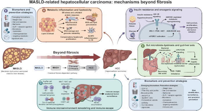
Figure 1: Evidence-graded framework for MASLD-related HCC beyond fibrosis. The central pathway shows progression from metabolic dysfunction-associated steatotic liver disease (MASLD) through metabolic dysfunction-associated steatohepatitis (MASH), fibrosis/cirrhosis, and hepatocellular carcinoma (HCC), while recognizing that HCC can occur without cirrhosis. (A) Metabolic inflammation and lipotoxicity include endoplasmic reticulum (ER) stress, reactive oxygen species (ROS), DNA injury, and NLRP3 inflammasome signaling. (B) Insulin resistance and oncogenic signaling include insulin-like growth factor-1 receptor (IGF-1R), insulin receptor substrate-1 (IRS-1), phosphoinositide 3-kinase (PI3K), protein kinase B (AKT), mechanistic target of rapamycin complex 1 (mTORC1), aldo-keto reductase family 1 member B1 (AKR1B1), advanced glycation end product–receptor for advanced glycation end product (AGE-RAGE), and metabolic reprogramming. (C) Gut dysbiosis and the gut–liver axis include lipopolysaccharide (LPS)–Toll-like receptor 4 (TLR4), farnesoid X receptor (FXR), and Takeda G protein-coupled receptor 5 (TGR5) signaling. (D) Immune remodeling includes Kupffer-cell states, mucosal-associated invariant T (MAIT) and natural killer T (NKT) cells, regulatory T (Treg) cells, CD8+ T cells, dendritic cell ferroptosis, and myeloid-derived suppressor cells (MDSCs). (E) Clinical translation distinguishes future-risk measures from early-detection tests and disease-modifying interventions; direct HCC prevention remains unproven for current metabolic therapies. Abbreviations: AGE, advanced glycation end products; AKR1B1, aldo-keto reductase family 1 member B1; AKT, protein kinase B; AFP, alpha-fetoprotein; CD8+, cluster of differentiation 8-positive; ELF, enhanced liver fibrosis; ER, endoplasmic reticulum; FIB-4, Fibrosis-4 index; FXR, farnesoid X receptor; GALAD, gender, age, AFP-L3, AFP, and des-gamma-carboxy prothrombin; HCC, hepatocellular carcinoma; IGF-1R, insulin-like growth factor 1 receptor; IRS-1, insulin receptor substrate 1; LPS, lipopolysaccharide; MAIT, mucosal-associated invariant T; MASLD, metabolic dysfunction-associated steatotic liver disease; MASH, metabolic dysfunction-associated steatohepatitis; MDSCs, myeloid-derived suppressor cells; MRE, magnetic resonance elastography; mTORC1, mechanistic target of rapamycin complex 1; NKT, natural killer T; NLRP3, NOD-like receptor family pyrin domain-containing 3; PI3K, phosphoinositide 3-kinase; PRS, polygenic risk score; RAGE, receptor for advanced glycation end products; ROS, reactive oxygen species; TGR5, Takeda G protein-coupled receptor 5; TLR4, Toll-like receptor 4; Treg, regulatory T cell; VCTE, vibration-controlled transient elastography. Please click here to view a larger version of this figure.
Disclosures
The authors declare no conflicts of interest. During revision, the authors used Figurelabs (https://chat.figurelabs.ai/) to assist in creating Figure 1. The authors independently verified the scientific claims and references and take full responsibility for the final content.
Acknowledgements
No funding was received for this work.
References
- Rinella ME, et al. A multisociety Delphi consensus statement on new fatty liver disease nomenclature. Hepatology. 2023;78(6):1966-1986.
- European Association for the Study of the Liver (EASL), European Association for the Study of Diabetes (EASD), European Association for the Study of Obesity (EASO). EASL-EASD-EASO Clinical Practice Guidelines on the management of metabolic dysfunction-associated steatotic liver disease. J Hepatol. 2024;81(3):492-542.
- Huang DQ, El-Serag HB, Loomba R. Global epidemiology of NAFLD-related hepatocellular carcinoma: trends, predictions, risk factors and prevention. Nat Rev Gastroenterol Hepatol. 2021;18(4):223-238.
- Simon TG, et al. Cancer risk in patients with biopsy-confirmed nonalcoholic fatty liver disease: a population-based cohort study. Hepatology. 2021;74(5):2410-2423.
- Kanwal F, et al. Risk of hepatocellular cancer in patients with non-alcoholic fatty liver disease. Gastroenterology. 2018;155(6):1828-1837.e2.
- Singal AG, et al. AASLD Practice Guidance on prevention, diagnosis, and treatment of hepatocellular carcinoma. Hepatology. 2023;78(6):1922-1965.
- Li D, Zhang X. From mechanisms to management: early detection and improved treatment of MASLD and its related hepatocellular carcinoma. Hepatol Commun. 2025;9(12):e0846.
- Lekakis V, Papatheodoridis GV. Natural history of metabolic dysfunction-associated steatotic liver disease. Eur J Intern Med. 2024;122:3-10.
- Orci LA, et al. Incidence of hepatocellular carcinoma in patients with nonalcoholic fatty liver disease: a systematic review, meta-analysis, and meta-regression. Clin Gastroenterol Hepatol. 2022;20(2):283-292.e10.
- Behari J, et al. Incidence of hepatocellular carcinoma in nonalcoholic fatty liver disease without cirrhosis or advanced liver fibrosis. Hepatol Commun. 2023;7(7):e00183.
- Cholankeril G, et al. Longitudinal changes in fibrosis markers and the risk of hepatocellular carcinoma in non-alcoholic fatty liver disease. J Hepatol. 2023;78(3):493-500.
- Ebrahimi F, et al. Familial coaggregation of MASLD with hepatocellular carcinoma and adverse liver outcomes: nationwide multigenerational cohort study. J Hepatol. 2023;79(6):1374-1384.
- Loomba R, et al. AGA Clinical Practice Update on screening and surveillance for hepatocellular carcinoma in patients with nonalcoholic fatty liver disease: expert review. Gastroenterology. 2020;158(6):1822-1830.
- Saha S, et al. Proteomic analysis reveals USP7 as a novel regulator of palmitic acid-induced hepatocellular carcinoma cell death. Cell Death Dis. 2022;13(6):563.
- Deschler S, et al. Polyunsaturated fatty acid-induced metabolic exhaustion and ferroptosis impair the antitumour function of MAIT cells in MASLD. J Hepatol. 2025;83(6):1364-1378.
- Benedé-Ubieto R, Cubero FJ, Nevzorova YA. Breaking the barriers: the role of gut homeostasis in metabolic dysfunction-associated steatotic liver disease. Gut Microbes. 2024;16(1):2331460.
- Che Z, et al. Regulatory role and biomarker potential of gut microbiota metabolites in the progression of metabolic dysfunction-associated steatotic liver disease to hepatocellular carcinoma. Clin Transl Gastroenterol. 2025;16(12):e00914.
- Caturano A, et al. Metabolic dysfunction-associated steatotic liver disease, insulin resistance and hepatocellular carcinoma: a deadly triad. Eur J Clin Invest. 2026;56(1):e70132.
- Bisht S, Sharma U, Gupta S. Role of insulin-like growth factor/receptor signaling in hepatocellular carcinoma. Hepatol Forum. 2026;7(1):59-68.
- Pei J, Wang B, Wang D. Current studies on molecular mechanisms of insulin resistance. J Diabetes Res. 2022;2022:1863429.
- Jørgensen SH, et al. Multi-layered proteomics identifies insulin-induced upregulation of the EphA2 receptor via the ERK pathway which is dependent on low IGF1R level. Sci Rep. 2024;14(1):28856.
- Xie C, et al. Lysosome-localized IRTKS condensates promote mTORC1 activity leading to MASLD and HCC. Cell Rep. 2026;45(2):116893.
- Syamprasad NP, et al. AKR1B1 drives hyperglycemia-induced metabolic reprogramming in MASLD-associated hepatocellular carcinoma. JHEP Rep. 2024;6(2):100974.
- Huang G, Li D. The macrophage opera: from metabolic prolog to oncogenic crescendo in metabolic dysfunction-associated steatotic liver disease. Front Med (Lausanne). 2026;13:1751290.
- Li N, et al. Tim-3 facilitates dendritic cell ferroptosis and impairs antitumor immunity in steatohepatitis-related hepatocellular carcinoma. J Hepatol. 2026;85(2):331-344.
- Wang H, Tsung A, Mishra L, Huang H. Regulatory T cell: a double-edged sword from metabolic-dysfunction-associated steatohepatitis to hepatocellular carcinoma. EBioMedicine. 2024;101:105031.
- Jin J, et al. Immunotherapy resistance in MASLD-related hepatocellular carcinoma: special immune microenvironment and gut microbiota. Int J Biol Sci. 2025;21(13):5666-5690.
- Fan R, et al. aMAP risk score predicts hepatocellular carcinoma development in patients with chronic hepatitis. J Hepatol. 2020;73(6):1368-1378.
- Kim GA, et al. A risk prediction model for hepatocellular carcinoma in non-alcoholic fatty liver disease without cirrhosis. Liver Int. 2024;44(3):738-748.
- Bianco C, et al. Non-invasive stratification of hepatocellular carcinoma risk in non-alcoholic fatty liver using polygenic risk scores. J Hepatol. 2021;74(4):775-782.
- Fujiwara N, et al. A blood-based prognostic liver secretome signature and long-term hepatocellular carcinoma risk in advanced liver fibrosis. Med. 2021;2(7):836-850.e10.
- Myojin Y, Hikita H. Growth differentiation factor 15: from stress response to clinical utility in chronic liver diseases. J Gastroenterol. 2026;61(2):117-130.
- Abdelhameed F, et al. Circulating angiopoietin-like protein 8 and steatotic liver disease related to metabolic dysfunction: an updated systematic review and meta-analysis. Front Endocrinol (Lausanne). 2025;16:1574842.
- Park KJ. Associations between apolipoprotein B/A1 ratio, lipoprotein(a), and the risk of metabolic-associated fatty liver diseases in a Korean population. Lab Med. 2023;54(6):633-637.
- Best J, et al. GALAD score detects early hepatocellular carcinoma in an international cohort of patients with nonalcoholic steatohepatitis. Clin Gastroenterol Hepatol. 2020;18(3):728-735.e4.
- Aminian A, et al. Association of bariatric surgery with major adverse liver and cardiovascular outcomes in patients with biopsy-proven nonalcoholic steatohepatitis. JAMA. 2021;326(20):2031-2042.
- Harrison SA, et al. A phase 3, randomized, controlled trial of resmetirom in NASH with liver fibrosis. N Engl J Med. 2024;390(6):497-509.
- Sanyal AJ, et al. Phase 3 trial of semaglutide in metabolic dysfunction-associated steatohepatitis. N Engl J Med. 2025;392(21):2089-2099.
- Loomba R, et al. Tirzepatide for metabolic dysfunction-associated steatohepatitis with liver fibrosis. N Engl J Med. 2024;391(4):299-310.
- Soong RS, et al. SGLT2 inhibitors and the risk of hepatocellular carcinoma in patients with MASLD and type 2 diabetes. Diabetes Metab. 2025;51(6):101698.
- Schwenk S, Kaplan DE. Chemoprevention of hepatocellular carcinoma. Hepatol Commun. 2025;9(11):e0836.