Domuzların araştırmalarda kullanımı son yıllarda artmıştır. Bununla birlikte, domuzlar zor hava yolu anatomisi ile karakterizedir. Endoskopik olarak yönlendirilen endotrakeal entübasyonun nasıl yapılacağını göstererek, mevcut protokol, hayvanların acı çekmesini ve gereksiz ölümleri önlemek için laboratuvar hayvanlarının güvenliğini daha da arttırmayı amaçlamaktadır.
Bu içeriği görüntülemek için JoVE aboneliğiniz gereklidir. Giriş yapın veya ücretsiz denemenizi bugün başlatın.
Yöntem makalesi
Domuzlarda Güvenli Hava Yolu Yönetimi için Standartlaştırılmış Bir Model Olarak Esnek Entübasyon Endoskobu Kullanılarak Endotrakeal Entübasyon
Bu makalede
Düzeltme bildirimi
Important: There has been an erratum issued for this article. View Erratum Notice
Özet
Özet
Endotrakeal entübasyon, güvenli bir hava yolu veya yüksek ventilasyon basınçları gerektiren çeşitli müdahaleler için domuz modellerinde translasyonel araştırmalar için genellikle temel bir gerekliliktir. Endotrakeal entübasyon, optimal koşullar altında yüksek bir başarı oranı elde etmek için minimum sayıda başarılı endotrakeal entübasyon gerektiren zorlu bir beceridir ve bu da anesteziyoloji dışı araştırmacılar için genellikle ulaşılamaz bir durumdur. Spesifik domuz hava yolu anatomisi nedeniyle, genellikle zor bir hava yolu varsayılabilir. Güvenli bir hava yolu kurmanın imkansızlığı, laboratuvar hayvanının yaralanmasına, olumsuz olaylarına veya ölümüne neden olabilir. Prospektif, randomize, kontrollü bir değerlendirme yaklaşımı kullanılarak, fiberoptik yardımlı endotrakeal entübasyonun daha uzun sürdüğü ancak oksijen satürasyonunda klinik olarak ilgili düşüşlere neden olmadan konvansiyonel entübasyondan daha yüksek bir ilk geçiş başarı oranına sahip olduğu gösterilmiştir. Bu model, endoskopik olarak yönlendirilen endotrakeal entübasyon için standartlaştırılmış bir rejim sunarak, özellikle doğrudan laringoskopi yoluyla endotrakeal entübasyon tekniğinde deneyimsiz araştırmacılar için güvenli bir hava yolu sağlar. Bu prosedürün hayvan ıstırabını ve gereksiz hayvan kayıplarını en aza indirmesi beklenmektedir.
Giriş
Endotrakeal entübasyon, güvenli bir hava yolu veya yüksek ventilasyon basınçları (kardiyopulmoner resüsitasyon sırasında ventilasyon1 veya akut solunum sıkıntısı sendromu2 gibi) gerektiren veya supraglottik hava yolu cihazları tarafından iç kompresyon yoluyla serebral kan akışının tehlikeye atılmamasını gerektiren çeşitli müdahaleler için domuz modellerinde translasyonel araştırma için genellikle temel bir gerekliliktir3 , domuzlarda beklenen zor bir hava yolu bağlamında zaman zaman alternatif olarak çoğaltılan 4,5.
Erişim kısıtlı. Bu içeriği görüntülemek için lütfen giriş yapın veya deneme sürümünü başlatın.
Protokol
Bu protokoldeki deneyler Devlet ve Kurumsal Hayvan Bakım Komitesi (Landesuntersuchungsamt Rheinland-Pfalz, Koblenz, Almanya; onay no. G20-1-135) tarafından onaylanmıştır. Deneyler ARRIVE yönergelerine uygun olarak gerçekleştirildi. Bu çalışmada genel olarak ortalama ağırlığı 30 kg ± 2 kg ve 12-16 haftalık olan 10 adet anestezi uygulanmış erkek domuz (Sus scrofa domestica) kullanılmıştır.
1. Hayvan hazırlığı
- Stresi en aza indirmek için hayvanlar için normal bir ortam sağlayın. Aspirasyon riskini azaltmak için planlanan deneyden 6 saat önce yiyecekleri durdurun, ancak suya erişime izin verin.
- Domu....
Erişim kısıtlı. Bu içeriği görüntülemek için lütfen giriş yapın veya deneme sürümünü başlatın.
Sonuçlar
Endotrakeal entübasyon 10 erkek domuza (yaş 12-16 hafta, ağırlık 30 kg ± 3 kg) prospektif, randomize, kontrollü bir çalışma ortamında yapıldı. Domuzlar iki gruba randomize edildi: biri konvansiyonel olarak laringoskopik olarak entübe edildi (CI grubu), diğer grup protokolde (FIE grubu) tanımlandığı gibi esnek bir entübasyon endoskobu ile entübe edildi. Grup ödevi mühürlü zarflar çekilerek yapıldı. Araştırmacı günlük olarak rastgele atandı.
Çalışma, anestezide 3 yıldan fazla deneyime s.......
Erişim kısıtlı. Bu içeriği görüntülemek için lütfen giriş yapın veya deneme sürümünü başlatın.
Tartışma
Önceki çalışmalarda, araştırma grubumuzdomuz modeli 2,27,32,33'ün translasyonel faydaları ile ilgili belirli ayrıntıları zaten açıklamıştır. Genel olarak, hayvanın stres seviyesini ve gereksiz ağrıyı azaltmak, herhangi bir çalışma protokolünün ayrılmaz bir parçası olmalı ve güvenilir bir şekilde tekrarlanabilir veriler üretmek için çok önemlidir. Bu nedenle, beklenen zor bir hava yolu ile domuz.......
Erişim kısıtlı. Bu içeriği görüntülemek için lütfen giriş yapın veya deneme sürümünü başlatın.
Açıklamalar
Esnek entübasyon endoskobu ve aksesuarları, üretici tarafından yalnızca araştırma amacıyla koşulsuz olarak sağlanmıştır. Yazarlar başka hiçbir finansal veya diğer çıkar çatışması olmadığını beyan ederler.
Teşekkürler
Yazarlar, mükemmel teknik desteği için Dagmar Dirvonskis'e teşekkür etmek istiyor.
....Erişim kısıtlı. Bu içeriği görüntülemek için lütfen giriş yapın veya deneme sürümünü başlatın.
Malzemeler
| Ad | Şirket | Katalog numarası | Yorumlar |
|---|---|---|---|
| Ambu aScope Regular | Ambu GmbH, Medizinprodukte, Bad Nauheim, Almanya | Tek kullanımlık fiber optik dış çap 5 mm | |
| Ambu aView Monitör | Ambu GmbH, Medizinprodukte, Bad Nauheim, Almanya | monitör | |
| Atracurium Hikma 50 mg / 5mL | Hikma Pharma GmbH, Martinsried | atracurium | |
| Azaperone (Stresnil) 40mg / mL | Lilly Deutschland GmbH, Bad Homburg, Almanya | azaperone | |
| BD Discardit II Spritze 2, 5, 10, 20 mL | Becton Dickinson S.A. Carretera, Mequinenza Fraga, İspanya | şırınga | |
| BD Luer Connecta | Becton Dickinson İnfüzyon Tedavisi, AB Helsingborg, Schweden | 3 musluk | |
| BD Microlance 3 20 G | Becton Dickinson S.A. Carretera, Mequinenza Fraga, İspanya | kanül | |
| Curafix i.v. klasikler | Lohmann & Rauscher International GmbH & Co. KG, Rengsdorf, Almanya | Kanül tutma pansuman | |
| Engströ m Bakım İstasyonu | GE Heathcare, Madison ABD | ventilatör | |
| Fentanil-Janssen 0.05 mg/mL | Janssen-Cilag GmbH, Neuss | fentanil | |
| Fü hrungsstab, Durchmesser 4.3 | Rü | endotrakeal tüp tanıtıcısı | |
| Windows için IBM SPSS Statistics, Sürüm 20 | Windows için IBM SPSS Statistics, Sürüm 20.0. Armonk, NY: IBM Corp.) | İstatistiksel yazılım | |
| Incetomat-line 150 cm | Fresenius, Kabi Deutschland, GmbH | perfusor hattı | |
| Intrafix Primeline | B. Braun Melsungen AG, Melsungen, Almanya | İnfüzyon hattı | |
| JOZA Einmal Nitril Untersuchungshandschuhe | JOZA, Mü nchen, Almanya | tek kullanımlık eldivenler | |
| Laringoskop, 45.48.50, KL 2000 | Medicon | Laringoskop sapı | |
| Littmann Classic III Stetoskop | 3M Deutschland GmbH, Neuss, Almanya | stetoskop | |
| Luer Lock | B.Braun Melsungen AG, Almanya | ||
| Msakat Vlieskompresse | Maim GmbH, Neuenkirchen, Almanya | Polar kompres dili sabitlemek için | |
| Masimo LNCS Adtx SpO2 sensor | Masimo Corporation Irvine, Ca 92618 ABD | kuyruk için doygunluk klipsi | |
| Masimo LNCS TC-I SpO2 kulak klipsi sensörü | Masimo Corporation Irvine, Ca 92618 ABD | Kulak için doygunluk klipsi | |
| Masimo Radical 7 | Masimo Corporation Irvine, Ca 92618 ABD | periferik oksijen satürasyonu | |
| Midazolam 15 mg / 3 mL | Hameln Pharma GmbH, Hameln, Almanya | midazolam | |
| Midmark Köpek Maskesi Diyaframlı Küçük Plastik FRSCM-0005 | Midmark Corp., Dayton, Ohio, ABD | köpek ventilasyon | |
| maskesi Octeniderm farblos | Schü lke & Mayr GmbH, Nordenstedt, Almanya | Alkollü dezenfektan | |
| Orijinal Perfüzör şırınga 50 mL | B.Braun Melsungen AG, Almanya | perfüzör şırınga | |
| Perfüzör FM Braun | B.Braun Melsungen AG, Almanya | şırınga pompası | |
| Propofol %2 20 mg/mL (50 mL şişeler) | Fresenius, Kabi Deutschland, GmbH | propofol | |
| RÜ SCH Fü hrungsstab fü r Endotrakealtubus (ID 5.6 mm) | Teleflex Medical Sdn. Bhd, Malezya | PVC kaplı boru kılavuz teli | |
| Rü Schelit Süper Güvenlik Şeffaf >ID 6/6.5 /7.0 mm | Teleflex Medical Sdn. Bhd, Malezya | endotrakeal tüp | |
| Paslanmaz Macintosh Grö ß e 4 | Welch Allyn69604 | laringoskop için bıçak | |
| Sterofundin | B.Braun Melsungen AG, Melsungen, Almanya | Dengeli elektrolit çözeltisi | |
| Ultrastop Antibeschlagmittel damlalıklı şişe 25 mL | Sigmapharm Arzneimittel GmbH, Wien, Avusturya | Buğu önleyici ajan | |
| Vasofix Safety 22 G-16 G | B.Braun Melsungen AG, Almanya | venöz kateter | |
| VBM Manşet Manometresi | VBM Medizintechnik GmbH, Sulz a.N., Almanya | manşet basınç göstergesi | |
| Zelette | Lohmann & Rauscher International GmbH & Co. KG, Rengsdorf, Almanya | Doku çubuğu |
Kaynaklar
- Kleinman, M. E., Oh, W., Stonestreet, B. S. Comparison of intravenous and endotracheal epinephrine during cardiopulmonary resuscitation in newborn piglets. Critical Care Medicine. 27 (12), 2748-2754 (1999).
- Rissel, R., et al.
Erişim kısıtlı. Bu içeriği görüntülemek için lütfen giriş yapın veya deneme sürümünü başlatın.
Yeniden basım ve izinler
Düzeltme
Formal Correction: Erratum: Endotracheal Intubation Using a Flexible Intubation Endoscope As a Standardized Model for Safe Airway Management in Swine
Posted by JoVE Editors on 4/03/2023. Citeable Link.
An erratum was issued for: Endotracheal Intubation Using a Flexible Intubation Endoscope As a Standardized Model for Safe Airway Management in Swine. The Protocol, Representative Results, and Discussion sections were updated.
In the Protocol, step 1.5 was updated from:
Disinfect the skin with a disinfectant (alcoholic) before inserting a peripheral vein cannula (22 G) into an ear vein. Spray the area, wipe once, then spray again, and allow the disinfectant to dry.
to:
Disinfect the skin with a disinfectant (alcoholic) before inserting a peripheral vein cannula (22 G) into an ear vein. Spray the area, wipe once, then spray again, and allow the disinfectant to dry. Secure the ear cannula with a band-aid (See Table of Materials).
In the Protocol, step 3.7 was updated from:
While maintaining the position of the endoscope, advance the endotracheal tube until it becomes visible in the camera image.
NOTE: If the endotracheal tube cannot be advanced through the glottic plane, there is a possibility that it has become caught on the arytenoid cartilage. In this case, the endotracheal tube must be withdrawn 1 cm and rotated by 90° before gently advancing again. If necessary, this maneuver can be repeated. Similar calibers of flexible intubation endoscope and endotracheal tube can minimize the risk of this issue occurring. If the endotracheal tube cannot be advanced despite this maneuver, it is likely that the subglottic narrowness-the narrowest part of the porcine larynx-cannot be passed. In this case, a smaller endotracheal tube size needs to be selected. Regular commercially available endotracheal tubes in sizes 6.5 cm or 7.0 cm ID should be able to pass the glottis as long as no anatomic abnormalities are present.
to:
While maintaining the position of the endoscope, advance the endotracheal tube until it becomes visible in the camera image.
NOTE: If the endotracheal tube cannot be advanced through the glottic plane, there is a possibility that it has become caught on the arytenoid cartilage. In this case, the endotracheal tube must be withdrawn 1 cm and rotated by 90° before gently advancing again. If necessary, this maneuver can be repeated. Similar calibers of flexible intubation endoscope and endotracheal tube can minimize the risk of this issue occurring. If the endotracheal tube cannot be advanced despite this maneuver, it is likely that the subglottic narrowness-the narrowest part of the porcine larynx-cannot be passed. In this case, a smaller endotracheal tube size needs to be selected. Regular commercially available endotracheal tubes in sizes 6.5 cm or 7.0 cm ID should be able to pass the glottis as long as no anatomic abnormalities are present. Endotracheal tube size requirements vary depending on the piglet size and breed.
In the Representative Results, the sixth paragraph was updated from:
Statistical analyses were performed using commercially available software (see Table of Materials). Normal distribution was examined using the Kolmogorov-Smirnoff test28. If a normal distribution was determined, group differences were analyzed using t-tests of independent samples29 or the Mann-Whitney U test30 for the non-parametric version. Data are presented as mean (± standard deviation). Correlations of ordinal-scale data were examined using Spearman's correlation coefficient31. A significance level of p < 0.05 was assumed.
to:
Statistical analyses were performed using commercially available software (see Table of Materials). Normal distribution was examined using the Kolmogorov-Smirnoff test28. If a normal distribution was determined, group differences were analyzed using t-tests of independent samples29 or the Mann-Whitney U test30 for the non-parametric version. Data are presented as mean (± standard deviation). Correlations of ordinal-scale data were examined using Spearman's correlation coefficient31. A significance level of p < 0.05 was assumed. All tests were performed with exploratory intention; therefore p-values are descriptive. Nevertheless, p < 0.05 was accepted as indicative of statistical significance.
In the Representative Results, the legend for figure 1 was updated from:
Figure 1: Number of intubation attempts in group comparison. For the group that was intubated using a flexible intubation endoscope, every intubation attempt was successful; in the group that was conventionally intubated, it took an average of 1.4 attempts before the endotracheal tube could be placed correctly. Error bars show the standard deviation. Please click here to view a larger version of this figure.
to:
Figure 1: Number of intubation attempts in group comparison. For the group that was intubated using a flexible intubation endoscope, every intubation attempt was successful; in the group that was conventionally intubated, it took an average of 1.4 attempts before the endotracheal tube could be placed correctly. Error bars show the standard deviation. n = 5 (for each group). Please click here to view a larger version of this figure.
In the Representative Results, figure 2 was updated from:
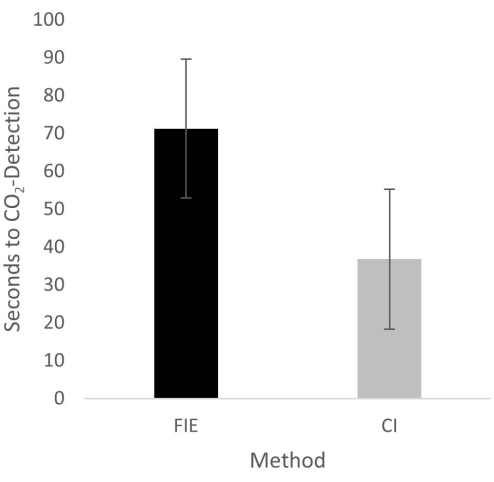
Figure 2: Time until CO2 detection in group comparison. For the group that was intubated using a flexible intubation endoscope, it took significantly longer until end-tidal CO2 could be detected, depicted as mean and standard deviation. Please click here to view a larger version of this figure.
to:
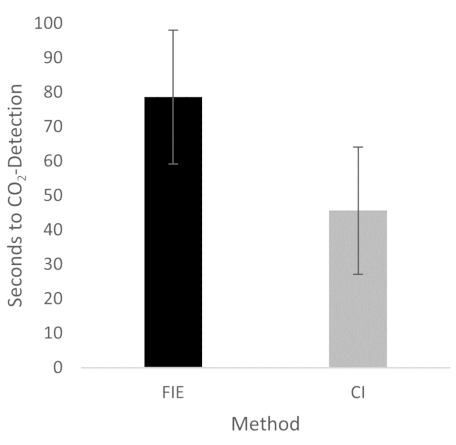
Figure 2: Time until CO2 detection in group comparison. For the group that was intubated using a flexible intubation endoscope, it took significantly longer until end-tidal CO2 could be detected, depicted as mean and standard deviation. n = 5 (for each group). Please click here to view a larger version of this figure.
In the Discussion, the fifth paragraph was updated from:
The increased duration had no clinical significance in this cohort. At no time was the termination criterion-a saturation of less than 93%-reached. This is shown in the results because a procedure change was unnecessary at any time. Prior adequate mask ventilation is a critical step to allow sufficient time for fiberoptic endotracheal tube placement to avoid rapid desaturation34. These results are consistent with previous studies comparing conventional intubation and endoscopically assisted intubations with inexperienced providers35.
to:
The increased duration had no clinical significance in this cohort. At no time was the termination criterion-a saturation of less than 93%-reached. This is shown in the results because a procedure change was unnecessary at any time. Prior adequate mask ventilation is a critical step to allow sufficient time for fiberoptic endotracheal tube placement to avoid rapid desaturation34. These results are consistent with previous studies comparing conventional intubation and endoscopically assisted intubations with inexperienced providers35. We attribute the prolonged duration of fiberoptic intubation to the fact that one must first reorient again after insertion, whereas with conventional intubation, one retains a view of the glottis. It is also important to avoid contact with the mucosa with the flexible intubation endoscope during advancement. This requires occasional corrective maneuvers. Last but not least, after successful placement, retraction of the relatively long endoscope is required, which increases the time to CO2 detection slightly.
