Bu protokol, N-laktoil-fenilalaninin diyabetik kardiyomiyopati, kardiyak lipid metabolizması ve kolesterol sentezi üzerindeki etkilerini, özellikle adenozin monofosfat-aktive protein kinaz alfa 1/3-hidroksi-3-metilglutaril-koenzim A redüktaz yoluna odaklanarak incelemek için in vivo ve in vitro yaklaşımları açıklamaktadır.
Bu içeriği görüntülemek için JoVE aboneliğiniz gereklidir. Giriş yapın veya ücretsiz denemenizi bugün başlatın.
Araştırma makalesi
N-Laktoil-fenilalanin, Lipid Metabolizmasını Düzenleyerek Diyabetik Kardiyomiyopati Fare Modelinde Kardiyak Fonksiyonu İyileştirir
Bu makalede
Özet
Özet
Diyabetik kardiyomiyopati (DCM), diyabetin en ciddi komplikasyonlarından biridir. Endojen bir metabolit olan N-laktoil-fenilalanin (Lac-Phe), kilo kaybını teşvik edebilir ve glikoz homeostazını iyileştirebilir. Bu çalışma, Lac-Phe'nin DCM üzerindeki terapötik etkisini araştırmayı ve altında yatan mekanizmayı daha ayrıntılı incelemeyi amaçlamıştır. DCM'li tip 2 diyabetik farelere Lac-Phe uygulanmıştır. Lac-Phe, kardiyak kontraktil fonksiyonu artırmış, patolojik remodelasyonu iyileştirmiş ve kalpte lipid birikimini azaltmıştır. In vitro ortamda, Lac-Phe'nin etkileri yüksek glikoz ve palmitik asit ile stimüle edilen H9c2 hücrelerinde incelenmiştir. Lac-Phe, H9c2 hücrelerinde kolesterol sentezini azaltarak lipid birikimini düşürmüştür. Mekanistik olarak Lac-Phe, adenozin monofosfatla aktive olan protein kinaz alfa 1'in (AMPKα1) fosforilasyonunu ve aktivasyonunu teşvik etmiştir. Ardından, aktive olan AMPKα1, aşağı akış hedefi olan 3-hidroksi-3-metilglutaril-koenzim A redüktazı (HMGCR) fosforile ederek inhibe etmiş; bu durum kolesterol sentezinin azalmasına ve nihayetinde H9c2 hücrelerindeki lipid metabolizması dengesizliğinin iyileşmesine yol açmıştır. Bu bulgular, Lac-Phe'nin DCM'deki H9c2 hücrelerinde lipid metabolizmasını AMPKα1/HMGCR yolu üzerinden iyileştirdiğini, böylece kardiyak kontraktil fonksiyonu artırdığını ve patolojik remodelasyonu hafiflettiğini göstermektedir. Lac-Phe, diyabetik kardiyomiyopati için potansiyel bir terapötik yaklaşım temsil edebilir.
Giriş
Diyabet, kalıcı hiperglisemi ile karakterize metabolik bir hastalıktır1. Son yirmi yılda diyabet, her yıl altı milyondan fazla insanın diyabet ve komplikasyonları nedeniyle ölmesiyle dünya çapında ilk on ölüm nedeninden biri haline gelmiştir2. Diyabete bağlı bir kardiyak patolojik durum olan diyabetik kardiyomiyopati (DCM), diyabetle ilişkili en ciddi komplikasyonlardan birini temsil eder3,4. Kalp yetmezliği için bağımsız bir risk faktörü olarak işlev görür ve kardiyak fibrozis ile kardiyomiyosit hipertrofisi dahil olmak üzere, nihayetinde hem diyastolik hem de sistolik disfonksiyona yol açan yapısal ve fonksiyonel miyokardiyal değişikliklerle karakterizedir5,6. β-blokerler, anjiyotensin dönüştürücü enzim inhibitörleri ve anjiyotensin II tip 1 reseptör blokerleri gibi birçok geleneksel kardiyovasküler tedavi şu anda DCM yönetiminde kullanılsa da7,8, klinik ortamlarda etkinlikleri suboptimal düzeyde kalmaktadır. DCM'nin altında yatan mekanizmalar derinlemesine çalışılmadığından9, şu anda bu hastalığı hedefleyen net bir tedavi stratejisi bulunmamaktadır10,11. DCM'nin klinik belirtileri, erken evrede asemptomatik diyastolik disfonksiyondan, geç evrede sistolik disfonksiyon ve klinik kalp yetmezliğine, hatta ölüme kadar ilerler12,13. Bu nedenle, bu durum için etkili tedavi seçeneklerinin araştırılması ve geliştirilmesine acil ihtiyaç vardır.
Dengesiz lipid metabolizması, DCM'nin temel bir özelliğidir14. DCM'de, kandaki serbest yağ asidi (FFA) seviyelerinin artması, kardiyomiyositler tarafından aşırı yağ asidi alımına yol açabilir15,16. Kardiyomiyositlerdeki yağ asidi metabolizması temel olarak yağ asidi oksidasyonuna bağlıdır. Zamanla, kardiyomiyositlerdeki yağ asidi oksidasyonu, alınan tüm yağ asitlerini tamamen metabolize etmek için yetersiz kalır ve fazla yağ asitleri ile bunların ara metabolitleri kardiyomiyositlerde birikir17. Bu aşırı lipid yükü lipotoksisiteyi tetikler, miyokardiyal enerji arzını bozar, kardiyak oksidatif stresi ve inflamatuar yanıtları teşvik ederek nihayetinde kardiyomiyositlerde ciddi hasara neden olur18,19. Bu nedenle, kardiyak lipid metabolizmasının aktif olarak düzenlenmesi, DCM için potansiyel bir terapötik hedef olabilir20.
N-laktoil-fenilalanin (Lac-Phe), egzersiz sonrası konsantrasyonu hızla artan endojen bir metabolittir21. Mekanistik olarak Lac-Phe, laktat ve fenilalaninin kondenzasyonu yoluylale sitozolik enzim karnozin dideptidaz 2 (CNDP2) tarafından sentezlenir. Bu biyosentetik yolak; makrofajlar, epitel hücreler ve fiziksel aktivite sonrası dolaşıma Lac-Phe salgılayan mezenkimal kök hücreler dahil olmak üzere çeşitli hücre tiplerine yaygın olarak dağılmıştır22. Lac-Phe'nin kronik uygulamalarının gıda alımını etkili bir şekilde azalttığı, obeziteyi ve vücut ağırlığını düşürdüğü ve glikoz homeostazını iyileştirdiği gösterilmiştir21. Yakın tarihli bir çalışma, Lac-Phe'nin omurilik yaralanması sonrası mikrogliya/makrofajların lipid metabolizma kapasitesini iyileştirebileceğini göstermiştir23. İlginç bir şekilde, çalışmalar tip 2 diyabet için birinci basamak bir ilaç olan metforminin hem akut hem de kronik uygulamasının Lac-Phe seviyelerini yükseltebildiğini ve Lac-Phe'nin metforminle ilişkili anti-obezite etkilerine aracılık ettiğini göstermektedir22. Ayrıca, Sha ve arkadaşları yakın zamanda Lac-Phe'nin mitokondriyal disfonksiyonu hafifleterek miyokardiyal hücre ölümünü azalttığını ve böylece transvers aortik konstriksiyon (TAC) kaynaklı kalp yetmezliğini iyileştirdiğini göstermiştir24. Uzamış hiperglisemi ve obezite, DCM'de lipid metabolizmasının disregülasyonuna katkıda bulunan önemli faktörlerdir25. Topluca, bu kanıtlar Lac-Phe'nin DCM üzerinde potansiyel bir terapötik etkiye sahip olabileceğini düşündürmektedir. Ancak, bugüne kadar Lac-Phe'nin spesifik olarak DCM'deki rolü araştırılmamıştır.
Bu çalışmada, diyabetik kardiyomiyopatisi olan farelere dışarıdan Lac-Phe uygulanmasının, kardiyak kontraktil fonksiyonu etkili bir şekilde iyileştirdiğini ve kardiyak patolojik yeniden şekillenmeyi azalttığını bulduk. Lac-Phe'nin, lipid metabolizması dengesizliğinin düzenlenmesi yoluyla DCM'li farelerde kalp fonksiyonlarını iyileştirdiğini saptadık. Lac-Phe, DCM'li farelerin kardiyomiyositlerinde lipid damlacığı (LD) birikimini azalttı ve kalpteki kolesterol içeriğini düşürdü. Mekanistik olarak Lac-Phe, adenozin monofosfat-aktive protein kinaz alfa 1'in (AMPKα1) fosforilasyonunu ve aktivasyonunu teşvik ederek, böylece 3-hidroksi-3-metilglutaril-koenzim A redüktazın (HMGCR) fosforilasyonunu artırdı. HMGCR fosforilasyonu, enzim aktivitesini azaltarak kolesterol üretiminin düşmesini sağlar ve böylece kardiyomiyositlerdeki lipid metabolizması dengesizliğini iyileştirir. Bu sonuçlar, Lac-Phe ile DCM'nin potansiyel tedavisi için teorik bir temel oluşturmaktadır.
Erişim kısıtlı. Bu içeriği görüntülemek için lütfen giriş yapın veya deneme sürümünü başlatın.
Protokol
The animal study protocol was approved by the Institutional Ethics Committee of Nanjing Drum Tower Hospital (Approval Number: 2025AE1065, 26 September 2025) and was conducted in accordance with the guidelines outlined in the Guidelines for the Care and Use of Laboratory Animals (8th Edition), published by the National Institutes of Health of the United States.
Animals
Thirty 5-week-old male C57BL/6J mice were used in this study. After 1 week of adaptive feeding, all mice were housed with access to food and water under an alternating 12 h day-night cycle at a temperature of 22°C ± 2°C and humidity of 40%–60%. An HFD combined with low-dose STZ was used to establish the DCM mouse model (Figure 1A). Four weeks after initiation of high-fat diet (HFD; 60% fat, 20% protein, and 20% carbohydrate) feeding, mice were fasted overnight and subsequently administered streptozotocin (STZ; 8 mg/mL dissolved in citrate buffer, pH 4.5) via intraperitoneal injection at a dosage of 35 mg/kg/day for three consecutive days. Control group mice were fed a control diet (10% fat, 20% protein, and 70% carbohydrate) and received equivalent volumes of citrate buffer. Following 15 additional weeks of high-fat diet maintenance, mice meeting both criteria, random blood glucose of ≥13.3 mmol/L and left ventricular ejection fraction (LVEF) of <55%, were defined as successfully established DCM models. These validated DCM mice were numbered and randomly assigned to the DCM and DCM + Lac-Phe groups using a random number table. Approximately half of the mice failed to develop the model; these animals were euthanized in accordance with animal ethics requirements. In brief, mice were divided into three groups: Control, DCM, and DCM + Lac-Phe (n = 10 for each group). All 10 mice per group underwent echocardiography, body weight measurement, and OGTT. Following the 28-day treatment period, hearts were collected for subsequent analyses. There were 10 mice in each group. Of these, three mice per group were used for transcriptome sequencing, three were used for transmission electron microscopy and histological staining, and three were used for protein expression analysis. The remaining mouse in each group was reserved as a backup. After successful establishment of the DCM model, Lac-Phe was dissolved in normal saline to a final concentration of 10 mg/mL. Mice in the DCM + Lac-Phe group received intraperitoneal Lac-Phe at 50 mg/kg/day in an injection volume of 5 mL/kg, once daily for 28 consecutive days. Mice in the Control and DCM groups were administered an equivalent volume of normal saline as vehicle control.
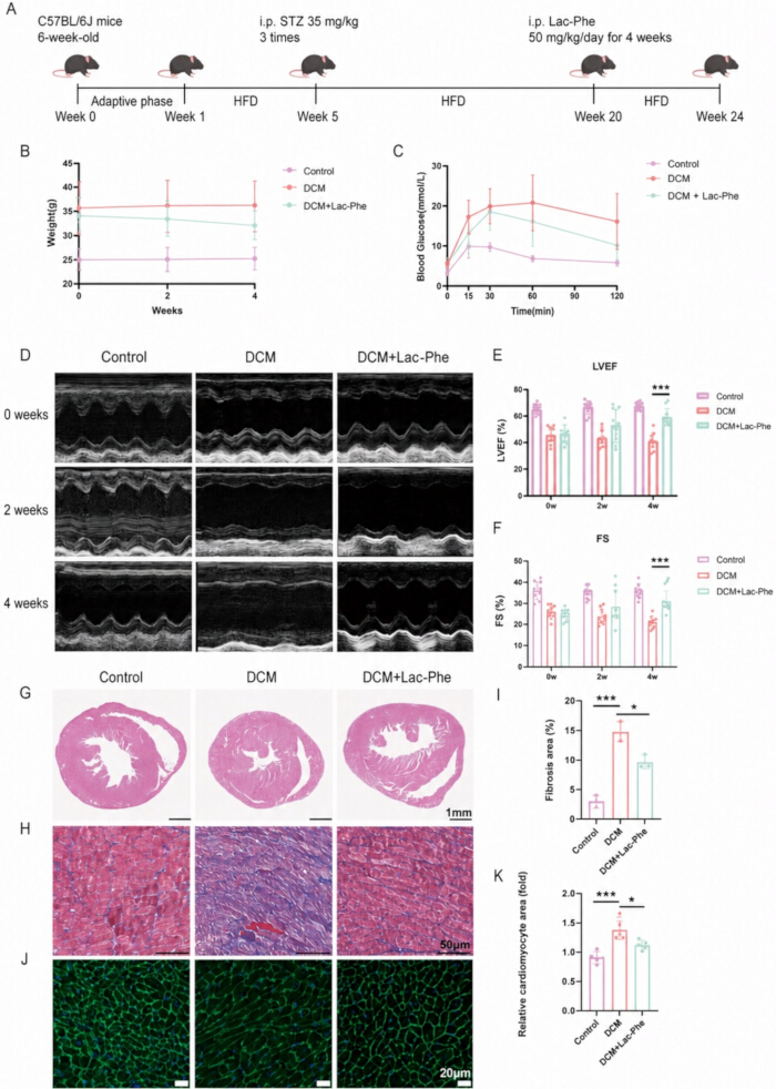
Figure 1. Lac-Phe improves cardiac function and pathological remodeling in mice with diabetic cardiomyopathy. (A) Schematic of the HFD/STZ-induced DCM mouse model and Lac-Phe treatment protocol. (B) Body weights of mice in the Control, DCM, and DCM + Lac-Phe groups during the 4-week treatment period; n = 10 per group. (C) Oral glucose tolerance test (OGTT) in the indicated groups; n = 10 per group. (D) Representative echocardiographic images at 0, 2, and 4 weeks of treatment. (E,F) Quantification of left ventricular ejection fraction (LVEF) (E) and fractional shortening (FS) (F); n = 10 per group. (G) Representative hematoxylin and eosin (H&E)-stained heart sections. Scale bar = 1 mm. (H,I) Representative Masson’s trichrome staining of heart sections (H) and quantification of fibrotic area (I); n = 3 per group. Scale bar = 50 μm. (J,K) Representative wheat germ agglutinin (WGA) staining of heart sections (J) and quantification of relative cardiomyocyte area (K); n = 3 per group. Scale bar = 20 μm. Data are presented as the mean ± standard deviation. P < 0.05, **P < 0.001. Please click here to view a larger version of this figure.
Cardiac function
Following anesthesia with isoflurane inhalation, mice were secured in a supine position on a temperature-controlled platform (37°C). Left ventricular dimensions and mitral valve blood flow velocity during diastole and systole were measured using an echocardiography machine equipped with a 40 MHz transducer, from which LVEF and shortening fraction (FS) were calculated. Three consecutive cardiac cycles were collected from each mouse.
Cell treatment
H9c2 cells were cultured in Dulbecco’s modified Eagle’s medium supplemented with 10% fetal bovine serum and 1% penicillin–streptomycin at 37°C in a humidified atmosphere containing 5% CO₂. Cells were passaged every 2–3 days upon reaching 70%–80% confluence. For experiments, cells were seeded in 10 cm dishes and then divided into three groups: Control, high glucose/palmitic acid (HG/PA), and HG/PA + Lac-Phe. Cells in the HG/PA and HG/PA + Lac-Phe groups were first exposed to 0.25 mM palmitic acid and 50 mM glucose for 24 h. Thereafter, cells in the HG/PA + Lac-Phe group were treated with 10 μg/mL Lac-Phe for an additional 24 h while HG/PA exposure was maintained; cells in the HG/PA group received an equivalent volume of PBS under the same HG/PA conditions. In the p-AMPK inhibition experiments, 10 μM AMPK-IN-3 (dissolved in DMSO) was added simultaneously with Lac-Phe for 24 h, and the corresponding control groups received an equal volume of DMSO. For the atorvastatin experiments, H9c2 cells under HG/PA conditions were treated with 10 μM atorvastatin for 24 h, either alone or in combination with 10 μg/mL Lac-Phe.
Histological staining
After 28 days of Lac-Phe administration, mouse hearts were harvested and immersion-fixed in 4% paraformaldehyde (pH 7.4) for 24 h. Fixed cardiac tissues were paraffin-embedded and sectioned into 6-μm-thick slices. Tissue sections were stained with hematoxylin and eosin, wheat germ agglutinin, and Masson’s trichrome. All staining procedures were performed strictly following the manufacturers’ protocols. Whole-slide imaging was performed at 20× magnification using a high-resolution brightfield microscope equipped with a digital camera system.
Western blot analysis
Protein concentration was determined using the Bradford assay. Tissue lysates were resolved by SDS-PAGE on 10% (w/v) acrylamide gels and electrophoretically transferred to polyvinylidene difluoride membranes. Nonspecific binding sites were blocked with 5% bovine serum albumin for 2 h at room temperature. Membranes were then incubated overnight at 4°C with primary antibodies (diluted 1:1,000), followed by six washes with TBST (5 min each) on a shaker. The membranes were then incubated with horseradish peroxidase-conjugated secondary antibodies (diluted 1:5,000) for 2 h at room temperature, followed by another six washes with TBST (5 min each). Protein bands were visualized using an enhanced chemiluminescence reagent kit. Quantification of western blot results was performed by densitometric analysis using ImageJ software. All antibodies used in this study are listed in the Table of Materials.
Transcriptomics
Total RNA was isolated from cardiac tissue of C57BL/6J mice using TRIzol Reagent and RNeasy mini kits. RNA quality was evaluated using a 2100 bioanalyzer, demonstrating high RNA purity and integrity. RNA-seq libraries were prepared according to the VAHTS Universal V10 RNA-seq Library Prep Kit and sequenced using an Illumina NovaSeq 6000 System (single-read, 150 bp). Differentially expressed genes were identified using DESeq2, with an adjusted P value < 0.05 and an absolute log₂ fold change > 1 considered statistically significant. Gene Ontology (GO) enrichment analysis was conducted on differentially expressed genes, and the significance of enrichment of differentially expressed genes in GO terms was calculated using the hypergeometric distribution algorithm.
Oil Red O staining and LD540 lipid staining
Cardiac tissue was embedded in OCT compound and then cut into 10-μm-thick sections. The frozen sections were equilibrated at room temperature for 30 min and fixed with 4% paraformaldehyde for 10 min. After washing with PBS, the sections were placed in 60% isopropyl alcohol for 15 s and covered with Oil Red O working solution for 30 min. The sections were then treated with 60% isopropyl alcohol for 15 s and washed three times with PBS. For LD540 lipid staining, H9c2 cells were washed with PBS and fixed with 4% paraformaldehyde for 10 min at room temperature. The cells were then incubated with 10 μM LD540 for 30 min at room temperature.
Transmission electron microscopy (TEM)
Fresh mouse heart tissues were cut into 1–2 mm3 pieces, fixed in electron microscope fixative at 4°C for 4 h, then washed with double-distilled water and immersed in ethanol solutions of progressively increasing concentrations. The samples were embedded, sectioned, and then observed under a transmission electron microscope.
Total cholesterol (TC) content
Total cholesterol (TC) levels in serum and H9c2 cell samples were measured using the Amplex Red Cholesterol and Cholesteryl Ester Assay Kit according to the manufacturer’s instructions.
Oral Glucose Tolerance Test (OGTT)
Mice were fasted for 14 h (from 19:00 to 9:00) and gavaged with glucose (20% stock, 2 g/kg). Blood glucose levels were determined using blood collected from the tail vein at 0, 15, 30, 60, and 120 min after gavage.
Cell counting kit-8 (CCK-8) measurement
CCK-8 was used to measure cell viability. H9c2 cells were seeded into 96-well plates at an initial density of 3 × 10⁴ cells/well. After treatment with different concentrations of Lac-Phe under HG/PA conditions for 24 h, 10 μL of the kit reagent was added to 100 μL of culture medium per well, followed by incubation at 37°C for 4 h. The absorbance was measured at 450 nm using a multimode microplate reader to obtain the optical density (OD) value.
Molecular docking
The crystal structures of Lac-Phe and AMPKα1 were downloaded from PubChem26. The molecular docking process was carried out using CB-DOCK227.
Statistical analysis
All echocardiographic measurements, histological staining, and image quantifications were performed by investigators blinded to group assignment. All data analyses were performed using Prism and SPSS 20.0. Each experiment was repeated at least three times, and the data were presented as the mean ± standard deviation. Homogeneity of variance was assessed using Levene’s test. Normality was confirmed by graphical inspection of residuals. Comparisons among multiple groups were conducted using one-way analysis of variance, followed by Tukey's post hoc test for pairwise comparisons. For OGTT data, two-way repeated-measures ANOVA was used, with group as the between-subjects factor and time as the within-subjects factor, followed by Bonferroni's post hoc test. P < 0.05 was considered statistically significant.
Erişim kısıtlı. Bu içeriği görüntülemek için lütfen giriş yapın veya deneme sürümünü başlatın.
Sonuçlar
Lac-Phe improves cardiac function in diabetic cardiomyopathy mice
We induced DCM in C57BL/6J mice using an HFD combined with STZ injections (Figure 1A) and randomly assigned the successfully established DCM mice to an untreated group (DCM group) and a Lac-Phe treatment group, with control C57BL/6J mice serving as the negative control. According to the study by Li et al.21, we selected 50 mg/kg as the therapeutic dose of Lac...
Erişim kısıtlı. Bu içeriği görüntülemek için lütfen giriş yapın veya deneme sürümünü başlatın.
Tartışma
Bu çalışmada, Lac-Phe'nin DCM üzerindeki terapötik etkisini destekleyen kanıtlar sunuyoruz. Lac-Phe, DCM'de kardiyak yeniden şekillenmeyi ve kardiyak disfonksiyonu iyileştirirken, kardiyomiyositlerde kolesterol üretimini azaltmış ve lipid metabolizması dengesizliğini düzeltmiştir. Mekanistik olarak Lac-Phe, AMPKα1 fosforilasyonunu artırmıştır. Sonrasında, artan AMPKα1 fosforilasyonu, downstream hedefi olan HMGCR'nin artan fosforilasyonu ve inhibisyonu ile ilişkilendirilmiş; bu durum kolesterol sentezinde azalmaya ve niha...
Erişim kısıtlı. Bu içeriği görüntülemek için lütfen giriş yapın veya deneme sürümünü başlatın.
Açıklamalar
Çıkar Çatışması:
Yazarların beyan edilecek bir çıkar çatışması yoktur.
Teşekkürler
Teknik destekleri için Nanjing Drum Tower Hospital Clinical College ve Nanjing Üniversitesi Tıp Fakültesi Bağlı Hastanesi'ndeki araştırmacılara ve personele içten teşekkürlerimizi sunarız. Bu araştırma, Çin Ulusal Doğa Bilimleri Vakfı (Hibe No. 81870291 ve 82300384), Çin Doktora Sonrası Bilim Vakfı (Hibe No. 2023M731625) ve Jiangsu Eyaleti Doğa Bilimleri Vakfı'nın (Hibe No. BK20220175) hibeleri ile finanse edilmiştir. Ayrıca, Nanjing Tech Üniversitesi İleri Sentez Enstitüsü tarafından sağlanan kaynaklar ve iş birliği desteği için teşekkür ederiz.
Erişim kısıtlı. Bu içeriği görüntülemek için lütfen giriş yapın veya deneme sürümünü başlatın.
Malzemeler
| Ad | Şirket | Katalog numarası | Yorumlar |
|---|---|---|---|
| AMPK-IN-3 (p-AMPK inhibitörü) | MedChemExpress (MCE) | HY-132921 | Lac-Phe ile işlem görmüş hücrelerde AMPK sinyalini inhibe etmek için AMPK inhibisyon deneyleri sırasında 10 μM konsantrasyonda kullanılmıştır. |
| Amplex Red Kolesterol ve Kolesteril Ester Analiz Kiti | Beyotime | S0211S | Serum ve H9c2 hücre örneklerindeki toplam kolesterol seviyelerini belirlemek için kullanılmıştır. |
| Anti-AMPKα1 | Cell Signaling Technology | 2532; RRID: AB_330331 | AMPKα1 western blot analizi için kullanılan primer antikor. |
| Anti-GAPDH | Cell Signaling Technology | 5174; RRID: AB_10622025 | GAPDH western blot analizi için kullanılan primer antikor. |
| Anti-HMGCR | Abcam | ab174830; RRID: AB_2749818 | HMGCR western blot analizi için kullanılan primer antikor. |
| Anti-p-AMPKα1 (Thr172) | Cell Signaling Technology | 2535; RRID: AB_331250 | Fosforile AMPKα1 western blot analizi için kullanılan primer antikor. |
| Anti-p-HMGCR (Ser872) | Abcam | ab124350 | Fosforile HMGCR western blot analizi için kullanılan primer antikor. |
| Anti-tavşan IgG, HRP-bağlı antikor | Cell Signaling Technology | 7074; RRID: AB_2099233 | Western blot tespiti için 1:5.000 dilüsyonda kullanılmıştır. |
| Anti-β-tubulin | Cell Signaling Technology | 2128; RRID: AB_823664 | β-tubulin western blot analizi için kullanılan primer antikor. |
| Atorvastatin | MedChemExpress (MCE) | HY-17379 | HG/PA ile işlem görmüş H9c2 hücrelerinde lipid birikimi, toplam kolesterol seviyeleri ve HMGCR fosforilasyonu üzerindeki etkilerini değerlendirmek için tek başına veya Lac-Phe ile kombinasyon halinde kullanılmıştır. |
| Sığır serum albümini | Sigma-Aldrich | A7906 | Western blot analizi sırasında nonspesifik bağlanmayı önlemek için %5 oranında kullanılmıştır. |
| Bradford analiz reaktifi/kiti | Thermo Fisher Scientific | 23200 | Western blot analizi öncesinde protein konsantrasyonunu belirlemek için kullanılmıştır. |
| C57BL/6J fareleri (5 haftalık, erkek) | Model Animal Research Center of Nanjing University | N/A | HFD/STZ ile indüklenen diyabetik kardiyomiyopati fare modelini oluşturmak için kullanılmıştır. |
| Hücre Sayım Kiti-8 (CCK-8) | Sharebio | SB-CCK8S | İşlem sonrası H9c2 hücre canlılığını ölçmek için kullanılmıştır. |
| Sitrat tamponu | Kurum içi hazırlanmıştır | N/A | pH 4,5'te hazırlanmış olup STZ'yi çözmek için ve ilgili taşıyıcı kontrol olarak kullanılmıştır. |
| Kontrol diyeti (%10 yağ, %20 protein, %70 karbonhidrat) | Research Diets, Inc. | D12450J | Kontrol grubu fareler için diyet olarak kullanılmıştır. |
| D-(+)-Glikoz | Sigma-Aldrich | N/A | HG/PA hücre işlemi ve oral glikoz tolerans testi için kullanılmıştır. |
| Dimetil sülfoksit (DMSO) | Sigma-Aldrich | D2650 | AMPK-IN-3 için çözücü ve ilgili taşıyıcı kontrol olarak kullanılmıştır. |
| Dulbecco’s modifiye Eagle’s medyumu | Gibco, Thermo Fisher Scientific | 11965092 | H9c2 hücreleri için bazal kültür medyumu olarak kullanılmıştır. |
| 40 MHz transdüserli ekokardiyografi cihazı | FUJIFILM VisualSonics | Vevo 3100 LT; 40 MHz transdüser | Sol ventrikül boyutlarını ve mitral kapak kan akış hızını ölçmek, LVEF ve FS değerlerini türetmek için kullanılmıştır. |
| Elektron mikroskobu fiksatörü | ServiceBio | G1102 | Taramalı elektron mikroskobu için taze fare kalp dokusunu fikse etmek için kullanılmıştır. |
| Etanol | Sigma-Aldrich | E7023 | Taramalı elektron mikroskobu örnek hazırlama sırasındadehidratasyon için kademeli olarak artan konsantrasyonlarda kullanılmıştır. |
| Fetal sığır serumu | Gibco, Thermo Fisher Scientific | 10099141 | H9c2 hücre kültür medyumu takviyesi olarak %10 oranında kullanılmıştır. |
| H9c2 hücre hattı | Sunncell | RRID: CVCL_0286 | HG/PA ve Lac-Phe işlem deneyleri için in vitro kardiyak hücre modeli olarak kullanılmıştır. |
| Hematoksilin ve eosin boyası | ServiceBio | G1005-1/2 | Kardiyak dokunun histolojik değerlendirmesi için kullanılmıştır. |
| Yüksek yağlı diyet (%60 kcal yağ) | Research Diets, Inc. | D12492 | Diyabetik kardiyomiyopati fare modelini oluşturmak için düşük doz STZ ile birlikte kullanılmıştır. |
| Yüksek çözünürlüklü parlak alan mikroskobu | Leica | DM2500 | Boyanmış kardiyak dokunun 20× büyütmede tüm lam görüntülemesi için kullanılmıştır. |
| ImageJ | National Institutes of Health | N/A | Western blot sonuçlarının densitometrik kantifikasyonu için kullanılmıştır. |
| İzofluran | Sigma-Aldrich | 792632 | Fare ekokardiyografisi sırasında anestezi için kullanılmıştır. |
| İzopropil alkol | Sigma-Aldrich | I9516 | Dondurulmuş kardiyak kesitlerin Oil Red O boyaması sırasında %60 oranında kullanılmıştır. |
| LD540 lipid boyası | Beyotime | C2050S-1 | Hücre içi lipidleri boyamak için 10 μM konsantrasyonda kullanılmıştır. |
| Masson’s trikrom boyası | ServiceBio | G1006 | Kardiyak dokunun histolojik boyanması için kullanılmıştır. |
| N-laktoil-fenilalanin (Lac-Phe) | MedChemExpress (MCE) | HY-146098 | DCM farelerine uygulanmış ve HG/PA'ya maruz bırakılmış H9c2 hücrelerini tedavi etmek için kullanılmıştır. |
| Oil Red O çalışma çözeltisi | ServiceBio | G1015-100ML | Dondurulmuş kardiyak doku kesitlerindeki lipidleri boyamak için kullanılmıştır. |
| Omni-ECL Verimli Işık Kemilüminesans Kiti | EpiZyme | SQ203L | Western blotting sonrası protein bantlarının kemilüminesans görselleştirmesi için kullanılmıştır. |
| Palmitik asit | Sigma-Aldrich | P0500 | HG/PA ile işlem görmüş H9c2 hücre modelini oluşturmak için yüksek glikoz ile birlikte 0,25 mM oranında kullanılmıştır. |
| Paraformaldehit | Sigma-Aldrich | P6148 | %4'lük bir çözelti olarak hazırlanmış ve histolojik veya lipid boyaması öncesinde kardiyak dokuyu ve hücreleri fikse etmek için kullanılmıştır. |
| Penisilin-streptomisin | Gibco, Thermo Fisher Scientific | 15140122 | H9c2 hücre kültür medyumunda %1 oranında kullanılmıştır. |
| Fosfat tamponlu salin (PBS) | Gibco, Thermo Fisher Scientific | 10010023 | Boyama prosedürleri sırasında yıkama için ve hücre tedavi deneylerinde ilgili taşıyıcı olarak kullanılmıştır. |
| Prism | GraphPad Software | Version 8.0 | Deneysel verilerin istatistiksel analizi için kullanılmıştır. |
| PVDF transfer membranı, 0,45 μm | Thermo Fisher Scientific | 88518 | Western blot analizi sırasında proteinlerin elektroforetik transferi için kullanılmıştır. |
| RNeasy Mini Kit | QIAGEN | 74104 | Fare kardiyak dokusundan toplam RNA izolasyonu/saflaştırması için kullanılmıştır. |
| Sodyum klorür çözeltisi, %0,9 (normal salin) | Sigma-Aldrich | S8776 | Lac-Phe'yi çözmek için ve Kontrol ve DCM fareleri için taşıyıcı kontrol olarak kullanılmıştır. |
| SPSS | IBM | Version 20.0 | Deneysel verilerin istatistiksel analizi için kullanılmıştır. |
| Streptozotosin (STZ) | Sigma-Aldrich | S0130 | Diyabetik kardiyomiyopati fare modelini oluşturmak için HFD beslemesi ile birlikte uygulanmıştır. |
| TBS Tween-20 Tamponu (20×) | Thermo Fisher Scientific | 28360 | Western blot analizi sırasında primer ve sekonder antikor inkübasyonundan sonra membranları yıkamak için kullanılmıştır. |
| Tissue-Tek O.C.T. Bileşiği | Sakura Finetek | 4583 | Oil Red O boyaması için dondurulmuş kesitler hazırlanmadan önce kardiyak dokuyu gömmek için kullanılmıştır. |
| Taramalı elektron mikroskobu | HITACHI | HT7800 | Fare kardiyak dokusundaki ultrastrüktürel değişiklikleri incelemek için kullanılmıştır. |
| TRIzol Reaktifi | Invitrogen | 15596026 | Fare kardiyak dokusundan toplam RNA izolasyonu için kullanılmıştır. |
| VAHTS Universal V10 RNA-seq Kütüphane Hazırlama Kiti | Vazyme | NR616-01 | Transkriptomik analiz için RNA-seq kütüphaneleri hazırlamak için kullanılmıştır. |
| Buğday germ aglutinin (WGA) | ServiceBio | W7024 | Kardiyak dokunun histolojik değerlendirmesi için kullanılmıştır. |
Kaynaklar
- Pan KL, Hsu YC, Chang ST, Chung CM, Lin CL. The role of cardiac fibrosis in diabetic cardiomyopathy: from pathophysiology to clinical diagnostic tools. Int J Mol Sci. 2023;24(10):8604.
- Saeedi P, et al. Global and regional diabetes prevalence estimates for 2019 and projections for 2030 and 2045: results from the International Diabetes Federation Diabetes Atlas, 9th edition. Diabetes Res Clin Pract. 2019;157:107843.
- Zou R, et al. PGAM5-mediated PHB2 dephosphorylation contributes to diabetic cardiomyopathy by disrupting mitochondrial quality surveillance. Research. 2022;2022:0001.
- Jia G, DeMarco VG, Sowers JR. Insulin resistance and hyperinsulinaemia in diabetic cardiomyopathy. Nat Rev Endocrinol. 2016;12(3):144–53.
- Low Wang CC, Hess CN, Hiatt WR, Goldfine AB. Clinical update: cardiovascular disease in diabetes mellitus: atherosclerotic cardiovascular disease and heart failure in type 2 diabetes mellitus—mechanisms, management, and clinical considerations. Circulation. 2016;133(24):2459–502.
- Liu P, Zhang Z, Chen H, Chen Q. Pyroptosis: mechanisms and links with diabetic cardiomyopathy. Ageing Res Rev. 2024;94:102182.
- Park JJ. Epidemiology, pathophysiology, diagnosis and treatment of heart failure in diabetes. Diabetes Metab J. 2021;45(2):146–57.
- Gilca GE, et al. Diabetic cardiomyopathy: current approach and potential diagnostic and therapeutic targets. J Diabetes Res. 2017;2017:1310265.
- Nakamura K, et al. Pathophysiology and treatment of diabetic cardiomyopathy and heart failure in patients with diabetes mellitus. Int J Mol Sci. 2022;23(7):3587.
- Han X, et al. Cardiomyocyte OTUD1 drives diabetic cardiomyopathy via directly deubiquitinating AMPKα2 and inducing mitochondrial dysfunction. Nat Commun. 2025;16(1):666.
- Jia G, Hill MA, Sowers JR. Diabetic cardiomyopathy: an update of mechanisms contributing to this clinical entity. Circ Res. 2018;122(4):624–38.
- Yao Y, Yang B, Shi J. TiO(x)(OH)(4-2x) nanosheets with catalytic antioxidative activity alleviate oxidative injury in diabetic cardiomyopathy. J Am Chem Soc. 2025;147(4):3885–95.
- Seferovic PM, et al. Diabetic myocardial disorder: a clinical consensus statement of the Heart Failure Association of the ESC and the ESC Working Group on Myocardial & Pericardial Diseases. Eur J Heart Fail. 2024;26(9):1893–903.
- Wang H, et al. Inhibition of fatty acid uptake by TGR5 prevents diabetic cardiomyopathy. Nat Metab. 2024;6(6):1161–77.
- Zhao Y, et al. Ferroptosis: roles and molecular mechanisms in diabetic cardiomyopathy. Front Endocrinol. 2023;14:1140644.
- Nakamura M. Lipotoxicity as a therapeutic target in obesity and diabetic cardiomyopathy. J Pharm Pharm Sci. 2024;27:12568.
- Nakamura M, et al. Glycogen synthase kinase-3α promotes fatty acid uptake and lipotoxic cardiomyopathy. Cell Metab. 2019;29(5):1119–34.e12.
- Ritchie RH, Abel ED. Basic mechanisms of diabetic heart disease. Circ Res. 2020;126(11):1501–25.
- Thakur M, Tupe RS. Lipoxin and glycation in SREBP signaling: insight into diabetic cardiomyopathy and associated lipotoxicity. Prostaglandins Other Lipid Mediat. 2023;164:106698.
- Dhar A, et al. A comprehensive review of the novel therapeutic targets for the treatment of diabetic cardiomyopathy. Ther Adv Cardiovasc Dis. 2023;17:17539447231210170.
- Li VL, et al. An exercise-inducible metabolite that suppresses feeding and obesity. Nature. 2022;606(7915):785–90.
- Xiao S, et al. Lac-Phe mediates the effects of metformin on food intake and body weight. Nat Metab. 2024;6(4):659–69.
- Ying W, et al. N-lactoyl-phenylalanine modulates lipid metabolism in microglia/macrophage via the AMPK-PGC1α-PPARγ pathway to promote recovery in mice with spinal cord injury. J Neuroinflammation. 2025;22(1):167.
- Sha L, et al. N-Lac-Phe ameliorates myocardial cell death by improving mitochondrial dysfunction, ultimately improving TAC-induced heart failure. Cardiovasc Drugs Ther. 2026;40:1391–401.
- Ke J, Pan J, Lin H, Gu J. Diabetic cardiomyopathy: a brief summary on lipid toxicity. ESC Heart Fail. 2023;10(2):776–90.
- PubChem. PubChem. Available from: https://pubchem.ncbi.nlm.nih.gov/. Accessed August 15, 2026.
- Liu Y, Yang X, Gan J, et al. CB-Dock2: improved protein-ligand blind docking by integrating cavity detection, docking and homologous template fitting. Nucleic Acids Res. 2022;50(W1):W159–W164.
- GeneCards. GeneCards: the Human Gene Database. Version 6.1, June 17, 2026.
- Daina A, Michielin O, Zoete V. SwissTargetPrediction: updated data and new features for efficient prediction of protein targets of small molecules. Nucleic Acids Res. 2019;47(W1):W357–W364.
- Jo Y, DeBose-Boyd RA. Post-translational regulation of HMG CoA reductase. Cold Spring Harb Perspect Biol. 2022;14(12):a041253.
- Steinberg GR, Hardie DG. New insights into activation and function of the AMPK. Nat Rev Mol Cell Biol. 2023;24(4):255–72.
- Istvan ES, Deisenhofer J. Structural mechanism for statin inhibition of HMG-CoA reductase. Science. 2001;292(5519):1160–4.
- Beaudry D, et al. Statin therapy in cardiovascular diseases other than atherosclerosis. Curr Atheroscler Rep. 2007;9(1):25–32.
- Koch CG. Statin therapy. Curr Pharm Des. 2012;18(38):6284–90.
- Dewanjee S, et al. Autophagy in the diabetic heart: a potential pharmacotherapeutic target in diabetic cardiomyopathy. Ageing Res Rev. 2021;68:101338.
- Madonna R, et al. Empagliflozin inhibits excessive autophagy through the AMPK/GSK3β signalling pathway in diabetic cardiomyopathy. Cardiovasc Res. 2023;119(5):1175–89.
- Madhavi YV, et al. Targeting AMPK in diabetes and diabetic complications: energy homeostasis, autophagy and mitochondrial health. Curr Med Chem. 2019;26(27):5207–29.
- Fang C, et al. The AMPK pathway in fatty liver disease. Front Physiol. 2022;13:970292.
- Liu S, et al. AICAR-induced activation of AMPK inhibits TSH/SREBP-2/HMGCR pathway in liver. PLoS One. 2015;10(5):e0124951.
- Zhou H, et al. Empagliflozin rescues diabetic myocardial microvascular injury via AMPK-mediated inhibition of mitochondrial fission. Redox Biol. 2018;15:335–46.
- Lu QB, et al. METRNL ameliorates diabetic cardiomyopathy via inactivation of cGAS/STING signaling dependent on LKB1/AMPK/ULK1-mediated autophagy. J Adv Res. 2023;51:161–79.
- Wang D, et al. FGF1(ΔHBS) prevents diabetic cardiomyopathy by maintaining mitochondrial homeostasis and reducing oxidative stress via AMPK/Nur77 suppression. Signal Transduct Target Ther. 2021;6(1):133.
- He L. Metformin and systemic metabolism. Trends Pharmacol Sci. 2020;41(11):868–81.
- Yang F, et al. Metformin inhibits the NLRP3 inflammasome via AMPK/mTOR-dependent effects in diabetic cardiomyopathy. Int J Biol Sci. 2019;15(5):1010–9.
- Regitz-Zagrosek V, Gebhard C. Gender medicine: effects of sex and gender on cardiovascular disease manifestation and outcomes. Nat Rev Cardiol. 2023;20(4):236–47.
- Evrony GD, Hinch AG, Luo C. Applications of single-cell DNA sequencing. Annu Rev Genomics Hum Genet. 2021;22:171–97.
- Verboom K, et al. Smarter single cell total RNA sequencing. Nucleic Acids Res. 2019;47(16):e93.
Erişim kısıtlı. Bu içeriği görüntülemek için lütfen giriş yapın veya deneme sürümünü başlatın.
Yeniden basım ve izinler
Daha fazla makale keşfet
Bu makale yayımlandı
Video yakında